ED ITORIA LS
Editorials
E ARLY A NDROGEN D EPRIVATION FOR P ROSTATE C ANCER ?
T
HE dependence of the growth of prostate cancer on androgens is well documented. Androgen ablation triggers a cascade of biologic events that ends in irreversible damage to the DNA of androgen-sensitive prostate-cancer cells.1 Such treatment, traditionally reserved for men with metastatic disease, results in major objective and subjective benefits in most patients. However, in approximately 50 percent of patients, disease progression occurs 12 to 18 months after the initiation of treatment, and as a result, survival rates have not increased over the past five decades.2 Androgen ablation controls the tumor only temporarily because prostate cancer consists of androgendependent and androgen-independent clones.1 Tumor progression after androgen ablation is due to the proliferation of androgen-independent cells.1 At present, there is no conclusive evidence that androgen-deprivation therapy improves survival. Undoubtedly, effective control of androgen-independent disease will be necessary for that to occur. A major unresolved issue regarding hormonal therapy is the optimal time to initiate treatment. In this issue of the Journal, Messing et al.3 report that men with microscopical nodal metastasis (stage T1bN1M0 or T2N1M0) who underwent radical prostatectomy and in whom hormonal therapy was begun immediately survived significantly longer than men who were initially treated by radical prostatectomy alone. To our knowledge, this is the first prospectively randomized therapeutic study in such patients. Overall, it was well designed, conducted, and analyzed. The treatment groups appear to be reasonably balanced with regard to patient and disease characteristics, thus supporting the likelihood that the estimate of the treatment effect was unbiased. Nevertheless, the main result is rather surprising: a fairly large difference in survival between groups was found within a relatively short period. The cancerspecific survival rate in the observation group was only 62 percent at seven years, as compared with a rate of approximately 80 percent in three contemporary series of patients with microscopical nodal metastases who were treated with radical prostatectomy alone.4-6 In an ongoing study by the European Organization for Research and Treatment of Cancer, 302 patients with stage D1 disease (which includes nodal but not extranodal metastases) who did not undergo radical prostatectomy were randomly assigned to receive either immediate or delayed androgen-deprivation therapy. With a median follow-up of six years, no differ-
ence in cancer-specific or overall survival has yet been found (Schroder FH: personal communication). The Mayo Clinic series, which represents the largest retrospective series, with a nonrandomized control group and almost three decades of follow-up,5 found that the survival advantage in favor of immediate androgendeprivation therapy was limited to DNA diploid tumors and became apparent only after 10 years. What explains this difference? It is possible that the effect of the treatment in the study by Messing et al. might have been overestimated purely by chance (a type I error). It seems very unlikely that other studies would have missed such a large effect, since the hazard ratios ranged from 3 to 12. One concern is that the study never realized its projected goal of 240 patients. This is important, because the outcome of patients with nodal metastases is extremely variable and can be affected by a number of known and possibly unknown prognostic factors.7-9 Such effects on the outcome of trials can be minimized by randomization, but the study by Messing et al. was relatively small and might have been affected by imbalances of factors that had not been identified at the time the study began. One factor that might have influenced the outcome is the lack of a central pathological review to assess the Gleason scores.5,7,8 The absence of a correlation between histologic grade and survival suggests that an imbalance could have been present but unrecognized. How will the knowledge that immediate androgendeprivation therapy may be beneficial in men with nodal metastases who have undergone radical prostatectomy affect the treatment of prostate cancer? At present, because of the shift toward earlier diagnosis with increased screening efforts, less than 5 percent of patients who undergo radical prostatectomy have positive nodes. Furthermore, as indicated by Messing et al., the patterns of treatment have changed during the past decade, particularly after their study ended. Patients with evidence of microscopical nodal involvement are now unlikely to undergo radical prostatectomy. Although this point does not diminish the merits of the study, it may limit its clinical impact. How should physicians treat patients with asymptomatic metastatic prostate cancer? No study has shown conclusively that survival in patients given early androgen-deprivation therapy is longer than when treatment is deferred until the time of symptomatic progression. No advantage was found in the large randomized study of patients with prostate cancer, with or without extranodal metastases, that was conducted by the Veterans Administration Co-operative Urological Research Group in the 1960s.10 A more recent Medical Research Council study11 reported a significant trend toward increased survival with early androgen-deprivation therapy, largely in patients without extranodal metastases. The incidence of spinal cord compression and pathologic fractures doubled in the deferred-treatVol ume 341
The New England Journal of Medicine Downloaded from nejm.org at NIH Libary on October 30, 2015. For personal use only. No other uses without permission. Copyright © 1999 Massachusetts Medical Society. All rights reserved.
Numb e r 2 4
·
1837
The Ne w E n g l a nd Jo u r n a l o f Me d ic i ne
ment group. The deferred-treatment group had 54 more deaths than the immediate-treatment group. However, 29 of these men never received hormonal therapy, indicating that treatment was often initiated too late or not at all.12 Data from other clinical trials indicate that the outcome among patients with metastatic disease who have never received hormonal therapy depends heavily on the extent of disease; this factor may have influenced the outcome of the Medical Research Council trial.13 We need more data on patients with metastatic disease before standards of practice can be clearly delineated. How should the results of this study affect the treatment of patients who have elevated serum prostatespecific antigen (PSA) levels after radical prostatectomy? In the study by Messing et al., 80 percent of the patients had undetectable serum levels of PSA at the time androgen-deprivation therapy was initiated. In a recent report, Pound et al.14 described the natural history of 304 patients with an elevated PSA level as the sole evidence of relapse after radical prostatectomy. The time at which the PSA level first rose after surgery, the Gleason score, and the length of time required for PSA levels to double all predicted the probability of distant metastasis. The algorithm that these authors used identifies patients at high risk for distant metastasis and death due to prostate cancer who should be enrolled in randomized clinical trials of androgen-deprivation therapy and nonhormonal treatments. It also identifies men with a high probability of a favorable outcome, for whom watchful waiting is the most appropriate approach. Although less extensively evaluated, the data on patients with a biochemical relapse (defined as detectable serum PSA levels) after radiation therapy appear to be similar to those obtained after surgery.15 In summary, the study by Messing et al. is important because it touches on critical issues concerning the treatment of prostate cancer. The most important message of this study is that although it suggests an advantage for early androgen-deprivation therapy, that conclusion is not definitive. It should provide the impetus for a vigorous exploration of the role of endocrine and nonendocrine approaches to the adjuvant treatment of prostate cancer. MARIO A. EISENBERGER, M.D. PATRICK C. WALSH, M.D. Johns Hopkins Medical Institutions Baltimore, MD 21287-2101
REFERENCES 1. Denmeade SR, Lin XS, Isaacs JT. Role of programmed (apoptotic) cell death during the progression and therapy for prostate cancer. Prostate 1996;28:251-65. [Erratum, Prostate 1996;28:414.] 2. Schröder FH. Endocrine treatment of prostate cancer. In: Walsh PC, Retik AB, Vaughan ED, Wein AJ, eds. Campbell’s urology. 7th ed. Vol. 3. Philadelphia: W.B. Saunders, 1998:2627-44. 3. Messing EM, Manola J, Sarosdy M, Wilding G, Crawford ED, Trump D. Immediate hormonal therapy compared with observation after radical
1838 ·
prostatectomy and pelvic lymphadenectomy in men with node-positive prostate cancer. N Engl J Med 1999;341:1781-8. 4. Cadeddu JA, Partin AW, Epstein JI, Walsh PC. Stage D1 (T1-3,N1-3,M0) prostate cancer: a case-controlled comparison of conservative treatment versus radical prostatectomy. Urology 1997;50:251-5. 5. Zincke H, Bergstralh EJ, Larson-Keller JJ, et al. Stage D1 prostate cancer treated by radical prostatectomy and adjuvant hormonal treatment: evidence for favorable survival in patients with DNA diploid tumors. Cancer 1992;70:Suppl:311-23. 6. deKernion JB, Neuwirth H, Stein A, et al. Prognosis of patients with stage D1 prostate cancer following radical prostatectomy with and without early endocrine therapy. J Urol 1990;144:700-3. 7. Scrignoli AR, Walsh PC, Steinberg GD, Steiner MS, Epstein JI. Prognostic factors in men with stage D1 prostate cancer: identification of patients less likely to have prolonged survival after radical prostatectomy. J Urol 1994;152:1077-81. 8. Cheng L, Bergstralh EJ, Cheville JC, et al. Cancer volume of lymph node metastasis predicts progression in prostate cancer. Am J Surg Pathol 1998;22:1491-500. 9. Bauer JJ, Connelly RR, Sesterhenn IA, et al. Biostatistical modeling using traditional variables and genetic biomarkers for predicting the risk of prostate carcinoma recurrence after radical prostatectomy. Cancer 1997;79: 952-62. 10. Byar DP. Proceedings: the Veterans Administration Co-operative Urological Research Group’s studies of cancer of the prostate. Cancer 1973;32: 1126-30. 11. The Medical Research Council Prostate Cancer Working Party Investigators Group. Immediate versus deferred treatment for advanced prostatic cancer: initial results of the Medical Research Council Trial. Br J Urol 1997;79:235-46. 12. Walsh PC. Immediate versus deferred treatment for advanced prostatic cancer: initial results of the Medical Research Council Trial. J Urol 1997; 158:1623-4. 13. Eisenberger MA, Crawford ED, Wolf M, et al. Prognostic factors in stage D2 prostate cancer: important implications for future trials: results of a cooperative intergroup study (INT-0036). Semin Oncol 1994;21:613-9. 14. Pound CR, Partin AW, Eisenberger MA, Chan DW, Pearson JD, Walsh PC. Natural history of progression after PSA elevation following radical prostatectomy. JAMA 1999;281:1591-7. 15. Pollack A, Zagars GK, Kavadi VS. Prostate specific antigen doubling time and disease relapse after radiotherapy for prostate cancer. Cancer 1994;74:670-8. ©1999, Massachusetts Medical Society.
R EDUCING C ARDIAC R ISK IN N ONCARDIAC S URGERY
“A
VOID hypotension.” That recommendation has appeared at the end of many preoperative consultation notes for patients undergoing major noncardiac surgery. This not-so-helpful advice has reflected the state of the science of perioperative cardiac risk reduction. A great deal of research provides insight into how to identify patients at moderate and high risk.1-4 Very little is known about strategies that might improve their outcomes. The era in which physicians can only guess at how to reduce a patient’s risk of perioperative cardiac complications seems to be ending, however, as demonstrated by the study by Poldermans et al.5 in this issue of the Journal. This randomized trial provides the first strong evidence that any intervention — medical or surgical — reduces the risk of short-term cardiac complications associated with vascular surgery. The results apply most directly to the patients at highest risk as they undergo the highest-risk vascular proce-
Dec em b er 9 , 19 9 9 The New England Journal of Medicine Downloaded from nejm.org at NIH Libary on October 30, 2015. For personal use only. No other uses without permission. Copyright © 1999 Massachusetts Medical Society. All rights reserved.
ED ITORIA LS
dures. Nevertheless, these findings justify rethinking current strategies of perioperative care for patients undergoing major noncardiac procedures in general.6-8 All this change may seem surprising, since it results from a study in which just 112 patients were randomly assigned to treatment groups and followed for only 30 days after surgery. The trial was actually designed to enroll a total of 266 patients, but it was halted early by an independent safety committee according to a predetermined rule for stopping the trial. Relatively few patients were required because the investigators studied a population at unusually high risk (patients with abnormal results on stress echocardiography with dobutamine) as they underwent elective abdominal aortic or infrainguinal arterial reconstruction. These researchers had previously found that such patients have a 28 percent rate of perioperative death from cardiac causes or nonfatal myocardial infarction9; 34 percent of the patients randomly assigned to standard care in the current study had these complications. However, patients randomly assigned to standard care plus perioperative treatment with bisoprolol, a selective b1-adrenergic–receptor antagonist, had a complication rate of only 3.4 percent. This extraordinary 91 percent reduction in the risk of cardiac events sounds too good to be true, and perhaps future research will show that to be the case. But the findings are consistent with data showing reductions in perioperative ischemia with beta-blockade10 and similar in direction to those of the only previous randomized, controlled study of beta-blockers in patients undergoing major noncardiac surgery.11 In that trial, Mangano et al. tested the effects of atenolol as compared with placebo in a lower-risk group of patients undergoing elective noncardiac surgery. Patients randomly assigned to receive atenolol had 55 percent lower mortality over two years, but the benefit became apparent only after the hospitalization. Why was the effect of beta-blockade in the study by Poldermans et al. so much greater — and seen so soon? The reason may be that, in this investigation, an extremely high base-line risk was predicted by both the characteristics of the patients and the nature of the procedures that they underwent. Consequently, the opportunity to improve the outcome was considerable. Most patients who undergo noncardiac surgery are at much lower risk, of course — thus raising the question of whether these findings can be generalized to other populations. Does beta-blockade reduce risk for patients who do not have positive results on noninvasive tests for ischemia? Can this strategy improve outcomes after nonvascular procedures, which carry a lower risk of complications? Is the minimal one-week period of preoperative treatment with a beta-blocker that was used in this study necessary to achieve full protection? Do other beta-blockers offer benefits similar to those of the b1-selective (cardioselective) an-
tagonist bisoprolol? These questions can and should be addressed in future research. In the meantime, the findings of this study have profound implications for the evaluation and treatment of patients undergoing major noncardiac surgery. Current guidelines encourage the use of noninvasive tests for ischemia in patients considered to have an intermediate risk of complications on the basis of clinical data.6-8 Coronary catheterization and revascularization are commonly performed in patients who have abnormal test results, despite a lack of data demonstrating that this strategy improves outcomes.12 It seems likely that the cumulative morbidity resulting from three sequential procedures (coronary angiography, coronary revascularization, and then a major vascular procedure) would be higher than the 3.4 percent rate of major cardiac complications in this study among patients given bisoprolol. If other investigations confirm similarly low rates of cardiac complications with betablocker therapy in such patients, the role of coronary angiography and revascularization before noncardiac surgery will be greatly diminished. A subtle but intriguing possibility raised by this study is that the role of noninvasive testing for ischemia may also be reduced in the future. Beta-blockers are generally safe and inexpensive, and they offer many long-term benefits for patients with coronary artery disease. Why not just give these drugs to patients whose risk of cardiac complications, as indicated by the clinical data, is intermediate or high? This strategy was suggested in 1996 by Bodenheimer,13 who recommended a decrease in emphasis on preoperative risk stratification by means of noninvasive tests. Instead, he advocated increased efforts to prevent, detect, and reduce postoperative ischemia. There are several important controversies still to be resolved. Nonetheless, I will suggest some possible themes for the next generation of guidelines for perioperative evaluation and risk reduction for patients undergoing major noncardiac surgery: Preoperative risk stratification should be based on clinical data. My colleagues and I recently described a simple, prospectively validated index for the prediction of cardiac risk in patients undergoing major noncardiac surgery.2 It assigns one point to each of six clinical factors: a high-risk surgical procedure, a history of ischemic heart disease, a history of congestive heart failure, a history of cerebrovascular disease, preoperative treatment with insulin, and a preoperative serum creatinine concentration greater than 2.0 mg per deciliter (177 µmol per liter).2 This Revised Cardiac Risk Index has proved to be more accurate than other published algorithms1,3,4 and has identified a larger percentage of patients as having intermediate or high risk. Exercise electrocardiography and other noninvasive tests for myocardial ischemia should not be used for perioperative risk stratification. These tests are cost effecVol ume 341
The New England Journal of Medicine Downloaded from nejm.org at NIH Libary on October 30, 2015. For personal use only. No other uses without permission. Copyright © 1999 Massachusetts Medical Society. All rights reserved.
Numb e r 2 4
·
1839
The Ne w E n g l a nd Jo u r n a l o f Me d ic i ne
tive for the outpatient care of patients with chest pain syndromes, because they help identify those with highrisk coronary disease who might benefit from revascularization.14,15 However, there is no evidence that the routine use of these tests can improve perioperative care. They may be an appropriate part of the preoperative evaluation of patients whose exercise tolerance is limited or whose clinical risk is unclear. Coronary revascularization before noncardiac surgery should be recommended only for patients with unstable myocardial ischemic syndromes or results indicating a high risk on noninvasive tests for ischemia. There are no data showing that coronary revascularization reduces complications among patients undergoing elective noncardiac surgery; hence, coronary revascularization should be reserved for patients in whom it would be considered appropriate as part of their routine longterm care. In the absence of major contraindications, therapeutic doses of beta-adrenergic antagonists should be given to patients with an intermediate or high risk of cardiac complications. Patients who are not already receiving beta-blockers should be given one of these agents. Even if the drug causes complications, such as fatigue or impotence, these side effects can be tolerated during the perioperative period. Patients who are already receiving a beta-blocker should be evaluated to ensure that therapeutic serum concentrations have been achieved. In summary, the study by Poldermans et al. suggests that, in the future, perioperative care will be characterized by fewer tests, fewer coronary revascularization procedures, more use of beta-blockers — and fewer complications. THOMAS H. LEE, M.D. Partners Community HealthCare Boston, MA 02199
REFERENCES 1. Goldman L, Caldera DL, Nussbaum SR, et al. Multifactorial index of cardiac risk in noncardiac surgical procedures. N Engl J Med 1977;297: 845-50. 2. Lee TH, Marcantonio ER, Mangione CM, et al. Derivation and prospective validation of a simple index for prediction of cardiac risk of major noncardiac surgery. Circulation 1999;100:1043-9. 3. Detsky AS, Abrams HB, McLaughlin JR, et al. Predicting cardiac complications in patients undergoing non-cardiac surgery. J Gen Intern Med 1986;1:211-9. 4. L’Italien GJ, Paul SD, Hendel RC, et al. Development and validation of a Bayesian model for perioperative cardiac risk assessment in a cohort of 1,081 vascular surgical candidates. J Am Coll Cardiol 1996;27:779-86. 5. Poldermans D, Boersma E, Bax JJ, et al. The effect of bisoprolol on perioperative mortality and myocardial infarction in high-risk patients undergoing vascular surgery. N Engl J Med 1999;341:1789-94. 6. Guidelines for perioperative cardiovascular evaluation for noncardiac surgery: report of the American College of Cardiology/American Heart Association Task Force on Practice Guidelines. Circulation 1996;93:1278317. 7. American College of Physicians. Guidelines for assessing and managing the perioperative risk from coronary artery disease associated with major noncardiac surgery. Ann Intern Med 1997;127:309-12. 8. Palda VA, Detsky AS. Perioperative assessment and management of risk from coronary artery disease. Ann Intern Med 1997;127:313-28.
1840 ·
9. Poldermans D, Amese M, Fioretti PM, et al. Improved cardiac risk stratification in major vascular surgery with dobutamine-atropine stress echocardiography. J Am Coll Cardiol 1995;26:648-53. 10. Stone JG, Foëx P, Sear JW, Johnson LL, Khambatta HJ, Triner L. Myocardial ischemia in untreated hypertensive patients: effect of a single small oral dose of a beta-adrenergic blocking agent. Anesthesiology 1988; 68:495-500. 11. Mangano DT, Layug EL, Wallace A, Tateo I. Effect of atenolol on mortality and cardiovascular morbidity after noncardiac surgery. N Engl J Med 1996;335:1713-20. [Erratum, N Engl J Med 1997;336:1039.] 12. Mangano DT, Goldman L. Preoperative assessment of patients with known or suspected coronary disease. N Engl J Med 1995;333:1750-6. 13. Bodenheimer MM. Noncardiac surgery in the cardiac patient: what is the question? Ann Intern Med 1996;124:763-6. 14. Kuntz KM, Fleischmann KE, Hunink MG, Douglas PS. Cost-effectiveness of diagnostic strategies for patients with chest pain. Ann Intern Med 1999;130:709-18. 15. Garber AM, Solomon NA. Cost-effectiveness of alternative test strategies for the diagnosis of coronary artery disease. Ann Intern Med 1999; 130:719-28. ©1999, Massachusetts Medical Society.
M ILTEFOSINE — T HE L ONG -A WAITED T HERAPY FOR V ISCERAL L EISHMANIASIS ?
M
ILTEFOSINE was originally developed as an antineoplastic drug, but it has the potential to become the first highly effective, orally administered drug for treating visceral leishmaniasis, a life-threatening parasitic disease. We do not know whether additional clinical trials and experience with miltefosine will support the encouraging findings of the phase 2 study described by Jha et al. in this issue of the Journal.1 However, the prospect of a new drug that is administered orally rather than parenterally is good news indeed, because the drug could markedly facilitate the treatment of patients. Although asymptomatic or subclinical infection is common in some settings, patients with clinically evident visceral leishmaniasis (or kala-azar, which is Hindi for “black sickness” or “black fever”) are typically heavily infected throughout the reticuloendothelial system.2 These patients have fever, cachexia, splenomegaly, and pancytopenia, which can be severe. Ultimately, the patients die of the disease or of complicating conditions if they are not treated appropriately. Worldwide, there are estimated to be approximately 500,000 cases of visceral leishmaniasis per year, and many of them are associated with epidemics, particularly in the Indian subcontinent and Sudan.2,3 The epidemics underscore the need for therapy that not only is highly effective and safe, even in patients who are critically ill from leishmaniasis and coexisting illnesses (e.g., tuberculosis or dysentery), but also is easily administered and affordable for treating large numbers of impoverished patients. The latest in the series of epidemics centered in northeastern India flared up in the 1970s, probably in part because of the discontinuation of insecticide
Decem b er 9 , 19 9 9 The New England Journal of Medicine Downloaded from nejm.org at NIH Libary on October 30, 2015. For personal use only. No other uses without permission. Copyright © 1999 Massachusetts Medical Society. All rights reserved.
ED ITOR IA LS
spraying for malaria, which also affected the phlebotomine sandflies that transmit leishmaniasis. The epidemic, which is caused by Leishmania donovani, continues to generate as many as hundreds of thousands of cases annually.2,3 In recent years, the treatment of patients with Indian visceral leishmaniasis has been complicated by both the large number of infected people and the declining effectiveness of conventional parenteral therapy with pentavalent antimonial compounds.2 Although the lipid formulations of amphotericin B are an important recent advance in treating visceral leishmaniasis,2 their high cost precludes their use where they are most needed, and they require intravenous administration. The availability of an affordable oral agent would benefit patients even in rural areas and could also serve as a control measure, because humans are the reservoir hosts of this infection in India. The excess mortality from an epidemic of L. donovani infection in the 1980s and 1990s in southern Sudan, which has been affected by a civil war, has been estimated to be about 100,000 deaths among 300,000 persons at risk.4 The availability of an oral agent such as miltefosine would have been particularly helpful during the height of the epidemic, when Médecins Sans Frontières–Holland treated patients, sometimes more than 1000 at a time, with daily injections of pentavalent antimony in outdoor clinics under shade trees.4 Although antimonial therapy has remained highly effective in Sudan, there are obvious logistic difficulties in providing a month-long course of parenteral therapy in such settings. An oral agent that is effective even for severely debilitated and critically ill patients could facilitate patient care in remote and difficult-to-serve areas of the world such as Sudan. Visceral leishmaniasis is also problematic in other settings. For example, in northeastern Brazil, where L. chagasi is the etiologic agent and domestic dogs are reservoir hosts, the disease is now found not only in rural areas but also in periurban shanty settlements. Visceral leishmaniasis has emerged as an AIDS-associated opportunistic infection, particularly in southern Europe, where it is caused by L. infantum and where 25 to 70 percent of the adults with visceral leishmaniasis are also infected with the human immunodeficiency virus (HIV) and 1.5 to 9.0 percent of patients with AIDS have newly acquired or reactivated visceral leishmaniasis.2,5 An oral agent for primary and maintenance treatment of patients with visceral leishmaniasis who also have HIV infection would represent an important step forward, particularly if the therapy were effective despite the patients’ immunosuppression and safe despite concomitant treatment of other conditions. Enter the candidate oral agent, miltefosine. Miltefosine was first investigated in vitro and in animal models of visceral leishmaniasis because of the hypothesis that alkylphospholipid derivatives might have
antileishmanial activity.6,7 On the basis of the success of this early work and the safety of the drug in patients with cancer, several phase 1 and 2 clinical trials have been conducted to assess the role of miltefosine as therapy for Indian visceral leishmaniasis.1,8,9 The first two trials included 30 and 45 patients.8,9 The report by Jha et al. 1 describes the third and largest trial published to date. This phase 2 trial, which was conducted in 1998 and 1999 in India, included 120 HIV-negative patients at least 12 years of age, 71 percent of whom were male, with mild-to-moderate visceral leishmaniasis. They were sequentially enrolled in four treatment groups, which received regimens that varied according to dose and schedule. The cure rate was high (95 percent overall), even among patients in whom antimonial therapy had failed. Twenty-nine of 30 patients (97 percent) were cured with the fourweek regimen of 100 mg of miltefosine per day, which is now being evaluated further in India in a phase 3 trial (in which the regimen is 50 mg of miltefosine twice daily, with patients who weigh less than 25 kg receiving 50 mg once daily). Unpublished data suggest that three weeks of therapy may be as effective as four weeks (Sundar S, Murray HW: personal communication). In the various clinical trials, the toxic effects associated with miltefosine have usually been tolerable and reversible, although the therapeutic window appears to be narrow. Gastrointestinal symptoms, such as vomiting and diarrhea, although common, have typically been brief and of only mild-to-moderate severity. Some patients have had reversible hepatotoxicity or nephrotoxicity. Although the toxicity associated with miltefosine sounds milder than that with some parenteral therapies, gastrointestinal symptoms could be of more consequence in severely ill patients, such as those who are malnourished or dehydrated, than they were in the patients in the clinical trials. The treatment of women is complicated by the fact that pregnancy is a contraindication to the use of miltefosine because it is a teratogen in animals. Will miltefosine continue to be highly effective and acceptably tolerated when more patients are treated? How broadly applicable will miltefosine therapy be for the diversity encompassed by human leishmaniasis, which includes several clinical syndromes, caused by about 21 leishmanial species in 88 countries?2,3 Will miltefosine become one more option for treating a particular type of patient, or will it become the drug of choice for most patients who require systemic antileishmanial therapy? We do not know yet. For Indian visceral leishmaniasis, the ongoing phase 3 trial will involve 300 HIV-negative adults and adolescents who will be treated with miltefosine. A phase 1 and 2 escalating-dose study in children is also under way. Studies of other leishmanial syndromes, including American cutaneous leishmaniasis, are in progress or are being planned. Studies of visceral leishmaniasis Vol ume 341
The New England Journal of Medicine Downloaded from nejm.org at NIH Libary on October 30, 2015. For personal use only. No other uses without permission. Copyright © 1999 Massachusetts Medical Society. All rights reserved.
Numb e r 2 4
·
1841
The Ne w E n g l a nd Jo u r n a l o f Me d ic i ne
outside India are still needed, as are clinical trials that include severely debilitated patients and patients infected with HIV. In vivo studies are encouraging, because they indicate that the activity of miltefosine against L. donovani does not require host T cells or mechanisms mediated by activated macrophages.10 Could miltefosine, the fruit of careful basic-science and clinical research, be the long-awaited orally administered drug for treating visceral leishmaniasis? It could be. Optimism tempered by caution is warranted. Miltefosine could join the list of agents that appeared promising but fell by the wayside. However, the best-case scenario is that miltefosine becomes a licensed antileishmanial agent (in 2001, at the earliest), is affordably priced so that it can benefit the patients who need it the most, proves effective and safe in actual use after licensure, and fundamentally changes our approach to treating visceral leishmaniasis and perhaps other leishmanial syndromes, such that parenteral therapy is rarely needed. Dare we also hope for a future in which effective prevention and control measures markedly reduce the need for antileishmanial therapy? BARBARA L. HERWALDT, M.D., M.P.H. Centers for Disease Control and Prevention Atlanta, GA 30341-3724
REFERENCES 1. Jha TK, Sundar S, Thakur CP, et al. Miltefosine, an oral agent, for the treatment of Indian visceral leishmaniasis. N Engl J Med 1999;341:1795800. 2. Herwaldt BL. Leishmaniasis. Lancet 1999;354:1191-9. 3. Desjeux P. Leishmaniasis: public health aspects and control. Clin Dermatol 1996;14:417-23. 4. Seaman J, Mercer AJ, Sondorp HE, Herwaldt BL. Epidemic visceral leishmaniasis in southern Sudan: treatment of severely debilitated patients under wartime conditions and with limited resources. Ann Intern Med 1996;124:664-72. 5. Report on the consultative meeting on Leishmania/HIV co-infection, Rome, 6–7 September, 1994. Geneva: World Health Organization, 1995. (WHO/LEISH/95.35.) 6. Kuhlencord A, Maniera T, Eibl H, Unger C. Hexadecylphosphocholine: oral treatment of visceral leishmaniasis in mice. Antimicrob Agents Chemother 1992;36:1630-4. 7. Croft SL, Snowdon D, Yardley V. The activities of four anticancer alkyllysophospholipids against Leishmania donovani, Trypanosoma cruzi and Trypanosoma brucei. J Antimicrob Chemother 1996;38:1041-7. 8. Sundar S, Rosenkaimer F, Makharia MK, et al. Trial of oral miltefosine for visceral leishmaniasis. Lancet 1998;352:1821-3. 9. Sundar S, Gupta LB, Makharia MK, et al. Oral treatment of visceral leishmaniasis with miltefosine. Ann Trop Med Parasitol 1999;93:589-97. 10. Murray HW, Delph-Etienne S. Visceral leishmanicidal activity of hexadecylphosphocholine (miltefosine) in mice deficient in T cells and activated macrophage microbicidal mechanisms. J Infect Dis (in press). ©1999, Massachusetts Medical Society.
1842 ·
L ESSONS
FROM
S ECRETIN
I
N this issue of the Journal, Sandler and colleagues report the negative results of a double-blind, placebo-controlled trial of a single intravenous dose of synthetic human secretin in children with autism or pervasive developmental disorder.1 Autistic disorder is a serious neuropsychiatric disorder with onset in the first years of life that is characterized by delayed and deviant social and communication skills, associated with various forms of unusual behavior (e.g., repetitive behavior and unusual responses to the environment).2 The term pervasive developmental disorder not otherwise specified refers to a condition with symptoms suggestive of autism but that does not meet the full criteria for autism.2 In the years immediately after the first description of autism in 1943,3 there was speculation that the condition might be a form of schizophrenia, that it was more frequent in families with higher socioeconomic status, and that it was not associated with other medical conditions. Subsequent research has clarified that autism and related conditions are distinctive disorders, are seen in all social classes, and are strongly associated with some medical conditions, notably seizure disorder, for which persons with autism are at increased risk.4,5 Recent work has strongly implicated genetic factors in causing the disease; it appears that several genes are probably involved, and several promising leads have been identified.6 Studies of treatments for autism and related conditions support the importance of structured behavioral and educational intervention.7 Although no pharmacologic agent has proved curative, the treatment of specific symptoms — for example, with neuroleptic drugs — can greatly aid the child’s ability to be helped by such programs.8 Despite better detection and improved services, autism is a major burden for children and their families. It affects 1 in approximately 2000 children2 and is associated with some degree of mental retardation in about 75 percent of cases. In slightly less than half of cases, affected persons never develop communicative speech.2 Understandably, parents often feel overwhelmed and devastated by this diagnosis. As noted by Sandler et al.,1 given the absence of a “cure,” it is not surprising that a great number of treatments have been proposed; new treatments, often accompanied by extravagant claims that they are responsible for marked improvement or cure, are reported regularly, although usually with minimal or inadequate data. In the case of secretin, the impetus for interest in this drug was the reports in the broadcast and print media about a young child with autism who improved dramatically after receiving this gastrointestinal peptide during a study of pancreatic function and the report of a small, uncontrolled case series.9 The widespread media attention and reports
Dec em b er 9 , 19 9 9 The New England Journal of Medicine Downloaded from nejm.org at NIH Libary on October 30, 2015. For personal use only. No other uses without permission. Copyright © 1999 Massachusetts Medical Society. All rights reserved.
ED ITORIA LS
of dramatic improvement and cure led many parents to seek secretin treatment for their children, and the ensuing frenzy led to a black market for the drug. The interest in secretin was remarkable, because it occurred in the absence of substantive data on its potential benefit or safety; secretin had been approved by the Food and Drug Administration only for singledose use in the diagnosis of certain gastrointestinal disorders. The safety of repeated administration of secretin, which in its original form was derived from pigs, was unclear, and the potential for sensitization after an initial infusion was a concern. Sandler et al. found no significant improvement in various outcome measures after a single infusion of secretin, as compared with placebo. In addition, they note that in both the secretin group and the placebo group there was a significant decrease in the severity of symptoms over time (i.e., as a result of the nonspecific but important effects of being involved in research).10 None of the children treated with secretin had treatment-limiting adverse effects in this study, nor did there seem to be a delayed beneficial effect of the secretin infusion. The authors also note the interest of many parents in continuing the use of the drug in their children, even after the families obtained the results of this study. The authors rightly note the limitations of their study. It will, of course, need replication and extension, although the emerging results from other trials of secretin for the treatment of autism appear to be similar.11 Lessons to be learned from the secretin phenomenon relate to the relation between medicine and the news media, as well as to the nature and treatment of autism. The extensive media attention when substantive supporting data were absent was clearly premature and unfortunate. Parents scrambled to obtain this “cure” for their children in the absence of data on safety and efficacy — aided, in some cases, by wellmeaning, if not well-informed, health care professionals. What makes an interesting television program may not, of course, be the same as what makes good science. Although important findings do sometimes emerge unexpectedly and dramatically, most of the time scientific progress is made slowly and incrementally, as investigators replicate and extend results of previous work. In autism, the progress in clarifying the role of genetic factors is one such example.12 Methodical and painstaking work, however, may not be particularly newsworthy. Will the media devote as much attention and energy to publicizing the negative results reported by Sandler and colleagues as to the apparent initial success of secretin? From a policy perspective, improved communication between journalists and investigators in the attempt to provide accurate and honest information to parents is an important but as yet often unachieved goal. Given the seriousness of autism, the willingness of
parents to pursue unproven or emerging treatments is understandable. Treatments that have been considered over the years include lysergic acid diethylamide, high-dose glucocorticoids, psychosurgery, and injections of sheep-brain extract. Clearly, it is important that parents know that some interventions have been proved to be effective and helpful; such treatments should not be lightly abandoned. Examples include intensive special education and attention to the child’s behavior to improve communication, speech, and other skills. The attempt to educate professionals by developing guidelines for the diagnosis and treatment of autism is welcome in this regard.8,13 Unfortunately, claims may be made on the basis of uncontrolled, single-case reports with all the attendant problems (e.g., ambiguities regarding diagnosis and the nature of the treatment and the fact that some children improve without intervention). Pursuing unproven treatments risks depleting the financial and psychosocial resources of families.14,15 It is important that physicians help families make informed decisions about treatment for autism. The nonspecific gains in behavioral and developmental functioning that can be realized as a result of being involved in research deserve particular mention. These gains highlight the importance of controlled research, as well as the potential of systematic attention in improving the lives of people with autism.10 FRED R. VOLKMAR, M.D. Yale University School of Medicine New Haven, CT 06520
REFERENCES 1. Sandler AD, Sutton KA, DeWeese J, Girardi MA, Sheppard V, Bodfish JW. Lack of benefit of a single dose of synthetic human secretin in the treatment of autism and pervasive developmental disorder. N Engl J Med 1999;341:1801-6. 2. American Psychiatric Association. Diagnostic and statistical manual. 4th ed. Washington, D.C.: APA Press, 1994. 3. Kanner L. Autistic disturbances of affective contact. Nerv Child 1943; 2:217-50. 4. Rapin I. Autism. N Engl J Med 1997;337:97-104. 5. Volkmar FR, Klin A, Cohen DJ. Diagnosis and classification of autism and related conditions: consensus and issues. In: Cohen DJ, Volkmar FR, eds. Handbook of autism and pervasive developmental disorders. New York: Wiley, 1997:5-40. 6. Rutter M, Bailey A, Simonoff E, Pickles A. Genetic influences and autism. In: Cohen DJ, Volkmar FR, eds. Handbook of autism and pervasive developmental disorders. New York: Wiley, 1997:370-87. 7. Harris SL, Handleman JS. Helping children with autism enter the mainstream. In: Cohen DJ, Volkmar FR, eds. Handbook of autism and pervasive developmental disorders. New York: Wiley, 1997:665-75. 8. Volkmar FR, Cook E, Pomeroy J, Realmuto G, Tanguay P. Practice parameters for the assessment and treatment of children and adolescents with autism and pervasive developmental disorders. J Am Acad Child Adolesc Psychiatry 1999;38:Suppl:32-54. 9. Horvath K, Stefanatos G, Sokolski KN, Wachtel R, Nabors L, Tildon JT. Improved social and language skills after secretin administration in patients with autism spectrum disorders. J Assoc Acad Minor Phys 1998; 9:9-15. 10. Shapiro AK, Shapiro E. The powerful placebo: from ancient priest to modern physician. Baltimore: Johns Hopkins University Press, 1997. 11. Owley T, Steele E, Corsello C, et al. A double-blind, placebo-controlled trial of secretin for the treatment of autistic disorder. Medscape General
Vol ume 341 The New England Journal of Medicine Downloaded from nejm.org at NIH Libary on October 30, 2015. For personal use only. No other uses without permission. Copyright © 1999 Massachusetts Medical Society. All rights reserved.
Numb e r 2 4
·
1843
The Ne w E n g l a nd Jo u r n a l o f Me d ic i ne
Medicine 1999 (http://www.medscape.com/medscape/GeneralMedicine/ journal/1999/v01.n10/mgm1006.owle/mgm1006.owle-01.html). 12. Rutter M. Autism: two-way interplay between research and clinical work. J Child Psychol Psychiatry 1999;40:169-88. 13. Filipek PA, Accardo PJ, Baranek G, et al. The screening and diagnosis of autistic spectrum disorders. J Autism Dev Disord 1999;29:43782.
1844 ·
14. Sparrow S, Zigler E. Evaluation of a patterning treatment for retarded children. Pediatrics 1978;62:137-50. 15. Mesibov GB. Facilitated communication: a warning for pediatric psychologists. J Pediatr Psychol 1995;20:127-30. ©1999, Massachusetts Medical Society.
Dec em b er 9 , 19 9 9 The New England Journal of Medicine Downloaded from nejm.org at NIH Libary on October 30, 2015. For personal use only. No other uses without permission. Copyright © 1999 Massachusetts Medical Society. All rights reserved.
OC C A S IONA L NOTES
Occasional Notes
D EATH
OF A
P RESIDENT
I
T was the best of times. The last war had ended a generation earlier, and a European war had just been avoided. Prosperity was visible. There were new medicines for frightening diseases. As snow blanketed the Virginia countryside, the young nation’s future seemed bright. It was the last month of the century: December 1799. But on a frigid afternoon, three physicians, gathered around a dying man, were not so optimistic. The man’s wife looked on as he gasped for air, constantly shifting position. His aide lay on the bed beside him, repositioning him, propping up his exhausted frame. Christopher Sheels, a slave valet, stood beside the dying man. A porcelain bleeding bowl rested nearby. After lighting a fire to warm him, slave housemaid Caroline Branham joined slave seamstress Charlotte and slave housemaid Molly (surnames unknown) just inside the doorway. The patient’s eyes were alert and comprehending. George Washington, who had recently retired as president of the United States, was preparing to die.1,2 Each physician knew him well. The 69-year-old, Edinburgh-trained James Craik had frequently visited the president’s Mount Vernon estate. He and Washington had fought together in the French and Indian Wars. Gustavus Richard Brown, also trained in Edinburgh, was a wealthy, 52-year-old physician from Port Tobacco, Maryland, who had just cofounded the Medical and Chirurgical Faculty of Maryland. Elisha Cullen Dick, a 37-year-old physician trained in Pennsylvania, was a former quarantine superintendent and board-of-health physician in Alexandria, Virginia. He knew the latest medical literature and was clinically aggressive. He had been appointed coroner the previous year. Craik, the first physician to arrive, at 9 a.m., obtained the medical history.1,2 On Friday, December 13, Washington had “taken a cold,” with mild hoarseness. At 2 the next morning, he awoke and had difficulty breathing. By 6 a.m., he was febrile, with throat pain and respiratory distress. Unable to swallow, he spoke with difficulty. His aide, Colonel Tobias Lear, sent for Craik and bloodletter George Rawlins. At about 7:30 a.m., Rawlins removed 12 to 14 oz (355 to 414 ml) of blood, with Washington requesting additional bloodletting. The mixture of molasses, vinegar, and butter Lear gave him brought on nearly fatal choking. Craik applied a blister of cantharides to Washington’s throat and removed approximately 18 oz (532 ml) of blood at 9:30 a.m., with a similar amount removed at 11 a.m. Washington repeatedly gargled sage
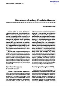
Figure 1. The Death of Washington, an 1896 Sketch in Oil by Howard Pyle (1853–1911). The men in the room are Tobias Lear (kneeling), James Craik (standing), and Gustavus Richard Brown (seated). Martha Washington sits at the foot of the bed. This little-known sketch was published in an 1897 book on George Washington by future president Woodrow Wilson.3 President Warren Harding’s son also researched the medical aspects of Washington’s death.4 Courtesy of the Boston Public Library Print Department.
tea with vinegar. Tilting his head back to drip the mixture down his throat, he nearly suffocated, unable to cough the fluid up. Still alert, he rose and walked about the bedroom, then sat upright in a chair for two hours. Returning to bed, he squirmed to find a comfortable position. Arriving at 3 p.m., Dick argued that further bleeding might weaken Washington. Craik nevertheless ordered a fourth bleeding, with the removal of 32 oz (946 ml) of blood. Brown arrived at 4 p.m., at which time calomel (mercurous chloride) and tartar emetic (antimony potassium tartrate) were administered. Awaiting a therapeutic effect (Fig. 1), the physicians might well have thought about Benjamin Rush, a medical colleague and friend of Washington whose professional fate was being decided that day. Craik had served with Rush in the Revolutionary War, Brown Vol ume 341
The New England Journal of Medicine Downloaded from nejm.org at NIH Libary on October 30, 2015. For personal use only. No other uses without permission. Copyright © 1999 Massachusetts Medical Society. All rights reserved.
Numb e r 2 4
·
1845
The Ne w E n g l a nd Jo u r n a l o f Me d ic i ne
had been his classmate in Edinburgh (class of 1768), and Dick had been his student in Pennsylvania. America’s most famous physician and a signer of the Declaration of Independence, Rush was fighting allegations of medical malpractice. The legal case concerned bloodletting, which Rush championed. Journalist William Cobbett had charged Rush with killing patients. Rush had sued. In their opening statements, the lawyers for the two men traded blood-tinged metaphors. Rush’s lawyer argued, “[A physician’s] reputation is a fabric delicate as air, the slightest gust of popular prejudice or caprice dissipates it. . . . Virtue, bleeding at every pore, calls for justice on her despoiler.”5 Cobbett’s lawyer quoted his client, “The times are ominous indeed, when quack to quack cries purge and bleed.”5 The verdict was scheduled for December 14, as Washington lay dying. After the fourth bloodletting, Washington’s condition improved, and he was able to swallow. He examined his will. Realizing that Sheels had been standing for hours, Washington motioned him to sit down. Around 5 p.m., Washington again sat up in a chair but soon returned to bed and was helped into an upright position. He continued to struggle for air, and his condition began to deteriorate. At 8 p.m., the physicians applied blisters of cantharides to his feet, arms, and legs and then applied wheat-bran cataplasms (poultices) to his throat. His condition deteriorated further. At around 10 p.m., Washington whispered burial instructions to Lear. At 10:20 p.m., George Washington died. Sheels, Branham, Charlotte, and Molly looked on. Craik closed his friend’s eyelids, while Dick stopped the bedroom clock. The body was carried downstairs and laid on a table in the unheated dining room. News of Washington’s death spread quickly. Symbolic funeral services held in hundreds of cities featured elaborate cortèges with empty coffins, riderless horses, and tolling bells.6 Newspapers published heroic poems by grieving women. People made pilgrimages to Mount Vernon. In France, Napoleon ordered the hanging of black crêpe from flags and standards, and the marquis de Fontanes delivered a stirring éloge (official eulogy) at the temple de Mars (Hôtel des Invalides).7 In the American capital, Reverend Richard Allen, minister of the African Methodist Episcopal Church, announced that Washington’s slaves would eventually be free. Americans dressed in black or wore mourning badges for months. But amid the sorrow there was controversy over Washington’s medical care. Rush’s victory in the bloodletting suit, on the day of Washington’s death, could not eliminate popular suspicion that overuse of bloodletting was harmful. Craik and Dick chose a preemptive strike. In an open letter to the nation,8 they attributed Washington’s death to “cynanche trachealis,” reviewing the on1846 ·
set and course of the illness and describing their treatment. Apparently the first justification of a medical practice to the American public, the explanation backfired. That Washington had died at the hands of his physicians was immediately suggested by his friends,9 as well as by American and British medical scholars10,11 and the press.12,13 Some 20th-century authors have charged that he was murdered.14-16 Why was 80 oz (2365 ml) of blood removed in 12 hours, and was such treatment helpful or harmful? The physicians, who did not provide a rationale for this treatment, were nevertheless using accepted “heroic” therapy. They understood that Washington’s condition was inflammatory (subsuming what we now know as infection) and that inflammation was associated with tissue swelling, which in turn was related to transudation. But they lacked modern antiinflammatory therapy. According to some 19th-century historians, Washington’s physicians might have reasoned that because bloodletting caused visible dermal vasoconstriction, it would also constrict the vessels associated with swelling in the windpipe17,18 and that the dehydrating effects of “purging” (with the use of calomel), diaphoresis (with sage tea and subemetic doses of antimony), and blistering (with cantharides) would potentiate the effect. This speculation may reflect the historians’ a posteriori reasoning.19,20 In any case, Washington’s blood eventually became viscous and flowed slowly,1 presumably reflecting dehydration and hypovolemia. Modern physicians would doubt the beneficial effects of such therapy on local inflammatory swelling and would worry that aggressive bleeding might cause weakness and worsen the hypoxia associated with partial airway obstruction; they would also worry that iatrogenic dehydration might lead to electrolyte imbalance. Lacking such modern concepts, Washington’s physicians may have reasoned that with death approaching, “heroic depletion” was their only option. What disorder led to Washington’s death? Dick rejected Craik’s diagnosis of “inflammatory quinsy” and proposed three alternatives: “stridula suffocatis,” “laryngea,” or “cynanche tracheitis [sic]”21; the third, as corrected, eventually prevailed. Cynanche trachealis (literally, “dog strangulation”) was a relatively new diagnostic entity at the time. Beginning in the late 1770s, Brown’s teacher, the great Edinburgh nosologist William Cullen, defined it as “inflammation of the glottis, larynx, or upper part of the trachea . . . a rare occurrence . . . [producing] such an obstruction of the passage of the air, as suffocates, and thereby proves suddenly fatal.”22 Cynanche could not have been unknown to Washington’s physicians. Dick and Craik had been discussing diagnostic possibilities during a “croup” epidemic that winter.21 Moreover, one of the earliest and most authoritative descriptions of cynanche was reported in 1770 by Brown’s own nephew, also named Gusta-
Dec em b er 9 , 19 9 9 The New England Journal of Medicine Downloaded from nejm.org at NIH Libary on October 30, 2015. For personal use only. No other uses without permission. Copyright © 1999 Massachusetts Medical Society. All rights reserved.
OC C A S IONA L NOTES
vus Brown,23 with subsequent reports by Dick’s teacher, Benjamin Rush.24,25 Brown, Craik, and Dick, who probably knew as much about cynanche as any three physicians in the United States, even summoned Brown’s nephew to Washington’s bedside.26 He lived in St. Mary’s County, Maryland, however, and failed to arrive in time. Although historians do not agree on the cause of Washington’s death, the signs and symptoms1,18,19 point to acute bacterial epiglottitis. This diagnosis, proposed first in 183827 and several times since,28,29 is consistent with current clinical and epidemiologic information.30-32 Medical reports during the period from 1776 to 1826 suggest that cynanche trachealis corresponded to the modern diagnosis of bacterial epiglottitis, but the term was probably also used to refer to some cases of laryngeal diphtheria and viral croup. Other suggested diagnoses seem less likely. Quinsy33 causes unilateral neck swelling, which Washington did not have, and is seen almost exclusively in children. Washington had probably been exposed to streptococci as a child34 and had also apparently had diphtheria.35 Laryngeal diphtheria was a slowly progressive disease largely confined to childhood, as it is now, and diphtheria was not prevalent in Virginia in 1799.28,35 Pneumonia, Ludwig’s angina, Vincent’s angina, and other proposed diagnoses have largely been ruled out.28,36-38 Could Washington have survived epiglottitis? Dick, overruled in his opposition to bloodletting, next argued for tracheotomy.21,39 In 1799, even elective tracheotomy, let alone tracheotomy performed on an emergency basis, was rarely undertaken. It is improbable that, at the time of Washington’s illness, tracheotomy had not been performed in the United States, as has been claimed,40 although a workable procedure had been described in surgical detail only the year before.41 Undoubtedly, the specter of failure with a grisly, painful (in the absence of anesthesia), and untried surgical experiment on the former president weighed heavily in Craik’s decision to veto this radical suggestion. One historian has defended Craik by arguing that tracheotomy with the patient in the supine position would have led to positional ball-and-valve airway closure and rapid death.42 But Dick’s later comments on tracheotomy specified the upright position.39 Tracheotomy may have been the only lifesaving option left, but it was not attempted. After Washington’s death, his physicians spent the night at Mount Vernon. In the morning, Dick measured the frozen corpse; it was 1.9 m (6 ft, 31/2 in.) long. Craik declined payment but recommended that Lear pay each of the other two physicians $40 (about $375 in 1999 dollars), after which they left. Several hours later, the last physician who had been summoned arrived. William Thornton, a physician trained in Edinburgh and a family friend, had been called
the previous evening, as Washington’s illness became critical. Thornton had rushed to Mount Vernon with the same idea as Dick: to perform an emergency tracheotomy. Too late, Thornton still hoped that Washington might be in a suspended state from which he could be aroused. After conducting a careful examination of the corpse, Thornton proposed that the body be thawed gradually, first in cool water and then with warm blankets and rubbing of the skin, with the subsequent performance of a tracheotomy, artificial respiration at the tracheotomy site, and transfusion of lamb’s blood.43 Although Martha Washington must have known that her husband had once revived a frozen slave thought to be dead, she refused this proposal. Thornton and Craik persuaded the family to encase the coffin in lead because of the risk of communicable disease.9,44 At the funeral, one of Washington’s closest friends, Bryan, Lord Fairfax, “caught” a cynanche-like disease. He attributed his survival to copious bloodletting.45 EPILOGUE
James Craik apparently never again spoke about the events of December 14. But he did have second thoughts about declining payment, submitting to the estate on December 24 a bill for the same fee Brown and Dick had received at his suggestion. He was also bequeathed Washington’s valuable tambour secretary and circular chair. Craik, who had named one of his sons George Washington, attended the death of Martha Washington two years later. He died in 1814. One of his grandsons, William Craik, became a U.S. congressman. Meeting over the holidays, Gustavus Richard Brown praised Elisha Cullen Dick and said that he wished they had heeded his advice about bloodletting.26,46 Dick, who initially talked of “putting [away] his lancet forever” to become a nurse,26 was less charitable to his colleagues, later criticizing Brown explicitly and Craik implicitly.21,39 Brown made no further comments about Washington’s treatment. Gustavus Brown died in 1801, and Gustavus Richard Brown in 1804. Dick seems never to have given up revisiting the events of December 14. Despite his strenuous arguments against bloodletting and in favor of tracheotomy, he later reversed himself, arguing that in patients with cynanche, bloodletting ad deliquium (to the point of syncope) was so effective it removed the need for tracheotomy.39 Later still, his preferred treatment regressed to a “strong toddy” with red pepper. Dick and Rush became national experts on bloodletting as a treatment for cynanche and other diseases, ignoring evidence against its use.47 Dick, who became mayor of Alexandria, Virginia, in 1804, remained devoted to Washington’s memory, spearheading both a movement to make his birthday a national holiday and the erection of a national monument. He died in 1825. Vol ume 341
The New England Journal of Medicine Downloaded from nejm.org at NIH Libary on October 30, 2015. For personal use only. No other uses without permission. Copyright © 1999 Massachusetts Medical Society. All rights reserved.
Numb e r 2 4
·
1847
The Ne w E n g l a nd Jo u r n a l o f Me d ic i ne
His grandson, James Alfred Pearce, became a U.S. senator. Benjamin Rush’s legal victory invited further attack. His tormentor, Cobbett, subsequently accused Rush’s pupil, Dick, of causing Washington’s death. Chased by Rush’s son, Cobbett escaped a duel and fled to England, where he became a member of Parliament. Rush, who gave the proceeds from his $5,000 judgment ($47,000 in 1999 dollars) to charity, died in 1813. He is remembered today as one of America’s greatest physicians, a father of psychiatry, and a founder of the liberal humanist tradition in American medicine. William Thornton pursued a career as an inventor and architect, designing the nation’s first Capitol and developing the city of Washington. He also directed the patent office, wrote a seminal work on teaching deaf–mute persons, codeveloped the first steamboat, and was involved in many social causes. He died in 1828. Washington’s will specified that on his wife’s death, the slaves he owned at Mount Vernon (about half the total number) were to be freed. Persuaded by family members that this provision of the will might provoke a slave to murder her, Martha Washington freed them all. Sadly, Sheels, Branham, Charlotte, and Molly had been owned by Mrs. Washington, who was prevented by inheritance laws from freeing them. On her death in 1802, they and the remaining slave families were dispersed according to those laws. Around 1830, as historian Jared Sparks prepared to write his biography of Washington,48 he tracked down Caroline Branham, who was at that time owned by Washington’s grandson. The elderly woman gave Sparks the last eyewitness account of George Washington’s death in exchange for the freedom of her enslaved grandson.49 As a free man, the grandson, Robert Robinson, left a menial job in a cracker bakery, educated himself, and moved to Alexandria, near Mount Vernon, where his grandmother had lived as Washington’s slave. Appointed minister of the Methodist Church on South Washington Street, he became an influential African-American leader. In considering the final illness of George Washington, it is worth remembering that he received prompt and expert medical care that reflected thencurrent concepts. In questioning his physicians’ treatment decisions, we should also reflect on the balance between art and science in medicine, especially in the context of modern therapy for diseases whose pathogenesis and natural history are poorly understood (e.g., atherosclerosis and diabetes mellitus). In 1999, the treatment of many medical conditions still lacks a sound scientific or empirical basis. Advances in science permit us to uncover pitfalls in prior medical practice but do not by themselves advance the art. Thus, physicians must not only continue to develop the science of medicine but also maintain and strengthen its problem-solving aspects and practice as an art. 1848 ·
The last 16 hours of Washington’s life must have been agonizing as he fought for air, unable to find a comfortable position. His chief concern was apparently that his physicians “enable him to die easy.”50 Though not a Christian, he must have been impatient to reach a “hereafter” with as little trouble as possible. According to Lear’s notes, at the very end, Washington settled back in bed and appeared calm. His last act in life was a medical one: he felt his own pulse, a practice that he had probably picked up in ministering to his slaves and family. Not even his physicians learned the result as his fingers slipped from his wrist and his breathing stopped. DAVID M. MORENS, M.D. University of Hawaii Honolulu, HI 96822 A bibliography (244 entries) on the death of George Washington and related subjects addressed in this report is available from the author on request. (Please provide a complete address, including e-mail address.)
I am indebted to Mary V. Thompson and Barbara McMillan of the Mount Vernon Ladies’ Association of the Union; Stephen J. Greenberg, Elizabeth Tunis, and the staff of the History of Medicine Division, National Library of Medicine; George K. Combs of the Lloyd House, Alexandria Library; Virginia M. Tanji of the University of Hawaii; Erlinda Tacadena of Tripler Army Medical Center; Ratna Soetjahja Morens; and numerous staff members at the Library of Congress for research assistance; to Robert J. Littman for translating important passages from the Latin; and to Philip K. Wilson and Peter R. Henriques for valuable suggestions on the manuscript.
REFERENCES 1. Lear T. Tobias Lear’s narrative accounts of the death of George Washington. In: Twohig D, Chase PD, Runge BH, et al., eds. The papers of George Washington. Retirement series 4: April–December 1799. Charlottesville: University Press of Virginia, 1999:542-55. 2. A comparative critique of Washington’s last illness. In: Carroll JA, Ashworth MW. George Washington. Vol. 7. First in peace. New York: Charles Scribner’s Sons, 1957:637-53. (Appendix VII-2.) 3. Wilson W. George Washington. New York: Harper & Brothers, 1897: (plate illustration follows page 304). 4. Harding WG II. Oral surgery and the presidents — a century of contrast. J Oral Surg 1974;32:490-3. 5. (Taken in shorthand by T. Carpenter.) A report of an action for a libel, brought by Dr. Benjamin Rush, against William Cobbett, in the Supreme Court of Pennsylvania, December term, 1799, for certain defamatory publications in a news-paper, entitled Porcupine’s Gazette, of which the said William Cobbett was editor. Philadelphia: W.W. Woodward, 1800. 6. George Washington’s invisible corpse and the beaver hat. In: Laderman G. The sacred remains: American attitudes toward death, 1799–1883. New Haven, Conn.: Yale University Press, 1996:15-21, 180. 7. Fontanes L-J-P. Éloge funèbre de Washington, prononcé dans le temple de Mars, le 20 pluviôse, an 8. Paris: H. Agasse, 1800. 8. Craik J, Dick EC. From “The Times [and District of Columbia Advertiser],” a newspaper printed in Alexandria (Virginia), dated in December, 1799. Med Repos 1800;3:311-2. 9. Boyd TM. Death of a hero, death of a friend: George Washington’s last hours. Virginia Cavalcade 1984;33:136-43. 10. Brickell J. Observations on the medical treatment of General Washington, in his last illness, addressed to his physicians Messrs. Craik and Dick. In: Porcupine P. The Rush-Light. No. II. New York: William Cobbett, February 28, 1800:81-5. 11. Reid J. Observations on the medical treatment of General Washington’s last illness. Med Phys J 1800;3(January–June):473-5. 12. A note to Dr. Dick. In: Porcupine P. The Rush-Light. No. II. New York: William Cobbett, February 28, 1800:85-6. 13. Porcupine P. The American Rush-Light; by the help of which, wayward and disaffected Britons may see a complete specimen of the baseness,
Dec em b er 9 , 19 9 9 The New England Journal of Medicine Downloaded from nejm.org at NIH Libary on October 30, 2015. For personal use only. No other uses without permission. Copyright © 1999 Massachusetts Medical Society. All rights reserved.
OC C A S IONA L NOTES
dishonesty, ingratitude, and perfidy of Republicans, and of the profligacy, injustice, and tyranny of Republican governments. London: J. Wright, 1800. 14. Lloyd JU. Who killed George Washington? Eclectic Med J 1923;83: 353-6, 403-8, 453-6. 15. Marx R. A medical profile of George Washington. American Heritage 1955;6:43-7, 106-7. 16. Pirrucello F. How the doctors killed George Washington. Chicago Tribune Magazine. February 20, 1977. 17. Jackson J. Memoir on the last sickness of General Washington and its treatment by the attendant physicians. (Privately printed), 1860. 18. Solis-Cohen S. Washington’s death and the doctors. Lippincott’s Monthly Magazine 1899;64:945-52. 19. Reflections on blood-letting. In: King LS. Medical thinking: a historical preface. Princeton, N.J.: Princeton University Press, 1982:227-44, 327. 20. Therapeutic change. In: Warner JH. The therapeutic perspective: medical practice, knowledge, and identity in America, 1820–1885. Cambridge, Mass: Harvard University Press, 1986:83-161. 21. Nydegger JA. The last illness of George Washington. Med Rec 1917; 92:1128. 22. Of the quinsy, or cynanche. In: Cullen W. First lines of the practice of physic. Vol. 1. Chapter 5. Edinburgh, Scotland: C. Elliott, 1784:278-306. 23. Brown G. Disputatio medica inauguralis de cynanche phlogistica. Edinburgi, Scotland: Balfour, Auld, et Smellie, 1770. 24. Rush B. (Unfound newspaper article on cynanche trachialis [sic]). In: Corner GW, ed. The autobiography of Benjamin Rush: his “Travels through Life” together with his Commonplace Book for 1789-1813. Westport, Conn.: Greenwood Press, 1948:82. 25. Observations on the cynanche trachealis. In: Rush B. Medical inquiries and observations. Vol. 2. 3rd ed. Philadelphia: Thomas and William Bradford et al., 1809:373-81. 26. Washington’s physicians, diseases and death. In: Blanton WB. Medicine in Virginia in the 18th century. Richmond, Va.: Garrett & Massie, 1931: 297-312. 27. Marsh H. Cases of acute inflammation confined to the epiglottis. Dublin J Med Sci 1838;13:1-23. 28. Wells WA. Last illness and death of Washington. Virginia Medical Monthly 1927;53:629-42. 29. Estes JW. George Washington and the doctors: treating America’s first superhero. Med Heritage 1985;1:44-57. 30. Frantz TD, Rasgon BM, Quesenberry CP Jr. Acute epiglottitis in adults: analysis of 129 cases. JAMA 1994;272:1358-60. 31. Berg S, Trollfors B, Nylén O, Hugosson S, Prellner K, Carenfelt C. Incidence, aetiology, and prognosis of acute epiglottitis in children and adults in Sweden. Scand J Infect Dis 1996;28:261-4. 32. Trollfors B, Nylén O, Carenfelt C, et al. Aetiology of acute epiglottitis in adults. Scand J Infect Dis 1998;30:49-51. 33. Of the death, funeral, and family. In: Prussing EE. The estate of George Washington, deceased. Boston: Little, Brown, 1927:17-23.
34. Katz AR, Morens DM. Severe streptococcal infections in historical perspective. Clin Infect Dis 1992;14:298-307. 35. Willius FA, Keys TE. The medical history of George Washington (1732-1799). II. Proceedings of Staff Meetings of the Mayo Clinic 1942; 17:107-12. 36. Brown MW. The famous controversy about Washington’s last illness. Med J Rec 1932;135:39-41. 37. Barker C. A case report. Yale J Biol Med 1936/1937;9:185-7. 38. George Washington: 1732-1799. In: Wold KC. Mr. President — how is your health? St. Paul, Minn.: Bruce Publishing, 1948:1-17. 39. Dick EC. Facts and observations relative to the disease of cynanche trachealis, or croup. Philadelphia Med Physical J 1809(May, Suppl 3):242-55. 40. Reece RL. George Washington: his death and his doctors. Minnesota Med 1966;49:1185-90. 41. Desault PJ. Mémoire sur la bronchotomie & sur les moyens d’y suppléer en certains cas. In: Bichat M-F-X, ed. Oeuvres chirurgicales de P.J. Desault, chirurgien en chef du grand Hospice d’Humanité, ci-devant HôtelDieu de Paris; ou tableau de sa doctrine & de sa pratique dans le traitement des maladies externes. Seconde Partie. Maladies des parties molles. Paris: C.Ve. Desault, 1798:212-53. 42. Scheidemandel HH. Did George Washington die of quinsy? Arch Otolaryngol 1976;102:519-21. 43. Thornton W. (Untitled manuscript catalogued as “Miscellaneous writing on sleep.”) Library of Congress, The Papers of William Thornton, document page 3057. 44. Idem. (Untitled letter to John Marshall, dated from the City of Washington, January 2, 1800.) Library of Congress, The Papers of William Thornton, document pages 378-9. 45. Fairfax B. (Untitled letter, probably to the Earl of Buchan, dated from Mount Eagle, near Alexandria, Virginia, 18 January 1800.) Original in the Bodleian Library, Oxford, England. Accession no. RM-100. Catalog no. PS-2261. (Photostat copy at the Mount Vernon Ladies’ Association of the Union, Mount Vernon, Va.) 46. Brown GR. (Letter designated “Dr. Brown to Dr. Craik,” January 2, 1800.) In: Ford WC, ed. Last illness and death: the writings of George Washington. Vol. 14. New York: G.P. Putnam’s Sons, 1893:243-67[257f ]. 47. Smith TW. Observations on the medical treatment of the croup. Philadelphia Medical Museum 1808;4:31-5. 48. Sparks J. The life of George Washington. Boston: Ferdinand Andrews, 1839. 49. The Custis family. In: Powell MG. The history of old Alexandria, Virginia, from July 13, 1749 to May 24, 1861. Book II. Richmond, Va.: William Byrd Press, 1928:241-9. 50. Corner GW, ed. The autobiography of Benjamin Rush: his “Travels through Life” together with his Commonplace Book for 1789-1813. Westport, Conn.: Greenwood Press, 1948:245-9. ©1999, Massachusetts Medical Society.
Vol ume 341 The New England Journal of Medicine Downloaded from nejm.org at NIH Libary on October 30, 2015. For personal use only. No other uses without permission. Copyright © 1999 Massachusetts Medical Society. All rights reserved.
Numb e r 2 4
·
1849
The Ne w E n g l a nd Jo u r n a l o f Me d ic i ne
INFORMATION FOR AUTHORS These guidelines are in accordance with the “Uniform Requirements for Manuscripts Submitted to Biomedical Journals.” (The complete document appears in N Engl J Med 1997;336:309-15.) MANUSCRIPTS Manuscripts containing original material are accepted for consideration if neither the article nor any part of its essential substance, tables, or figures has been or will be published or submitted elsewhere before appearing in the Journal. This restriction does not apply to abstracts or press reports published in connection with scientific meetings. Authors should submit to the Editor copies of any published papers or other manuscripts in preparation or submitted elsewhere that are related to the manuscript to be considered by the Journal. The Journal discourages the submission of more than one article dealing with related aspects of the same study. Submit an original manuscript with one set of original figures and two copies of the complete manuscript. Manuscripts must be no longer than 3000 words. Please supply a word count (not including abstract or references). Use standard-sized paper, and triple-space throughout. Address all submissions to the Editor, New England Journal of Medicine, 10 Shattuck St., Boston, MA 02115-6094. A covering letter signed by all authors should identify the person (with the address and telephone number) responsible for negotiations concerning the manuscript; the letter should make it clear that the final manuscript has been seen and approved by all authors and that they have taken due care to ensure the integrity of the work. At least one person’s name must accompany a group name — e.g., Thelma J. Smith, for the Boston Porphyria Group. As stated in the Uniform Requirements (see above), credit for authorship requires substantial contributions to: (a) conception and design, or analysis and interpretation of data; and (b) the drafting of the article or critical revision for important intellectual content. If more than 12 authors are listed for a multicenter trial, or more than 8 for a study from a single institution, each author must sign a statement attesting that he or she fulfills the authorship criteria of the Uniform Requirements. No more than 12 names will be listed under the title; other names will appear in a footnote. Acknowledgments will be limited to a column of Journal space, and those acknowledged will be listed only once. (See editorial, Nov. 21, 1991, issue.) LETTERS TO THE EDITOR See the Journal correspondence section. CONFLICT OF INTEREST Authors of research articles should disclose at the time of submission any financial arrangement they may have with a company whose product figures prominently in the submitted manuscript or with a company making a competing product. Such information will be held in confidence while the paper is under review and will not influence the editorial decision, but if the article is accepted for publication, the editors will usually discuss with the authors the manner in which such information is to be communicated to the reader. Because the essence of reviews and editorials is selection and interpretation of the literature, the Journal expects that authors of such articles will not have any financial interest in a company (or its competitor) that makes a product discussed in the article. COPYRIGHT Authors agree to execute copyright transfer forms as requested. Copyright in any contribution is owned by the Massachusetts Medical Society. The Society and its licensees have the right to use, reproduce, transmit, derivate, publish, and distribute the contribution, in the Journal or otherwise, in any form or medium. Authors will not use or authorize the use of the contribution without the Society’s written consent, except as may be allowed by U.S. fair use law. UNITS OF MEASUREMENT Authors should express all measurements in conventional units, with Système International (SI) units given in parentheses throughout the text. Figures and tables should use conventional units, with conversion factors given in legends or footnotes. In accordance with the Uniform Requirements, however, manuscripts containing only SI units will not be returned for that reason. TITLES AND AUTHORS’ NAMES With the manuscript, provide a page giving the title of the paper; titles should be concise and descrip-
1850 ·
tive (not declarative). Also include a running head of fewer than 40 letter spaces; the name(s) of the author(s), including the first name(s) and no more than two graduate degrees; the name of the department and institution in which the work was done; the institutional affiliation of each author; and the name and address of the author to whom reprint requests should be addressed. Any grant support that requires acknowledgment should be mentioned on this page. ABSTRACTS Provide on a separate page an abstract of not more than 250 words. This abstract should consist of four paragraphs, labeled Background, Methods, Results, and Conclusions. They should briefly describe, respectively, the problem being addressed in the study, how the study was performed, the salient results, and what the authors conclude from the results. KEY WORDS Three to 10 key words or short phrases should be added to the bottom of the abstract page; these will help us index the article and may be published with the Abstract. Use terms from the Medical Subject Headings from Index Medicus. REFERENCES References must be triple-spaced and numbered consecutively as they are cited. References first cited in tables or figure legends must be numbered so that they will be in sequence with references cited in the text. The style of references is that of Index Medicus. List all authors when there are six or fewer; when there are seven or more, list the first three, then “et al.” The following is a sample reference: 1. Lahita R, Kluger J, Drayer DE, Koffler D, Reidenberg MM. Antibodies to nuclear antigens in patients treated with procainamide or acetylprocainamide. N Engl J Med 1979;301:1382-5.
Numbered references to personal communications, unpublished data, and manuscripts either “in preparation” or “submitted for publication” are unacceptable (see “Permissions”). If essential, such material may be incorporated in the appropriate place in the text. TABLES Double-space tables and provide a title for each. If an article is accepted, the Journal will arrange to deposit extensive tables of important data with the National Auxiliary Publications Service (NAPS); we will pay for the deposit and add an appropriate footnote to the text. This service makes available microfiche or photocopies of tables at moderate charges to those who request them. ILLUSTRATIONS Figures should be professionally designed. Symbols, lettering, and numbering should be clear and large enough to remain legible after the figure has been reduced to fit the width of a single column. The back of each figure should include the sequence number, the name of the author, and the proper orientation (e.g., “top”). Do not mount the figure on cardboard. Photomicrographs should be cropped to a width of 8 cm, and electron photomicrographs should have internal scale markers. If photographs of patients are used, either the subjects should not be identifiable or their pictures must be accompanied by written permission to use the figure. Permission forms are available from the Editor. Legends for illustrations should be triple-spaced on a separate sheet and should not appear on the illustrations. Color illustrations are encouraged. Send both transparencies and prints for this purpose. ABBREVIATIONS Except for units of measurement, abbreviations are discouraged. Consult Scientific Style and Format: The CBE Manual for Authors, Editors, and Publishers (Sixth edition. New York: Cambridge University Press, 1994) for lists of standard abbreviations. The first time an abbreviation appears it should be preceded by the words for which it stands. DRUG NAMES Generic names should generally be used. When proprietary brands are used in research, include the brand name in parentheses in the Methods section. PERMISSIONS Materials taken from other sources must be accompanied by a written statement from both author and publisher giving permission to the Journal for reproduction. Obtain permission in writing from at least one author of papers still in press, unpublished data, and personal communications. REVIEW AND ACTION Manuscripts are examined by the editorial staff and are usually sent to outside reviewers. We encourage authors to suggest the names of possible reviewers, but we reserve the right of final selection. Only one copy of rejected manuscripts will be returned, usually within six weeks. Decisions about potentially acceptable manuscripts may take longer.
Dec em b er 9 , 19 9 9 The New England Journal of Medicine Downloaded from nejm.org at NIH Libary on October 30, 2015. For personal use only. No other uses without permission. Copyright © 1999 Massachusetts Medical Society. All rights reserved.