Advances in Biological Research 11 (4): 202-211, 2017 ISSN 1992-0067 © IDOSI Publications, 2017 DOI: 10.5829/idosi.abr.2017.202.211
Ebola Virus Disease: Recent Advances 1
1
Isha Mishra, 1Raghav Mishra, 2Sanjiv Mishra, 2Anuradha Mishra and 3Prashant Dhakad
Department of Pharmacy, Shri Ram Murti Smarak College of Engineering and Technology, Bareilly, Uttar Pradesh, India (243202) 2 SRM State Ayurvedic College and Hospital, Bareilly, Uttar Pradesh, India (243001) 3 Department of Pharmacy, School of Medical and Allied Sciences, Galgotias University, Greater Noida, Uttar Pradesh, India (201308)
Abstract: The multifaceted nature of Ebola sickness and its flare-ups has highlighted the need to enquire the characteristics of Ebola virus Disease including ebb and flow comprehension of the transmission progression and the impact of control intercessions against its transmission. As indicated by the accessible information, boundaries to counteract and control the illness in influenced nations incorporate faltering and disordered wellbeing frameworks, substandard clean conditions, poor individual hygiene practices and false convictions and disgrace identified with Ebola Virus Disease. This review is correlative to past surveys and centers to review the episodes, method of transmission, clinical signs, treatment and aversion of Ebola Virus Disease. Key words: Ebola virus
Outbreak
Classification
Virus reservoir
INTRODUCTION
Symptoms
Treatment
explanation has been advanced, these outbreaks have been credited to interruption into or impedance with already little frequented territories, because of population pressure and agricultural developments [1]. Ebola virus (EBOV) and Marburg virus (MARV), members of the Filoviridae virus family are among the most virulent pathogens in humans [3]. The Zaire species of Ebola virus is the causative agent of the 2014-2015 epidemic in West Africa, in which the case casualty rate has been accounted for to be as high as 70 percent rates, in prior flare- ups have reached 80 to 90 percent [4]. Marburg virus has brought about a comparable ailment in a littler number of outbreaks in Central Africa [5].
During the past 20 years or more, there have been a number of severe eruptions of several diseases apparently "new" to medication. [1] Around 41 new developing infectious life forms or strains have bounced from their animal hosts into people and it is almost certain that other beforehand obscure irresistible diseases will rise into the human population sooner rather than later. An emerging disease is an infectious disease that has recently showed up in a population, or is quickly expanding in occurrence or geographic range. There are roughly 1, 407 living beings (fungi, bacteria, parasites, protozoa and viruses) that can infect humans. Approximately 58% of these are thought to be animal diseases and the vast majority of these have an Old- World origin as an aftereffect of man’s advancement of agriculture and animal domestication [2]. Great cases are the underlying episodes of Kyasanur forest disease, O'nyong nyong fever, Bolivian hemorrhagic fever, Marburg and Ebola fevers. To some extent their acknowledgement can be accounted for by improved communications, both general and medical (through WHO (World Health Organization) and other international channels), but in almost every case where an
Fig. 1: Ebola Zaire Filovirus [2]
Corresponding Author: Isha Mishra, Department of Pharmacy, Shri Ram Murti Smarak College of Engineering and Technology, Bareilly, Uttar Pradesh, India (243202).
202
Advan. Biol. Res., 11 (4): 202-211, 2017
Ebola virus is a non- segmented, negative- sense, single stranded RNA virus that looks like Rhabdoviruses (like rabies) and Paramyxoviruses (like measles, mumps) in its genome organization and replication mechanism. It is a member of the family Filoviridae, taken from the Latin word "filum" meaning string- like, based upon their filamentous structure. Ebola virus illness brings the most alarming of infectious disease syndromes to mind. Perpetually, Ebola is a extreme ailment caused by Ebola virus. It is highly infectious, rapidly fatal, with a demise rate of up to 90%, however can be prevented. It is spread through direct contact with body fluids like blood, salivation, urine, sperm and so forth of an infected person and by contact with contaminated surfaces or equipment, including linen soiled by body fluids from an infected person. The Ebola virus can be relatively easily disposed of with heat, alcohol-based products and sodium hypochlorite (bleach) or calcium hypochlorite (bleaching powder) at appropriate concentrations [6]
lower case- casualty rate (around 30 percent) than is typical for the Zaire and Sudan viruses. Sequencing has demonstrated that the agent is most firmly identified with the Ivory Coast species. The fifth Ebola species, the Reston virus, contrasts particularly from the others, since it is obviously maintained in an animal reservoir in the Philippines and has not been found in Africa [8-18]. The Ebola Reston virus was found when it brought about an outbreak of lethal infection in macaques imported into the United States in 1989 [6]. Outbreak of Ebola Virus Disease: Ebola first appeared in 1976 in two concurrent outbreaks, in Nzara, Sudan and in Yambuku, Democratic Republic of Congo. The last was in a village situated closed to the Ebola River, from which the disease takes its name [1]. The initially perceived filovirus outbreak was in Europe with the Marburg virus in 1967. This occurred in Germany and Yugoslavia among vaccine workers handling tissue specimens from imported African green monkeys. Thirty-one individuals were infected, with a 23% fatality rate. The virus was named after the German town of Marburg, where the German outbreak occurred [2]. In 1990, a mysterious episode of fatal illness occurred in Philippine imported Cynomolgus crab-eating macaque monkeys at a primate holding facility in Reston. Researchers in the end decided this was a strain of EBOV of asian origin and they assigned it as the Reston Ebola virus (REBOV). REBOV is the only species of EBOV that does not bring about disease in humans and it has given major insights to the pathogenesis of the lethal Ebola virus strains [19-21]. Repeated episodes and new strains of EBOV include the Ivory Coast ebola virus, named for a solitary human case that occurred in 1994. [22]. Outbreaks have additionally destroyed mountain gorilla populations and numerous EBOV outbreaks have occurred in Gabon somewhat around 1994 and 1996 as well as small outbreaks in other parts of Africa [23-26]. In 2007 another strain of EBOV rose in Western Uganda in the township of Bundibugyo [27]. This denoted the discovery of a fifth strain of the virus, the Bundibugyo Ebola virus. This outbreak kept going 2 months, with 149 speculated cases and 37 deaths. On March 21, 2014, an extensive outbreak initiated in Guinea, Liberia, Nigeria and Sierra Leone. This was the first expansive outbreak in West Africa [28].
Classification: The Ebola fever is caused by members of a family of filoviruses called Ebola virus and there are five distinct species: Bundibugyo ebolavirus, Tai Forest ebolavirus, Reston ebolavirus, Sudan ebolavirus and Zaire ebolavirus [6] Before, Ebola and Marburg viruses were classified as "hemorrhagic fever viruses", based upon their clinical symptoms, which include coagulation defects, bleeding and shock [7] The accompanying four species cause disease in humans: The Zaire virus, since it was initially perceived in 1976, has caused various expansive outbreaks in Central Africa, with death rates ranging from 55 to 88 percent. It is the causative agent of the 2014-2015 West African Ebola epidemics. The Sudan virus has been connected with a casefatality rate of around 50 percent in four epidemics: two in Sudan in the 1970s, one in Uganda in 2000 and another in Sudan in 2004. The Ivory Coast virus has only been identified as the cause of illness in one individual and that individual survived the exposure happened when an ethologist performed a necropsy on a chimpanzee discovered dead in the Tai Forest, where marked decrease in the great ape population had been observed. The Bundibugyo virus rose in Uganda in 2007, creating an outbreak of Ebola virus disease with a 203
Advan. Biol. Res., 11 (4): 202-211, 2017
Fig. 2: Classification of Ebola virus During EBOV’s known history, a few secondary cases have entered South Africa (with one health worker demise), Europe and most as of late the U.S. The current outbreak of Ebola Virus Disease (EVD) in West Africa (Guinea Conakry, Liberia and Sierra Leone) is creating anxiety, fear and frenzy for communities in these three countries, the deadliest in years with the larger part of passing’s happening in Guinea and Sierra Leone. It is spreading speedier then health services can cope, with numerous individuals escaping administrations, refusing to turn bodies over to internment groups and government frameworks neglecting to react when called upon. Individuals escape to regions they regard safe, conveying the virus and its consequences with them. Furthermore, profoundly established cultural practices, for example those around death and burial, can add to transmission. The WHO is leading the response to the outbreak, including tending to zones of social mobilization and risk communication. This study was done to better comprehend the local beliefs and practices likely to enhance or hinder efforts to react to the outbreak in Liberia [29].
Ebola virus sequences, not infectious virus, have been recognized in samples collected from bats in Central Africa [33] Of 24 plants and 19 vertebrate species experimentally inoculated with EBOV, only bats showed no clinical indications of disease, which is considered confirmation that these bats are a reservoirs species of EBOV [30]. Mode of Transmission: Infections with Ebola virus are intense. There is no carrier state. Since the natural reservoir of the virus is obscure, the way in which the virus first shows up in a human at the start of an outbreak has not been resolved. In any case, researchers have hypothesized that the first patient becomes infected through contact with an infected animal [34]. In Africa, infection has been reported through the handling of infected chimpanzees, gorillas, fruit bats, monkeys, forest antelope and porcupines found sick or dead or in the rainforest [5]. Epidemics of Ebola virus disease are for the most part through to start when an individual becomes infected through contact with the meat or body fluids of an infected animal. Once the patient turns out to be sick or dies, the virus then spreads to other people who come into direct contact with the infected individual’s blood, skin or other body fluids. Considers in laboratory primates have found that animals can be infected with Ebola virus through droplet inoculation of virus into the animal mouth or eyes suggesting that human infection can came about because of the accidental transfer of virus to these locales from contaminated hands.
Virus Reservoir: The natural reservoir for Ebola has not recognized until today; however, bats are considered to the most likely candidate species [30] Fruit bats of the Pteropodidae family are thought to be the natural host of the Ebola virus [31] Three types of fruit bats (Hypsignathus monstrous, Epomops franqueti and Myonycteris torquata) were found to conceivably carry the virus without getting ill [30] Marburg virus has been isolated forwardly from bats captured in Uganda [32] only 204
Advan. Biol. Res., 11 (4): 202-211, 2017
Fig. 3: Mode of Transmission of Ebola virus [36] Symptoms: Approximately 95% of the patients show up signs within 21 days after exposure which is the suggested period for follow-up of contacts [8]. In general, blood samples begin to be tried positive by polymerase chain reaction based diagnostics one day before symptoms onset. Typical features include fever, profound weakness, diarrhea, abdominal pain, cramping, nausea and vomiting for 3-5 days and perhaps persevering for up to a week [48]. Some days later, ordinarily starts five to seven days after the primary symptoms victims can begin to bleed through the eyes, nose, or mouth. A hemorrhagic rash can develop on the whole body, which also bleeds. Bleeding happens most commonly in the gastrointestinal tract and may exhibit as melena, petechiae, conjunctival hemorrhage, hematuria, easy bruising, or intraperitoneal bleeding. Mucous membrane bleeding, failure of venipuncture sites and excessive clot formation have additionally been described [49]. Muscle pain and swelling of the pharynx likewise happen in most victims. The most well known signs and symptoms reported from West Africa during the current outbreak from indication onset to the time the case was detected include: fever (87%), fatigue (76%), vomiting (68%), diarrhea (66%) and loss of appetite (65%). Clinical symptoms and chemical laboratory tests confirm multi-organ association. Most common hematological changes are leucopenia and lymphopenia, with a particular diminished neutrophil count and an increase in liver enzymes. With progression of the disease, EVD patients develop thrombocytopenia, lengthening of the pro-thrombin time and activated partial
Ebola is spread through direct contact (through broken skin or unprotected mucous membranes in, for instance, the eyes, nose or mouth) with Blood or body fluids (counting however not restricted to feces, saliva, sweat, urine, vomit, breast milk and semen) of a person who is sick with Ebola, Objects (like needles and syringes) that have been defiled with the virus, Infected fruit bats or primates (apes and monkeys) and Perhaps from contact with semen from a man who has recovered from Ebola (for example, by having oral, vaginal, or anal sex) Ebola is not spread through the air or by water, or in general, by food. Nonetheless, in Africa, Ebola might be spread as an aftereffect of handling bushmeat (wild animals hunted for food) and contact with infected bats. There is no proof that mosquitoes or other insects can transmit Ebola virus. Only a few species of mammals (for example: humans, monkeys and apes) have shown the capacity to become infected with and spread Ebola virus [35]. Clinical Manifestations: Perceiving the signs of EVD is challenging [37]. The incubation period and duration of the disease course of Ebola virus disease fluctuates between 2-21 days with an average of 4-10 days [38]. 205
Advan. Biol. Res., 11 (4): 202-211, 2017 Table 1: Summary of Organ System involved in EBOLA S.No.
Organ System
Tissue Damage
Cellular Targets
Clinical Symptoms
1
Central Nervous System [39]
Vascular endothelium
Endothelial cells
Enhanced vascular permeability through damage to endothelium and macrophage intervened vascular permeability, hypotension
2
Genitourinary tract [40]
Renal proximal tubular epithelium, bladder, epididymides, testes
Epithelial cells, interstitial cells of Leydig, endothelial cells
Intense kidney injury, tubular necrosis, renal failure
3
Gastrointestinal System [41-42]
Tongue epithelium, esophagus, small and large intestine, salivary ducts, gastric glands, duodenal Brunner’s glands
Epithelial cells, glandular epithelial cells
Nausea, vomiting, diarrhea, dehydration, hypovolemia, hypotension
4
Endocrine System [43]
Adrenal glands: focal degeneration and necrosis of cortical cells in zona glomerulosa and zona fasciculate
Adrenal cortical cells, endothelial cells lining sinusoids, fibroblast-like cells between adrenal cortical cells of the zona glomerulosa and fibroblastic tissue of the capsule
Adrenal inadequacy, shock, hypotension
5
Pulmonary System [44]
Alveolar capillaries, respiratory epithelium of trachea and larynx
Alveolar and interstitial macrophages, bronchial and bronchiolar epithelial cells, pneumocytes
Tachypnea, Dyspnea, Respiratory Failure
6
Hepatic System [45-46]
Hepatocellular vacuolar change, degeneration, necrosis
Hepatocytes, Kupffer cells, sinusoidal endothelial cells
Hepatic inadequacy, hepatic failure, coagulopathies, bleeding
7
Immunologic System [47]
Dendritic cells, monocytes, macrophages
Immune dysregulation, Immunosuppression
Fig. 4: Symptoms of Ebola 206
Advan. Biol. Res., 11 (4): 202-211, 2017
thromboplastin time. The extending of the clotting time together with the observed increase in fibrin degradation products suggest a consumptive coagulopathy due to disseminated intravascular coagulation. These patterns are the same for all countries [8]. These symptoms advance over the time and patients experience the ill effects of dehydration, stupor, confusion, hypotension and multi-organ failure, leading to fulminant shock and eventually death. Fatal cases have a tendency to grow early clinical signs during the infection and death frequently occurs between the sixth and sixteenth days of illness [50]. Le Patients die due to shock, haemorrhage and multi-organ failure. If patients recover, clinical improvement emerges simultaneously with the development of the antibody response. In lethal cases the antibody response sometimes remains absent. Long-term complications of EVD have not been studied broadly, but available literature suggests that patients recovered from EVD could grow long-term symptoms and disorders such as recurrent hepatitis, myelitis, prolonged hair loss, psychosis and uveitis [51].
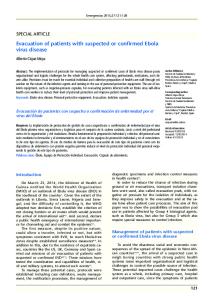
hypovolemia, electrolyte abnormalities, hematologic abnormalities, refractory shock, hypoxia, hemorrhage, septic shock and multi-organ failure. The backbone of treatment for Ebola virus disease includes supportive care to keep up sufficient cardiovascular capacity while the resistant framework assembles a versatile reaction to dispose of the disease. In addition, several experimental antiviral therapies have been utilized as a part of patients with Ebola virus disease during the 2014 episode in West Africa. The adequacy of these agents is misty and is an active area of examination. Furthermore, the accessibility of these medications is limited [53]. Prevention and Control: The prevention of Ebola in Africa presents numerous challenges. Since the identity and location of the natural reservoir of Ebola virus are unknown, there are few built up primary prevention measures [9]. General Approach Practice careful hygiene. For instance, wash your hands with soap and water or an alcohol-based hand sanitizer. Avoid contact with blood and body fluids. Do not handle items that may have come in contact with an infected individual’s blood or body fluids (such as clothes, bedding, needles and medical equipment). Avoid funeral or burial customs that require handling the body of someone who has passed on from Ebola. Avoid contact with bats and nonhuman primates or blood, fluids and crude meat prepared from these animals. Avoid facilities in West Africa where Ebola patients are being dealt with. The U.S. embassy or consulate is regularly ready to give counsel on offices. Monitor your well being after you return for 21 days and look for medical care instantly on the off chance that you create indications of Ebola. Wear suitable personal protective equipment (PPE). Practice proper infection control and sterilization measures. Isolate patients with Ebola from other patients. Avoid direct contact with the assortments of individuals who have passed on from Ebola. Notify health officials on the off chance that you have had coordinate contact with the blood or body liquids of a man debilitated with Ebola [10].
Treatment: Severely ill patients require escalated supportive care. An extensive variety of studies in vitro and several animal models have been produced for EBOV and MARV; nonetheless, right now neither a licensed vaccine nor an approved treatment is available [52]. The accompanying basic interventions, when used early, can significantly improve the chances of survival: Providing intravenous fluids and adjusting electrolytes (body salts). Maintaining oxygen status and blood pressure. Treating other infections in the event that they happen. Experimental treatments for Ebola are in process, yet they have not yet been completely tested for wellbeing or viability. Recovery from Ebola relies on upon good supportive care and the patient’s immune response. Individual who recover from Ebola develop antibodies that keep going for no less than 10 years, possibly longer. It isn’t known whether individuals who recuperate are invulnerable forever or in the event that they can get infected with an alternative species of Ebola. A few people who have recovered from Ebola have grown long-term complications, for example, joint and vision problems [10]. Clinical management ought to concentrate on supportive care of complications, such as 207
Advan. Biol. Res., 11 (4): 202-211, 2017 Table 2: Experimental therapies for EBOLA virus Agent
Organization
Classification
Mechanism
Route
Phase
Zmapp [54]
Zmapp Biopharmaceutial
Anti-EBOV monoclonal antibody
Adsorbs on the protein of EBOV surface; lessens the capacity of the virus to identify the target cells
Intravenous
In phase I clinical trails
TKM-Ebola [55]
Tekmira Pharmaceuticals Corp.
siRNAs
Influences 3 of Ebola’s 7proteins (L, VP24 and VP35)
Oral
In phase I clinical trails
Brincidofovir [56]
Chimerix
Nucleoside analogue
Blocks DNA synthesis, inhibits viral replication
Oral
Phase II (Ebola); Phase III (CMV and ADV)
Jk-05
Chinese Academy of Military Medical Science Institute of Microbiology Epidemiology
Small molecular antiviral drug
Acts on RNA viral polymerase to inhibit viral replication
Oral
Approved in phase I clinical trails
BCX 4430 [57]
BioCryst Pharmaceuticals
Nucleoside analogue
Restrains the viral RNA polymerase function by incorporating into into new viral RNA chains and bringing about chain termination.
Oral or Intramuscular
Priority in phase I clinical trails
cAd3-EBO [58-59]
GlaxoSmithKline; The United States National Institute of Allergy and Infectious Diseases
Vaccine
Provokes an immune response against EBOV by expressing glycoproteins.
Intramuscular
In phase I clinical trails
rVSV–ZEBOV [60] Newlink Genetics; The Public Health Agency of Canada
Vaccine
Incites a neutralizing immune response to the Ebola virus by expressing glycoproteins
Oral and Intravenous
In phase I clinical trails
CONCLUSION
the effectively feeble human services and general wellbeing frameworks in the influenced nations, additionally triggers expanded mindfulness in countries at risk for EVD import cases. Given the ongoing outbreak, countries and clinical centers ought to know about the potential for admission of an EBOV infected individual.
Rapid and wide geographic spread of the current EBOV outbreak is reasons behind expanded alertness of clinicians managing returning travelers from the outbreak areas. EVD is an agonizing update that an outbreak anywhere can be a risk all over the place. Since Ebola virus is basically transmitted through contact with the body fluids of symptomatic patients, the infection spread can be halted by an early diagnosis, contact tracing, patient isolation and care, infection control and safe entombment. In spite of the fact that humans and great apes are EBOV end hosts, dogs and pigs were infected in African village outbreaks, recommending that other interim or amplifying hosts may exist. Fruit bats are one natural store for EBOV and additionally an assortment of other profoundly virulent viruses and the true viral load and range of these chose bat species needs further research. Specific antiviral EVD treatment techniques are still in the experimental stage. The current EVD outbreak focuses on
ACKNOWLEDGEMENTS The author would like to extend sincere appreciation to the Department of Pharmacy, SRMSCET, Bareilly for providing literature survey facility to carry out the work. Funding: This research did not receive any specific grant from funding agencies in the public, commercial, or notfor-profit sectors. REFERENCES 1.
208
Pattyn, S.R., 1978. Ebola Virus Haemorrhagic Fever, Elsevier/ North-Holland Biomedical Press, New York.
Advan. Biol. Res., 11 (4): 202-211, 2017
2. 3. 4.
5.
6.
7.
8.
9.
10.
11.
12.
13.
14.
Hatfill, S.J., T. Nordin and G.L. Shapiro, 2014. Ebola Virus Disease. JPANDS., 19(4): 101-114. Feldmann, H. and T.W. Geisbert, 2011. Ebola Haemorrhagic Fever. Lancet., 377(9768): 849-862. Bray, M. and F.A. Murphy, 2007. Filovirus Research: Knowledge expands to meet a growing threat. J. Infect. Dis., 196(Suppl 2): S438-S443. Kortepeter, M.G., D.G. Bausch and M. Bray, 2011. Basic clinical and laboratory features of filoviral Hemorrhagic Fever. J. Infect. Dis., 204(Suppl 3): S810-S816. Ebola outbreak, 2014. The impact of ebola outbreak on the demography of Africa. http://www.who.int/csr/disease/ebola/en/ (accessed 12.08.14). Mahanty, S. and M. Bray, 2004. Pathogenesis of Filoviral Haemorrhagic Fevers. Lancet Infect. Dis., 4(8): 487-498. WHO Ebola Response Team, 2014. Ebola Virus Disease in West Africa- The First 9 months of the epidemic and forward projections. N. Engl. J. Med., 371: 1481-1495. Courbot, M.C.G., C.Y. Lu, J.J.L. Soukate, E. Leroy and S. Baize, 1997. Isolation and partial molecular characterisation of a strain of Ebola virus during a recent epidemic of viral haemorrhagic fever in Gabon. Lancet., 349(9046): 181. Johnson, K.M., J.V. Lange, P.A. Webb and F.A. Murphy, 1977. Isolation and partial characterisation of a new virus causing acute haemorrhagic fever in Zaire. Lancet, 1: 569-571. Khan, A.S., F.K. Tshioko, D.L. Heymann, B. Le Guenno, P. Nabeth and B. Kerstiens. 1999. The reemergence of Ebola hemorrhagic fever, Democratic Republic of the Congo, 1995. Commission de Lutte contre les Epidemies a Kikwit. J. Infect Dis., 179(Suppl 1): S76-S86. World Health Organization, 2014. Unprecedented number of medical staff infected with Ebola. http://www.who.int/mediacentre/news/ebola/25august-2014/en/# (accessed on 25.08.14). Baize, S., D. Pannetier, L. Oestereich, T. Rieger, L. Koivogui and N. Magassouba, 2014. Emergence of Zaire Ebola virus disease in Guinea. N. Engl. J. Med., 371(15): 1418-1425. Ebola haemorrhagic fever in Sudan, 1976, 1978. Report of a WHO/International Study Team. Bull. World Health Organ., 56(2): 247-270.
15. Baron, R.C., J.B. McCormick and O.A. Zubeir, 1983. Ebola virus disease in southern Sudan: Hospital dissemination and intrafamilial spread. Bull. World Health Organ., 61(6): 997-1003. 16. Centers for Disease Control and Prevention (CDC), 2001. Outbreak of Ebola hemorrhagic fever Uganda, August 2000-January 2001. Morb Mortal Wkly Rep. (MMWR), 50(5): 73-88. 17. Sanchez, A., M. Lukwiya, D. Bausch, S. Mahanty, A.J. Sanchez, K.D. Wagoner, 2004. Analysis of human peripheral blood samples from fatal and nonfatal cases of Ebola (Sudan) hemorrhagic fever: Cellular responses, virus load and nitric oxide levels. J. Virol., 78(19): 10370-10377. 18. Onyango, C.O., M.L. Opoka and T.G. Ksiazek, 2004. Laboratory diagnosis of Ebola hemorrhagic fever during an outbreak in Yambio, Sudan, 2004. J. Infect. Dis., 196(Suppl 2): S193-S198. 19. Jahrling, P.B., T.W. Geisbert, D.W. Dalgard, E.D. Johnson, T.G. Ksiazek and W.C. Hall, 1990. Preliminary report: Isolation of Ebola virus from monkeys imported to USA. Lancet., 335(8688): 502-505. 20. Le Guenno, B., P. Formenty, M. Wyers, P. Gounon, F. Walker and C. Boesch, 1995. Isolation and partial characterization of a new strain of Ebola. Lancet., 345(8960): 1271-1274. 21. Rollin, P.E., R.J. Williams, D.S. Bressler, S. Pearson, M. Cottingham and G. Pucak, 1999. Ebola (subtype Reston) virus among quarantined nonhuman primates recently imported from the Philippines to the United States. J. Infect. Dis., 179(Suppl 1): S108-S114. 22. Formenty, P., C. Hatz, B. Le Guenno, A. Stoll, P. Rogenmoser and A. Widmer, 1999. Human infection due to Ebola virus, subtype Cote d’Ivoire: Clinical and Biologic Presentation. J. Infect. Dis., 179(Suppl 1): S48-S53. 23. Georges, A.J., E.M. Leroy, A.A. Renaut, C.T. Benissan, R.J. Nabias and M.T. Ngoc, 1999. Ebola hemorrhagic fever outbreaks in Gabon, 1994-1997: Epidemiologic and health control issues. J. Infect. Dis., 179(Suppl 1): S65-S75. 24. Leroy, E.M., P. Rouquet, P. Formenty, S. Souquiere, A. Kilbourne and J.M. Froment, 2002. Multiple Ebola virus transmission events and rapid decline of Central African wildlife. Science, 303(5658): 387-390. 209
Advan. Biol. Res., 11 (4): 202-211, 2017
25. Wittmann, T.J., R. Biek, A. Hassanin, P. Rouquet, P. Reed and P. Yaba, 2007. Isolates of Zaire ebolavirus from wild apes reveal genetic lineage and recombinants. Proc. Natl. Acad. Sci. USA, 104(43): 17123-17127. 26. Towner, J.S., T.K. Sealy, M.L. Khristova, C.G. Albarino, S. Conlan and S.A. Reeder, 2008. Newly discovered Ebola virus associated with hemorrhagic fever outbreak in Uganda. PLoS Pathog., 4(11): 1-6. 27. Dixon, M.G. and I.J. Schafer, 2014. Ebola viral disease outbreak- West Africa. Morb. Mortal Wkly Rep. (MMWR), 63(25): 548-551. 28. Swanepoel, R., P.A. Leman, F.J. Burt, N.A. Zachariades, L.E. Braack and T.G. Ksiazek, 1996. Experimental inoculation of plants and animals with Ebola virus. Emerg. Infect. Dis., 2(4): 321-325. 29. Omidian, P., K. Techoungue and J. Monger, 2014. Medical anthropology study of the ebola virus disease (EVD) Outbreak in Liberia/West Africa. WHO Field Report. Monrovia Liberia, pp: 1-20. 30. Gebretadik, F.A., M.F. Seifu and B.K. Glew, 2015. Review on Ebola virus disease: Its outbreak and current Status. Epidemiol., 5(4): 1-8. 31. Zulane, L.S., 2014. Key features of Ebola hemorrhagic fever: A Review. Asian Pac. J. Trop. Biomed., 4(11): 841-844. 32. Towner, J.S., B.R. Amman, T.K. Sealy, S.A.R. Carroll, J.A. Comer and A. Kemp, 2009. Isolation of genetically diverse Marburg virus from Egyptian fruit bats. PLoS Pathogen., 5(7): 1-9. 33. Leroy, E.M., B. Kumulungui, X. Pourrut, P. Rouquet, A. Hassanin and P. Yaba, 2005. Fruit bats as reservoirs of ebola virus. Nature, 438(7068): 575-576. 34. Centers for disease control and prevention, 2014. Ebola (Ebola virus d iseas e). http://www.cdc.gov/vhf/ebola. (accessed on 22.08.16). 35. Rewar, S. and D. Mirdha, 2014. Transmission of ebola virus disease: An overview. Ann. Glob. Health, 80(6): 444-451. 36. Mishra, B., 2014. The threat of Ebola: An update. Indian J. Med. Microbio., 32(4): 364-370. 37. Moghadam, S.R.J., O. Negar, S. Bayrami, S.J. Moghadam and S.A.S. Alinaghi, 2015. Ebola virus disease: A review literature. Asian Pac. J. Trop. Biomed., 5(4): 260-267. 38. Rajak, H., D.K. Jain, A. Singh, A.K. Sharma and A. Dixit, 2015. Ebola virus Disease: Past, present and Future. Asian Pac. J. Trop. Biomed., 5(5): 337-343.
39. Kang, S.S. and D.B. McGavern, 2010. Microbial induction of vascular pathology in the CNS. J. Neuroimmune Pharmacol., 5: 370-386. 40. Connor Jr, M.J., C. Kraft and A.K. Mehta, 2015. Successful delivery of RRT in Ebola Virus Disease. J. Am. Soc. Nephrol., 26(1): 31-37. 41. Jahrling, P.B., T.W. Geisbert, N.K. Jaax, M.A. Hanes, T.G. Ksiazek and C.J. Peters, 1996. Experimental infection of cynomolgus macaques with EbolaReston filoviruses from the 1989–1990 U.S. epizootic. Arch. Virol. Suppl., 11: 115-134. 42. Bah, E.I., M.C. Lamah, T. Fletcher, S.T. Jacob, D.M. Brett-Major and A. A. Sall, 2015. Clinical presentation of patients with Ebola Virus Disease in Conakry, Guinea. N Engl J. Med., 372(1): 40-47. 43. Davis, K.J., A.O. Anderson, T.W. Geisbert, K.E. Steele, J.B. Geisbert and P. Vogel, 1997. Pathology of experimental Ebola virus infection in African green monkeys. Involvement of fibroblastic reticular cells. Arch. Pathol. Lab. Med., 121: 805-819. 44. West, T.E. and A. Von Saint Andre-von Arnim, 2014. Clinical presentation and management of severe Ebola Virus disease. Ann. Am. Thorac. Soc., 11(9): 1341-1350. 45. Tandon, B.N. and S.K. Acharya, 1987. Viral diseases involving the liver. Baillieres Clin Gastroenterol., 1: 211-230. 46. Martines, R.B., D.L. Ng, P.W. Greer, P.E. Rollin and S.R. Zaki, 2015. Tissue and cellular tropism, pathology and pathogenesis of Ebola and Marburg Viruses. J. Pathol., 235(2): 153-174. 47. McElroy, A.K., B.R. Erickson, T.D. Flietstra, P.E. Rollin, S.T. Nichol and J.S. Towner, 2014. Biomarker correlates of survival in pediatric patients with ebola virus disease. Emerg. Infect. Dis., 20(10): 1683-1690. 48. McElroy, A.K., B.R. Erickson, T.D. Flietstra, P.E. Rollin, S.T. Nichol and J.S. Towner, 2014. Ebola hemorrhagic fever: Novel biomarker correlates of clinical outcome. J. Infect Dis., 210(4): 558-566. 49. Sarwar, U.N., S. Sitar and J.E. Ledgerwood, 2011. Filovirus emergence and vaccine development: A perspective for health care practitioners in travel medicine. Travel Med Infect Dis., 9(3): 126-134. 50. Wiwanitkit, V., 2014. Ebola virus infection: What should be known? N. Am. J. Med. Sci., 6(11): 549-552. 51. Goeijenbier, M., J.J. Van Kampen, C.B. Reusken, M.P. Koopmans and E.C. Van Gorp, 2014. Ebola virus disease: A review on epidemiology, symptoms, treatment and pathogenesis. Neth. J. Med., 72(9): 442-448. 210
Advan. Biol. Res., 11 (4): 202-211, 2017
52. Geisbert, T.W. and H. Feldmann, 2011. Recombinant vesicular stomatitis virus based vaccines against Ebola and Marburg virus infections. J. Infect. Dis., 204(Suppl 3): S1075-S1081. 53. Kalra, S., D. Kelkar, S.C. Galwankar, T.J. Papadimos, S.P. Stawicki and B. Arquilla, 2014. The emergence of ebola as a global health security threat: From “lessons learned” to coordinated multilateral containment efforts. J. Glob. Infect. Dis., 6(4): 164-177. 54. Davidson, E., C. Bryan, R.H. Fong, T. Barnes, J.M. Pfaff and M. Mabila, 2015. Mechanism of Binding to Ebola Virus Glycoprotein by the ZMapp, ZMAb and MB-003 Cocktail Antibodies. J. Virol., 89(21): 10982-10992. 55. Thi, E.P., C.E. Mire, A.C.H. Lee, J.B. Geisbert, J.Z. Zhou and K.N. Agans, 2015. Lipid nanoparticle siRNA treatment of Ebola-virus-Makona-infected nonhuman primates. Nature, 521(7552): 362-365. 56. Florescu, D.F., A.C. Kalil, A.L. Hewlett, A.J. Schuh, U. Stroher and T.M. Uyeki, 2015. Administration of Brincidofovir and Convalescent Plasma in a Patient with Ebola Virus Disease. Clin Infect Dis., 61: 969-973.
57. Warren, T.K., J. Wells, R.G. Panchal, K.S. Stuthman, N.L. Garza and S.A. Van Tongeren, 2014. Protection against filovirus diseases by a novel broad-spectrum nucleoside analogue BCX4430. Nature, 508(7496): 402-405. 58. Stanley, D.A., A.N. Honko, C. Asiedu, J.C. Trefry, A.W. Lau-Kilby and J.C. Johnson, 2014. Chimpanzee adenovirus vaccine generates acute and durable protective immunity against ebolavirus challenge. Nat Med., 20: 1126-1129. 59. Ledgerwood, J.E., A.D. De Zure, D.A. Stanley, L. Novik, M.E. Enama and N.M. Berkowitz, 2014. Chimpanzee adenovirus vector Ebola vaccinePreliminary report. N. Engl. J. Med., 373: 775-776. 60. Jones, S.M., H. Feldmann, J.B. Geisbert, L. Fernando, A. Grolla and H.R. Klenk, 2005. Live attenuated recombinant vaccine protects nonhuman primates against Ebola and Marburg viruses. Nat. Med., 11(7): 786-790.
211