CopertinaVol.34-n.2-ingl:Layout 1 08/06/15 14:45 Pagina 1
Rivista Italiana di Colon-Proctologia Founded in 1982
Vol. 34 - N. 2 June 2015
ISSN 1973-4905
INSTRUCTIONS FOR AUTHORS The manuscripts including tables and illustrations must be submitted to Pelviperineology only via the Isubmit system www.isubmit.it. This enables a rapid and effective peer review. Full upload instructions and support are available online from the submission site. In http://www.pelviperineology.org/pelviperineology authors instructions. html please find the updated guidelines for the Authors.
Post vesico-vaginal fistula repair incontinence - A new hypothesis and classification potentially guide prevention and cure P. PETROS, G. WILLIAMS, A. BROWNING
53
MR imaging of vaginal morphology, paravaginal attachments and ligaments. Normal features V. PILONI
Poste Italiane s.p.a. Spedizione in Abb. Post. - 70% - DCB Padova
Letters to the Editor – Re Native surgery and pelvic floor surgery, P. PETROS – Correspondence on the cardinal ligament, R. DE CARO, M. NEUMAN
ng
fu
67 ‘Taxe Perçue’ ‘Tassa Riscossa’ - Padova C.M.P.
Chronic pelvic pain syndrome in women. Review and preliminary results with low-energy extracorporeal shock wave therapy A. MENEGHINI, M. TREVISAN, E. LAMPROPOULOU, N. MENEGHINI
2005
pu na n Di s
60
AHI v e INKON TI N AN S
DE
RN 05 20
Vaginal evisceration in a patient with post hysterectomy vault prolapse managed conservatively with a vaginal ring pessary A. CHRYSOSTOMOU
CERR
I-
52
IF KT RU
EG
The spectrum of anorectal malformations: a congenital disease for the general surgeons P. MIDRIO
ST
er him
51
N KO
si D
ul es Wa n i t a I n d on
48
R
Opinions and evidence on management of pelvic organ prolapse. Review and consensus statement (POP Working Group) F. LA TORRE, F. PUCCIANI, G. DODI, G. GIULIANI, A. FRASSON, D. COLETTA AND P. PETROS
PE LG IK
39
P
On seeking PubMed status for the journal Pelviperineology (PPj) THE EDITORS
ia
38
E
Contents
as a r P an
gg
CopertinaVol.34-n.2-ingl:Layout 1 08/06/15 14:45 Pagina 3
Frontespizio n. 34-2:Adjustable 08/06/15 11:19 Pagina 37
Vol.
34
Rivista Italiana di Colon-Proctologia Founded in 1982
N. 2 June 2015
PELVIPERINEOLOGY A multidisciplinary pelvic floor journal www.pelviperineology.org
Editors GIUSEPPE DODI, Colorectal Surgeon, Italy - ANDRI NIEUWOUDT, Gynaecologist, Nederland PETER PETROS, Gynaecologist, Australia - AKIN SIVASLIOGLU, Urogynecologist, Turkey FLORIAN WAGENLEHNER, Urologist, Germany Editorial Board BURGHARD ABENDSTEIN, Gynaecologist, Austria ROBERTO ANGIOLI, Gynaecologist, Italy JACQUES BECO, Gynaecologist, Belgium CORNEL PETRE BRATILA, Gynaecologist, Romania SHUQING DING, Colorectal Surgeon, P .R. China PIERRE GADONNEIX, Urogynaecologist, France KLAUS GOESCHEN, Urogynaecologist, Germany DARREN M. GOLD, Colorectal Surgeon, Australia DANIELE GRASSI, Urologist, Italy ALDO INFANTINO, Colorectal Surgeon, Italy WOLFRAM JAEGER, Gynaecologist, Germany DIRK G. KIEBACK, Gynaecologist, Germany FILIPPO LA TORRE, Colorectal Surgeon, Italy NUCELIO LEMOS, Gynaecologist, Brazil BERNHARD LIEDL, Urologist, Germany ANDRI MULLER -FUNOGEA, Gynaecologist, Germany MENAHEM NEUMAN, Urogynaecologist, Israel OSCAR CONTRERAS ORTIZ, Gynaecologist, Argentina
PAULO PALMA, Urologist, Brazil FRANCESCO PESCE, Urologist, Italy MARC POSSOVER, Gynaecologist, Switzerland FILIPPO PUCCIANI, Colorectal Surgeon, Italy RICHARD REID, Gynaecologist, Australia GIULIO SANTORO, Colorectal Surgeon, Italy YUKI SEKIGUCHI, Urologist, Japan SALVATORE SIRACUSANO, Urologist, Italy MARCO SOLIGO, Gynaecologist, Italy JEAN PIERRE SPINOSA, Gynaecologist, Switzerland MICHAEL SWASH, Neurologist, UK VINCENT TSE, Urologist, Australia PETER VON THEOBALD, Gynaecologist, Reunion Island, France PAWEL WIECZOREK, Radiologist, Poland QINGKAI WU, Urogynecologist, P. R. China RUI ZHANG, Urogynaecologist, P. R. China CARL ZIMMERMAN, Gynaecologist, USA Sections
Aesthetic gynecology - RED ALINSOD (USA) Andrology - ANDREA GAROLLA (Italy) Chronic pelvic pain - MAREK JANTOS (Australia) EZIO VICENTI (Italy) Imaging - VITTORIO PILONI (Italy) Medical Informatics - MAURIZIO SPELLA (Italy) Pediatric Surgery - PAOLA MIDRIO (Italy)
Pelvic floor Rehabilitation - DONATELLA GIRAUDO (Italy), GIANFRANCO LAMBERTI (Italy) Psychology - SIBYLLA VERDI HUGHES (Italy) Sacral Neurostimulation - MARIA ANGELA CERRUTO (Italy) Sexology - OSCAR HORKY (Australia) Statistics - CARLO SCHIEVANO, (Italy)
Official Journal of the: International Society for Pelviperineology (www.pelviperineology.com) Pelvic Reconstructive Surgery and Incontinence Association (Turkey) Perhimpunan Disfungsi Dasar Panggul Wanita Indonesia Romanian Uro-Gyn Society
Editorial Office: LUISA MARCATO e-mail:
[email protected] Quarterly journal of scientific information registered at the Tribunale di Padova, Italy n. 741 dated 23-10-1982 and 26-05-2004 Editorial Director: GIUSEPPE DODI Printer “Tipografia Veneta” Via E. Dalla Costa, 6 - 35129 Padova - e-mail:
[email protected]
On seeking:Adjustable 08/06/15 14:48 Pagina 38
Editorial
On seeking PubMed status for the journal Pelviperineology (PPj) THE EDITORS St. Vincent’s Hospital Clinical School, Academic Department of Surgery, University of NSW, Sydney
A major claim for the PPJ’s request for PubMed status is that our policy, which encourages innovation, has allowed publication of major discoveries in innovative papers often rejected by other pelvic floor journals, in particular, those concerning the Integral Theory System (ITS). Innovation emphasis we see as the only way to overcome a major weakness in the peer review system; comfort with the familiar and discomfort outside its narrow field of knowledge. Many consider the ITS to be the next pelvic floor paradigm. In encouraging publication of the ITS scientific studies, PPj has, in some way, virtually morphed into the de facto ‘Home Journal” of the ITS. In 2007, the first editorial of Pelviperineology1 described the historical origins of the English version of the journal. PPj evolved from AAVIS (Australasian Association of Vaginal and Incontinence Surgeons) founded in 1996. From its inception, AAVIS was a multidisciplinary pelvic floor society of gynaecologists, urologists and coloproctologists, providing a support group for these surgeons who were the first in the world to adopt the Integral Theory paradigm of Petros and Ulmsten. They were also the first surgeons as a group to perform the tension free suburethral intravaginal slingplasty (TVT/IVS) procedures which the Editorial described as “the beginning of a revolution in pelvic medicine. Advances in our understanding of anatomy and physiology and the development of surgical prostheses have provided new options for pelvic surgeons”. The editorial continued, “Pelviperineology will seek to explore the integrated pelvis and publish articles from the four corners of the world. We hope this journal can be free of politics and so rise above the self-interest of any particular group. We will try to achieve this by being open to diverse views and consider alternative solutions when we can find them. We hope you can join us on this journey”. PPj has been a haven for the ‘diverse views’ of the 2007 Editorial. It has been a veritable lifeline for publications associated with the Integral Theory (IT). Many original IT articles concerning conditions which grossly affect patients’ quality of life were published for the first time in PPJ. Many of these ground breaking discoveries which include surgical cure of non-sphincteric fecal incontinence, obstructive defecation syndrome, chronic pelvic pain, obstructive micturition had been rejected by mainstream pelvic
38
journals such as Neurourology and Urodynamics, Disease of Colon and Rectum, International Urogynecology Journal, European Journal of Urology and so on. One can only hypothesize why. Perhaps it is because these pelvic floor journals place great store on the ”Peer Review” system which falls down in works which involve a change in thinking. This process was explained by Thomas Kuhn2 in his “Structure of Scientific revolutions”, as follows: “Normal science, for example, often suppresses fundamental novelties because they are necessarily subversive of its basic commitments”. And later3 “When the profession can no longer evade anomalies that subvert the existing tradition of scientific practice- then begin the extraordinary investigations that lead the profession to a new basis for the practice of science”. With regard to “the fundamental novelties being necessarily subversive of its basic commitments”,2 pelvic symptoms to date have been treated according to the Urodynamic paradigm, which states that other than urinary stress incontinence, most symptoms of pelvic pain, bladder & bowel dysfunction are considered as being incurable. However, this paradigm was invalidated in 2006 by the Cochrane Report.4 A large part of “the extraordinary investigations that lead the profession to a new basis for the practice of science” have been made possible only because of the mission statement expounded in the 2007 Editorial,1 ‘this by being open to diverse views and consider alternative solutions when we can find them’. This continues to be so, as evidenced by the large number of articles validating the Integral Theory’s predictions between 2007 to this day. In the process, as well as being open to ‘diverse views’ on pelvic floor, PPj has become the de facto ‘home journal’ for what many consider to be the next pelvic floor paradigm, the Integral Theory System. REFERENCES 1. Editorial. Pelviperineology 2007; 26: 3. 2. Kuhn T. The Structure of Scientific Revolutions, 3rd Ed University of Chicago Press, 1996, p 5. 3. Kuhn T. The Structure of Scientific Revolutions, 3rd Ed University of Chicago Press, 1996, p 6. 4. Glazener CMA, Lapidan MC. Urodynamic investigations for the management of urinary incontinence in children and adults (Cochrane Review), The Cochrane Library, 2006; Issue 1.
Pelviperineology 2015; 34: 38 http://www.pelviperineology.org
Opinions and evidence:ingynious 04/06/15 15:10 Pagina 39
Consensus
Opinions and evidence on management of pelvic organ prolapse. Review and consensus statement (POP Working Group) FILIPPO LA TORRE1, FILIPPO PUCCIANI2, GIUSEPPE DODI3, GIUSEPPE GIULIANI1, ALVISE FRASSON3, DIEGO COLETTA1 and PETER PETROS4 Coordinator POP Working Group, Università La Sapienza di Roma Università di Firenze 3 Università di Padova 4 University of N.S.W., Sydney 1 2
Abstract: Pelvic organ prolapse is a global health concern affecting adult women of all ages. POP can be defined as a downward descent of female pelvic organs, including the bladder, uterus, post-hysterectomy vaginal cuff and the small or large bowel, resulting in protrusion of the vagina, uterus, or both. Its development is multifactorial, with vaginal childbirth, advancing age, and increasing body-mass index as the most consistent risk factors. Vaginal delivery, hysterectomy, chronic straining, normal ageing, and abnormalities of connective tissue or connective-tissue repair predispose some women to disruption, stretching, or dysfunction of the levator ani complex, connective-tissue attachments of the vagina, or both, resulting in prolapse. Patients generally present with several complaints, including bladder, bowel, and pelvic symptoms. No guidelines exist regarding the management and treatment of these disorders. This paper is a reduced version of the original Consensus Statement of an Italian POP Working Group whose intention was to give guidance and support for the approaches to problems of the pelvic floor, to suggest recognized guidelines and to stimulate further studies of the topic. Contents: 1) Male/female pelvic anatomy; 2) Pelvic Organ Prolapse (POP): Literature update; 3) The Integral Theory; 4) POP and faecal incontinence; 5) POP and obstructed defecation; 6) How to evaluate POP; 7) The role of imaging; 8) The minimum/correct work-up for POP evaluation; 9) The urogynecological view; 10) The role of conservative treatment; 11) The surgeon role in front of POP; 12) Sacrocolpopexy and rectopexy; 13) The pexies are the gold standard for any POP repair? 14) POP repair after the FDA warning; 15) The shrinkage/erosion of implanted material: complications evaluation and management. Keywords: Pelvic organ prolapse; Incontinence; Obstructed defecation; Mesh, Integral Theory; TFS.
1. MALE/FEMALE PELVIC ANATOMY (Updates & limits of our knowledge)
2. PELVIC ORGAN PROLAPSE (POP): LITERATURE UPDATE (last 10 years)
The Pelvic Floor is composed of organs, muscle, fascia and ligaments, interconnected with each other and the bony pelvis by an extensive fibro-elastic network containing virtual anatomical spaces.1 The pelvic floor is composed of levator ani, coccygeus muscles with their fascia, perineal membrane, superficial perineal muscles, deep perineal muscles and perineal body. Three kinds of fascia can be described: visceral, parietal and endo-pelvic which is attached to the tendinous arcs at the pelvic side wall. The levator ani muscles ileococcygeous, pubo-rectalis and pubo-coccygeous (further divided in pubo-perinealis, pubo-vaginalis, and the pubo-analis) (Table 1)2,3 are composed mostly of type I striated muscle fibers. [Level of Evidence [LE] 2A, Grade of Recommendation [GR] B]. The perineal membrane is a triangularshaped fibro-muscular structure, attached to the pubic bones anteriorly.4,5 The deep and superficial transverse perinei have a supporting function, bulbo-spongiosus and ischio-cavernosus muscles sexual functions. The arcus tendineus levator ani and the arcus tendineus fascia pelvis attach muscles to the pelvic side wall. Central and peripheral nervous systems regulate all functions.6 [LE 1B, GR A]. The peripheral nervous system supplies the pelvic floor with: • branches of the sacral plexus: the pudendal nerve (coursing inferior to the pelvic floor) • levator ani nerve (coursing superior to the pelvic floor) • parasympathetic pelvic splanchnic nerves (nervi erigentes) • hypogastric nerve (sympathetic).1
Vaginal delivery poses the strongest risk factor for POP.7 Abnormalities of connective tissue predispose to pelvic organ prolapse (POP); excess straining is thought may cause pudendal nerve neuropathy,8 associated with POP.9 Increased MMP-1 immunohistochemical expression in utero-sacral ligaments is associated with urogenital prolapse.10 Elastin metabolism studies suggest increased degradation but also abnormal synthesis in woman with POP.11 High-risk pedigrees and linkage analysis showed evidence for significant genome-wide linkage on several chromosomes.12,13
Pelviperineology 2015; 34: 39-48 http://www.pelviperineology.org
3. THE INTEGRAL THEORY: A MUSCULO-ELASTIC THEORY OF PELVIC FLOOR FUNCTION AND DYSFUNCTION In according with Petros,14,15 POP and its symptoms such as urinary stress, urge, abnormal bowel, bladder emptying, some forms of pelvic pain and fecal incontinence are caused by laxity in the vagina or its supporting ligaments, a result of altered connective tissue. The main etiologies were childbirth related laxity compounded by ageing. The vagina is suspended like a suspension bridge, with the ligaments above and the muscles below (Fig. 1). The muscle forces (arrows) contract against the suspensory ligaments to give the bridge form and strength. Because the ligaments and vagina are the ultimate supports of the bladder and rectum (Fig. 1-2) anything which damages these structures can also affect the structure and function of bladder and rectum.
39
Opinions and evidence:ingynious 04/06/15 15:10 Pagina 40
Filippo La Torre, Filippo Pucciani, Giuseppe Dodi, Giuseppe Giuliani, Alvise Frasson, Diego Coletta and Peter Petros
Figure 1. – Integral Theory. View of pelvis from above and behind. Arrows: muscle forces. Ligaments: ATFP= arcus tendineus fascia pelvis; CL=cardinal ligament; USL=uterosacral ligament; PUL=pubourethral ligament; PB=perineal body; LP= levator plate; LMA=longitudinal muscle of anus; PCM=anterior pubococcygus muscle; PRM= puborectalis muscle; Circular broken lines = pelvic brim.
Figure 2. – Pathogenesis of rectocele. Perineal body (PB) components including deep transverse perineal muscles (DTP) are stretched laterally. The anus (A) and rectum protrude into the vagina. OF=obturator fossa. Surgery: TFS tape penetrates DTP and approximates the separated PB entities to form a neo central tendon to reduce rectocele and descending perineal syndrome.
Moreover, uterine prolapse can be caused by the elongated of cardinal ligament and of utero-sacral ligament. While cystocele can be the result of failed tension of cardinal ligament and arcus tendineus fascia pelvis support. Failed utero-sacral ligament may cause ‘posterior fornix syndrome’ (urgency, pelvic pain, nocturia, evacuation disorders). Failed perineal body can cause rectocele and manually assisted defecation and can contribute to Descending Perineal Syndrome.
rectal intussusception.25 Other than the proposals of the Integral Theory, the role of POP, rectal intussusception and pelvic floor dyssynergia in inducing OD is not known, so it is impossible to suggest the best surgical approach for correction of OD/POP. 6. HOW TO EVALUATE PELVIC ORGAN PROLAPSE
7-31% of women with POP have faecal incontinence (FI).8,16 Pathophysiology of POP and FI is vaginal delivery, advancing age, increased body-mass index, hysterectomy, chronic straining, normal ageing, abnormalities of connective tissue, connective-tissue repair.8 [LE 5, GR C]. FI and POP share common risk factors17 [LE 2, GR C]. 2.1% of women with descending perineum have some sign of genital descent with significant correlation between the Jorge incontinence score and degree of genital relaxation (rs 0.85, P < 0.001)18 [LE 3, GR C]. 50% of patients with rectal prolapse also experience FI19 and 38% have POP.
There is no universally accepted anamnestic-clinical method for evaluating POP. The ICS includes urogenital and rectal prolapses26 others the genitalia.16 Useful validated questionnaires for QOL are the Australian Pelvic Floor Questionnaire.27 [LE 1, GR B]. The Pelvic Floor Impact Questionnaire (PFIQ-7),28 Pelvic Floor Distress Inventory (PFDI-20)28 and Pelvic Organ Prolapse/Urinary Incontinence Sexual Questionnaire (PISQ-12)29 [LE 1, GR B] and a novel software scoring program30 [LE 1, GR B]. The POP-Q system attempts to overcome perceived deficiencies of the Baden and Walker halfway system.32 However the POP-Q itself has been questioned recently, in that it is complex, not easy to administer or teach and not useful for detection of recto-anal intussusception or rectal prolapse.
5. POP AND OBSTRUCTED DEFECATION
7. THE ROLE OF IMAGING
18-25% of women with POP report obstructed defecation (OD)20-21 and 32% of women with OD have POP.22 The pathophysiological mechanisms of OD-POP are unknown23 [LE 1, GR A]. The crux of the matter can be defined with the following questions: 1. Does posterior vaginal compartment anatomy correlate with ano-rectal function? 2. Does restoring the anatomy of the posterior vaginal compartment improve defecatory function? 3. What is the best surgical approach to restoration of posterior vaginal compartment anatomy and defecatory function? Other than those proposed by the Integral Theory, there are no answers to these three questions. [LE 3, GR C]. Breaks of the recto-vaginal septum cause high rectocele.24 Derangement of uterosacral ligaments starts recto-
Different types of imaging are used in according with the pelvic floor’s dysfunctions. Pelvic floor imaging is based essentially on: • Ultrasound evaluation (US) • Fluoroscopy (voiding cystourethrography, defecography, cystoproctography cystocolpodefecography) • Pelvic floor MRI. The most diffuse imaging modality of pelvic floor is ultrasound:33-37 • Transperineal ultrasonography (TPUS-called also translabial ultrasound or perineal ultrasound’) • Transvaginal ultrasonography (TVS) • Endoanal ultrasonography (EAUS). With TPUS and TVS it is possible to diagnose1 levator ani damage, avulsion defects, abnormal levator ani contractility and enlarged levator hiatus (ballooning), urethral mo-
4. POP AND FAECAL INCONTINENCE
40
Opinions and evidence:ingynious 04/06/15 15:10 Pagina 41
Opinions and evidence on management of pelvic organ prolapse. Review and consensus statement (POP Working Group)
bility, urethral vascularity, funneling, bladder neck descent, bladder wall thickness. EAUS is the gold standard to assess anal sphincter integrity. Fluoroscopy assessments are:38 voiding cystourethrography (VCUG), with or without urodynamic testing; evacuation proctography; cystoproctography and cystocolpoproctography. With the VCUG it is possible to study bladder: position (e.g. Cystocele), relation to the pubic symphysis, mobility, diverticula and fistulas. Evacuation proctography is indicated for suspicion of rectal intussusception, rectal prolapse, rectocele or pelvic dyssynergia. MRI38 is non invasive with no ionizing radiation. Its disadvantages are high cost, need for specialist radiological interpretation, absence of seated position. In our opinion, US remain the diagnostic procedure of choice to study any POP dysfunction because it is minimally invasive, cost-effective and gives rapid diagnosis.
(normal active, overactive, underactive and non-functioning), puborvesical muscle or avulsion injury, perineal descent during the valsalva the perineum shows a downward movement, low anal canal resting tone, inward scar or fistula within the vagina, rectocele and rectal intussusception. The examination must be conducted in any position which better displays the prolapse. In POP-Q staging, the hymen is the fixed point of reference for prolapse: anterior vaginal wall, uterus (cervix), apex of vagina (vaginal vault or cuff scar after hysterectomy), posterior vaginal wall.31 • Stage 0: No prolapse is demonstrated • Stage I: Most distal portion of the prolapse is more than 1 cm above the level of the hymen • Stage II: Most distal portion of the prolapse is 1 cm or less proximal to or distal to the plane of the hymen • Stage III: The most distal portion of the prolapse is more than 1 cm below the plane of the hymen • Stage IV: Complete eversion of the total length of the lower genital tract is demonstrated.
8. THE MINIMUM/CORRECT WORK-UP FOR POP EVALUATION
What kind of investigations are usually used in clinical practice of urogynecologic patients? Other than the Integral Theory System Questionnaire (ITSQ), there is no evidence that the use of questionnaires has any impact on treatment outcomes [LE 3, GR B]. Voiding diaries assist symptom quantification [LE 3-GR B]. There is a poor correlation between UI symptoms and urodynamic findings. The most diffuse imaging modality is ultrasound.33-36 Fluoroscopy has indications38 as does dynamic-MRI. What are the most common diseases in urogynecological clinical practice? At first evaluation, these are USI (72%), POP (61%), detrusor overactivity (13%-40%), bladder oversensitivity (10-13%) and voiding dysfunctions.
The first step of a diagnostic workup is a detailed history. Physical examination, while important, is quite poor for identification of many common pelvic floor problems.39 Also useful are scoring systems, imaging (endoanal US dynamic cystocolpoproctography (DCP), dynamic MRI), functional testing (ano-rectal manometry, pudendal nerve terminal motor latency testing and anorectal electromyography). Scoring Systems: Clinical practice relies scores and questionnaires: Australian Pelvic Floor Questionnaire, Pelvic organ prolapse quantification POP-Q, Baden Walker halfway assessment (still in general clinical use), Pelvic Floor Impact Questionnaire (PFIQ-7), Pelvic Floor Distress Inventory (PFDI-20), Pelvic Organ Prolapse/Urinary Incontinence Sexual Questionnaire (PISQ-12), Integral Theory System Questionnaire (ITSQ) and the Three Axial Perineal Evaluation (TAPE) score. The imaging assessment: increasingly is based on ultrasound. Since defecatory disorders are associated with POP, defecography evaluation is extended by opacifying the small bowel, vagina, and the urinary bladder.40 Functional tests: are anorectal manometry, pudendal nerve terminal motor latency testing (PNTMLT) and electromyography. 9. THE UROGYNECOLOGICAL VIEW: THE PARTICULAR POINT OF VIEW IN FRONT OF MAIN PROBLEM Symptoms linked to the bladder storage are USI, frequency, nocturia, urgency, emptying problems. Other symptoms are dyspareunia,vaginal laxity, vaginal bulging pelvic pressure, splinting/digitation, pain, acute or chronic, bladder, urethral pain, vulva or vaginal pain, pelvic or perineal or pudendal pain. What are the signs to search during the examination of a patients with symptoms of urogynecologic clinical practice? The first steps history taking and clinical evaluation;4244 examining with a full bladder for urine on coughing (stress incontinence), a cotton swab test for bladder neck hypermobility.45 Vaginal examination seeks anomalies of vulva (e.g. cysts, infections, tumors, atrophic changes), urethra (e.g. mucosal prolapse, urethral caruncle and diverticulum), vagina (length, mobility, scarring), pain, and estrogenization, scars (e.g. perianal, peri-vulval), muscle function
10. THE ROLE OF CONSERVATIVE TREATMENT Women are not aware of prolapse until their bulge extends beyond their introitus.46 Initial management is conservative47 pessary and pelvic floor muscle exercises48 typically for patients > 65 years 49-50 [LE 1, GR A]. With pessaries patients experiences significant improvement (P=0.045, Wilcoxon signed rank test) [LE 5, GR C]. There is little empirical evidence available regarding PFR effectiveness.51 Many patients abandon their exercise regimen over time.52,53 PFMT effects on urinary and fecal incontinence is different because the long-term success rate is well defined in both diseases (67%54 and 53%,55 respectively). PFR is recommended as the first-line treatment for stress, urge, or mixed incontinence in women of all ages.56 Rehabilitative treatment may be considered a first-line option for patients with faecal incontinence not responding to dietary modification or medication.57 OD treated by conservative/rehabilitative treatment can result in long-term success rate of 50% [LE 1, GR A].58,59,60 11. THE SURGEON’S ROLE IN TREATMENT OF POP Surgery for POP can be approached vaginally, abdominally, laparoscopically, robotically:61,62 anterior colporrhaphy, with or without synthetic graft; vaginal hysterectomy with uterosacral; posterior native tissue colporrhaphy; posthysterectomy apical prolapse with abdominal sacrocolpopexy. Anterior native tissue colporrhaphy has recurrence rates, up to 50%. Current evidence does not clearly support this approach to anterior compartment repair.63-67 The graft material most commonly in use for cystocele repair is polypropylene mesh, Amid Type 1.68,69 The poste-
41
Opinions and evidence:ingynious 04/06/15 15:10 Pagina 42
Filippo La Torre, Filippo Pucciani, Giuseppe Dodi, Giuseppe Giuliani, Alvise Frasson, Diego Coletta and Peter Petros
rior compartment is more successfully repaired with native tissue colporrhaphy with 80% cure rates. Mesh in the posterior compartment is not supported by current evidence.70-72 Apical prolapse rarely occurs in isolation; repair is often combined one or both other compartments. Transvaginal uterosacral ligament suspension can be performed either as an intra-peritoneal or extra-peritoneal vaginal procedure. A meta-analysis of transvaginal uterosacral ligament suspension reported successful apical outcome in 98%, median follow-up of 25 months.73 Ureteric injury/kinking, was reported in 11%.74-76 Success for the vaginal cuff is reported at 95% at 2 years.77 The McCall culdoplasty anchors the distal uterosacral ligament pedicles to the vaginal vault.78 Sacrospinous ligament vault suspension inserts sutures into the sacrospinous ligament.79,80 The Manchester repair is another option. The Gynecare Prolift reported 1 year success rates between 82 and 86%.81 Colpocleisis is an obliterative vaginal prolapse procedure performed with an aggressive perineorrhaphy.82 Abdominal sacrocolpopexy can be performed open, laparoscopically or with the aid of a robotic device. This approach maintains adequate vaginal length and sexual function. Reported success rates for all compartments are 78–100%, with mesh exposure in 3.4%.83,84 Short-term results are encouraging with 88% success at 1 year, but no long-term data regarding durability are available.
Rectopexy: two alternative perineal approach are described for external rectal prolapse: the Delorme and Altemeir procedure. Rectopexy consists of mobilization and fixation of rectum to the sacral promontory with suture or mesh.91 A Cochrane review of 12 randomized trials with 380 patients showed no better outcomes for one treatment over another [LE 1a, GR A].92 Ventral and posterior rectopexy associated with sigmoid resection have less postoperative constipation and with better outcomes regarding ODS. Recurrence rate after abdominal rectum mobilization-only does not differ with others types of procedures and this procedure has a recurrence rate of 28.9% at 10 years of FU93 [LE 2b, GR B]. According to Bordeianou,91 patients with complete rectal prolapse and constipation are candidates for sigmoid resection [LE 5, GR c]. In patients with preoperative findings of low resting pressure on anorectal manometry at the moment of rectopexy the division of lateral ligaments is recommended; it reduce frequency of defecation, doubling total and segmental colonic transit times94 [LE 1b, GR A]. Laparoscopic rectopexy has less post operative morbidity and shorter hospital stay94-96 [LE 1a, GR A] but there are limitations [LE 3b, GR B].97 To date there is not sufficient evidence to utilize robotic surgery for this type of procedure.91
12. SACROCOLPOPEXY AND RECTOPEXY
13. THE PEXIES ARE THE GOLD STANDARD FOR ANY POP REPAIR? HAVE WE A CORRECT ANSWER?
Sacrocolpopexy is considered the choice of treatment for [LE 2a, GR B]:85-87 • apical compartment disorders in associations or not with others concomitant defects as rectocele, enterocele or complete rectal prolapse; • apical defects in young woman and patient who wish to remain sexually active. Sacrocolpopexy use synthetic mesh or biologic mesh as xenografts (porcine dermis or bovine tissues) and allografts (cadaveric fascia) meshes to correct apical and/or advanced anterior wall prolapse.113 Recurrence rates of abdominal sacrocolpopexy (ASP) range from 0% to 22%85,86 [LE 2a, GR B]. When compared to sacrospinous ligament fixation (SSLF) and uterosacral ligament suspension (USLS), ASP has greater durability, lower rate of recurrence of vault prolapse and less dyspareunia compared with vaginal sacrospinous colpopexy71 [LE 1a, GR A]. Xenograft mesh has greater probability of operation failure than polypropylene mesh [LE 1b, GR A].88 Polypropylene mesh has an erosion risk that ranges from 3.4% to 10.5% after ASP; polyester mesh use has an increased risk of mesh erosion [LE 1b, GR A].89 Laparoscopic and robotic assisted rectopexy have lower blood loss quicker recovery, less pain and shorter hospital stay [LE 1a, GR A]. Robotic Sacrocolpopexy has a longer operation time and is more expensive.71 The last review of Cochrane comparing laparoscopic sacral-colpopexy with open and robotic techniques showed no decisive outcomes71 [LE 1a, GR A]. Women with prolapse can present with contemporaneous urinary incontinence, obstructed defecation and sexual dysfunction.85 In a multicenter randomized controlled trial of prophylactic Burch retropubic-urethropexy at the time of ASC, patients after Burch urethropexy showed significantly decreased risk of SUI post operatively90 [LE 1b, GR A]. Concomitant correction of rectocele may improve the symptoms of obstructed defecation [LE 1b, GR A].71
42
Up to now this question has no answer. Which surgical option should be chosen? Laparoscopic and laparotomic pexies have the lowest morbidity and recurrence rate.107,113,130-133 Despite the FDA report, transvaginal surgery with mesh can be safely performed in elderly.85,98 Another very interesting procedure is the TFS technique described by Petros.99 It is a very minimal method which reaches high level of cure of symptoms but without a powerful statistical evidence up to now. As per the TFS technique, the placement of a TFS sling through the uterosacral ligaments to suspend the rectum from above and through the two parts of perineal body to support it from below is reported to have great results but without level 1 evidence.100 Last, even if the encircling of the anus with a prosthesis101 surrounding the sphincter has high recurrence rate, it could be useful in elderly patients with rectal prolapse who can’t undergo major surgery. [LE 4, GR C] 14. POP REPAIR AFTER THE FDA WARNING. WHAT IS OUR SURGICAL APPROACH AND WHAT HAPPENED AFTER THE WARNING? To date are there any recommendations on the use of meshes? Regardless of the medical-legal controversies, the use of prostheses remains an appropriate treatment for many patients.102,103 Some recommendations are the following [LE 3, GR C]: • before using meshes it is fundamental to inform patients on risks, benefits, surgical and non-surgical alternatives102-105 • the routine use of biological material is not advisable as it seem to have no real benefit106-108 • heavier weight prostheses are reported to shrink more often than lower weight ones108
Opinions and evidence:ingynious 04/06/15 15:10 Pagina 43
Opinions and evidence on management of pelvic organ prolapse. Review and consensus statement (POP Working Group)
• In vaginal surgery macroporous monofilament polypropylene should be the choice while polyester prostheses frequently have been linked to erosion complications108 • due to the pressure of industries there is a huge number of different prostheses, and the surgeon is required to have a specific skill for each different product109 • a careful patient selection is crucial as individual factors may compromise the outcome (for example smoking, diabetes) • new products must not be assumed to have an equal or improved safety and efficacy until long term data are available102 • it is of paramount importance to continue to collect follow-up data, with the aim of reviewing long term outcomes102 What is still lacking? Multicentre randomised controlled trials with a longer follow-up and a sufficient power are required to evaluate and compare the different surgical procedures. 15. SHRINKAGE/EROSION OF IMPLANTED MATERIAL. COMPLICATIONS EVALUATION AND MANAGEMENT. WHICH ARE THE MORE COMMON COMPLICATIONS? Erosion is different from an extrusion which is the gradual passage of mesh out of the epithelium. The rate of erosions after vaginal surgery ranges between 5 and 19% and occurs in 3% of laparotomic sacrocolpopexies.103-105 Other adverse events are vaginal or pelvic pain (4-11%), dyspareunia(1-3%), rectal injuries ( 0.05). Overall, the lower third vaginal morphology was the less easily identifiable structure (visibility score, 2); the uterosacral ligaments and the paraurethral ligaments were the most frequently depicted attachments (visibility score, 3 and 4, respectively); the distance of the perineal body to the hymen was the most consistent reference landmark (mean +3 mm, range -2 to + 5 mm, visibility score 4). A failure rate of up to 40% in the depiction of uterosacral, cardinal and round ligaments occurred in both groups. Conclusions: nulliparous women and women after cesarean delivery do not differ significantly in their vaginal and paravaginal anatomy. Although MR mapping seems a promising tool, failure to depict any support structure in singular cases cannot be considered evidence of abnormality. Keywords: Female pelvic MRI; Vaginal and paravaginal MR anatomy; Endopelvic fascia; MR imaging of normal parametrium and paracolpium.
INTRODUCTION MR imaging, the newest technique used to evaluate pelvic floor anatomy, can provide detailed visualization of minute structures and could be helpful in the evaluation of the vaginal and paravaginal supporting anatomy, both of which are potentially involved in determining pelvic organ prolapse (POP). Compared with the more traditional techniques such as fluoroscopy and ultrasonography, MRI has several advantages including lack of ionizing radiation and superior contrast resolution of soft tissues. Unfortunately, to our knowledge little attention has been given in the literature to the issue of the MR imaging of vaginal and paravaginal anatomy as it appears in women before vaginal delivery. In addition, it seems likely that the vagina itself largely varies in shape in the healthy population. The aim of the present paper was to revitalize interest in this issue and highlight the role of MRI for proper identification of vaginal and paravaginal anatomy. With this purpose, MR series obtained in a group of nulliparous women with no evidence of pelvic organ prolapse were compared with those of a group of women who received cesarean section at delivery. MATERIALS AND METHODS Between January 10 and December 30, 2014 we reviewed the MR series of pelvic examinations performed at the diagnostic Imaging Centre of the Iniziativa Medica institute, Monselice (Padua), Italy in 25 nulliparous women aged 28-35 years, mean 31.3 years (group A) and in 8 women aged 31-40 years, mean 34.1 years (group B) who had had at least one cesarean delivery. Clues for the examination included characterization of known or suspected benign pathology such as uterine fibroid, ovarian cyst and search for endometriosic foci. Before the examination, the subjects answered a set of standardized questions on their history of urinary symptoms, bowel habit, and sexual activity, if any. Subjects with symptoms/signs of pelvic organ prolapse at physical examination, evacuation dysfunctions or lower urinary tract (LUT) symptoms, and those with history of prior pelvic surgery were excluded. Patients were Pelviperineology 2015; 34: 53-59 http://www.pelviperineology.org
imaged with a 1.5 T superconductive, horizontally oriented, magnet system (Philips Medical System, Achieva model, The Netherlands) equipped with high-speed gradients and a surface phased-array coil (Body SENSE XL Torso) wrapped around their pelvis. The typical examination was usually conducted on the following lines: T2-weighted images were obtained in all three planes (sagittal, axial and coronal) to provide a complete evaluation of pelvic floor anatomy using fast recovery spin echo pulse sequence (TR/TE, 3704/90 ms; FA, 90°; FOV, 320 cm; BW, 253.0; slice thickness, 4 mm; interslice gap, 1 mm; matrix size, 444 x 310; ETL, 18 and four excitations; scan time, 2.24 min). When needed, for better depiction of paravaginal anatomy, ligaments and levator ani muscle attachments, a proton density (PD) and a short tau inversion recovery (STIR) pulse sequences were also obtained in the axial and coronal plane. Occasionally, using specially adjusted oblique planes was also found useful. A single radiologist (P.V.) used a standardized approach for image analysis which followed the basic principles described by Tunn, Chou and coworkers.1-3 With regard to the terminology, the vaginal canal was used to denote the fibromuscular conduit that extends from the vulva to the the cervix of the deep uterus at approximately a 90 degree angle and about 60 degrees to the horizontal; the term vaginal wall included the vaginal mucosa, submucosa, and muscularis; and the term endopelvic fascia indicated those tissues between the vaginal muscularis and adjacent organs or the pelvic side walls.4 Special attention was given to visibility, signal intensity, and identification of vaginal wall structure and morphology at the De Lancey level I, II, and III.5 In addition, the MR features of fascial condensations in the expected sites including the perineal membrane, uterosacral, cardinal and round ligaments, paraurethral ligaments, as well as the attachments of levator ani muscle to the inside of lateral vaginal wall and to the internal obturator muscle were noted. Also, based on the symphysis pubis as anatomic landmark, the hymen plane was identified on sagittal MR images by drawing a line from the most posterior inferior point of the pubic bone through the external urethral orifice and the external vaginal opening (Figure 1). As such, with
53
MR imaging of vaginal morph:ingynious 05/06/15 10:09 Pagina 54
a
b
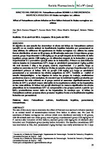
Figure 1. – (a): Method for tracing the hymen plane on T2-weighted TSE midsagittal MR images of female pelvis (a): a line is drawn (dotted line) starting at the lowermost posterior point of the pubic bone through the external urethral and vaginal orifices. The midpubic line (continuous line) is depicted for comparison. (b): The hymen plane is used as reference to measure the position of the perineal body and the distal vaginal angle. BL= bladder; UT= uterus; SP= Symphysis Pubis; MPL= midpubic line; HP= Hymen plane.
the plane of hymen being defined as zero, the location of the perineal body, expressed as millimeters above (negative numbers) or below (positive numbers) the hymen, was calculated. Moreover, the distal longitudinal vaginal axis relative to a horizontal reference line, referred to as the “distal vaginal angle” and the total vaginal length, defined as the greatest depth of the vagina,6 were measured. Finally, the MR anatomy appearance was characterized on individual pictures with regard to its (a) overall image quality, defined as the sharpness with which the single structure was depicted; (b) organ definition, defined as the ability to distinguish the various components as distinct anatomic structures; and (c) visibility score, defined as the frequency with which the presence (or absence) of the structure was visualized for each scanning level. To quantify them, a 4-point grading scale was used according to El Sayed7 when collecting data as follows: 1, not visible; 2, poorly visible; 3, moderately visible; 4, easily visible. Data analysis was performed with SPSS 5.1 (SPSS Inc, Chicago, III). Paired Student t tests were applied, with a significance level determined at P < 0.05 to assess the difference between group A and group B subjects. Values of various measurements were given as mean and standard deviation (SD). RESULTS The vaginal length varied from 68 to 84 mm and the mean was 77.3 mm (SD ± 3.2 mm) in nulliparae; in comparison, the length varied from 71.4 to 86.03 mm with a mean of 74.3 mm (SD ± 5.2 mm) in women after cesarean delivery, P > 0.05. The average distal vaginal angle was 70.1 degrees, range 58.2-77.4 degrees in nulliparous and 74.04 degrees, range 61.1-76.2 degrees in the cesarean group, P > 0.05. The average distance of the apex of the perineal body to the hymene plane was + 3.2 ± 2.4 mm, range -4 to +6 mm in nulliparous and + 2.4 ± 1.8 mm, range -1 to + 7 mm in the cesarean group, P > 0.05. Vaginal Morphology: at the DeLancey level I, the crosssectional vaginal configuration assumed a typical linear, horizontally oriented shape in over 91% of cases with a minimal (max 5°) obliquity toward the right or left side in the remaining 9% of cases. By contrast, a typical butterfly or H-shape of the vaginal morphology was seen in 74% and a W-shape in 26% of cases at the DeLancey level II showing a symmetric insertion to the inside of LA muscle and to the outer lateral margin of rectum (also called posterior vaginal sulcus), as opposed to a U-shape at the DeLancey level III in 82% of cases, respectively. Overall, the enface vaginal morphology was depicted at best in the midcoronal MR images as a rectangular structure of low signal intensity with thin bilateral, more or less symmetric linear
54
a
b
c
d
Figure 2. – (a) Normal vaginal morphology at MRI as seen on axial, coronal and sagittal planes: T2-weighted TSE axial images taken at DeLancey level I (a), Level II (b), and level III (c) for evidence of the three different vaginal shapes (dotted e line); (d) coronal STIR midvaginal image showing the vaginal apex and the site at which the suspensory vaginal support structures converge on both sides (dotted line); (e) sagittal T2 weighted TSE image showing the paracolpium as an hyperintense structure beyond the anterior and posterior vaginal walls. A vaginal angle of 72 degrees is formed by the intersection of the reference line (dotted line) with the distal vaginal axis (continuous line). V= vagina; BL= bladder; SL= suspensory ligaments; SP= symphysis pubis.
stripes originating from its upper lateral corner, interpreted as the suspensory vaginal ligaments. On the other hand, the midsagittal MR images were ideal for determining vaginal axis inclination, total vaginal length, and perineal body’s position with respect to the hymeneal plane (Figure 2). Vaginal Walls: most commonly, on T2-weighted axial images the structure of the vagina had a two-layered, 2-3 mm thick consistent appearance showing homogeneous hypointense signal intensity which represented the combined anterior and posterior walls faced together with their virtual internal lumen showing a high-signal-intensity due to mucous or secretion in the center (see Figure 2 b). Occasionally, three vaginal wall layers could also be identified from internal to external as follows: a low-signal-intensity inner layer, an intermediate-signal-intensity middle layer, and a low-signal-intensity outer layer. These correspond to layers of squamous keratinized epithelium, lamina propria of loose connective tissue, and a muscular layer, respectively.8 T2-weighted sagittal images allowed easier depiction and interpretation of the vaginal wall layers probably because of more favorable contrast and spatial resolution with adjacent structures. Parametrium and paracolpium: the uterosacral and round ligaments were seen alternatively on axial and/or coronal MR images with a variable frequency of 71% and 58% of cases as thin linear structures of low signal intensity (Figure 3) extending from the upper part of the cervix to the sides of the sacrum, and from the angles of the uterus downward, laterally and forward through the inguinal canal to the labia majora, respectively. Despite their superior visibility rate (up to 79% of cases), the cardinal ligaments couldn’t be recognized as isolated structures; rather, their identity was synonimus with the visible accompanying vas-
MR imaging of vaginal morph:ingynious 05/06/15 10:09 Pagina 55
MR imaging of vaginal morphology, paravaginal attachments and ligaments. Normal features
a
b
Figure 5. – (a) Paraurethral supporting structures seen on axial T2weighted TSE MR images: periurethral ligament (continuous line) and paraurethral ligament (dotted line); (b) pubourethral ligament (dotted line). Peri= periurethral ligaments; Para= paraurethral ligaments; PuboU= pubourethral ligaments.
Figure 3. – Axial T2-weighted TSE image taken at the level of uterine cervix showing the supporting structures of the uterosacral ligaments as thin curvilinear hypointense stripes coursing backward (short dotted line) and a portion of the round ligaments (long dotted line) coursing forward; the site of the cardinal ligaments is inferred by the presence of vessels, limphatics and nerves (long continuous line). C= cervix; BL= bladder; R= rectum.
Figure 6. – Insertion of the pubococcygeus to the obturator internus muscle through the endopelvic fascia (dotted line) as seen on axial oblique proton density (PD) pulse sequence. ATFP= arcus tendineus fasciae pelvis; U= urethra; V= vagina; PCm= pubococcygeus muscle.
Figure 4. – The paracolpium on axial T2-weighted TSE image is depicted as an hyperintense structure surrounding the vaginal walls (continuous line). Parac= paracolpium; U= urethra; OIm= obturator internus muscle; PRm= puborectalis muscle; R= rectum.
cular supply, nerves and fat forming the parametrium. Overall, a failure rate of up to 40% in the depiction of the uterosacral and round ligaments at MR imaging was registered in both groups. The paracolpium was identified as a hyperintense structure surrounding the vaginal wall anteriorly, laterally and posteriorly with variable thickness. Its high signal intensity is considered a result of a combination of connective tissue and venous plexus which is bulkier around the upper third of the vagina (Figure 4). Paraurethral attachments: three components were consistently recognized on axial MR images (Figure 5) in all but three cases, as follows: the periurethral ligaments as a thin hypointense arcuate structure coursing ventrally to the urethra and connecting the medial aspect of the puborectalis muscle of one side to the other; the paraurethral ligaments as a slightly oblique, hypointense thin structure originating
at the 4 and 8 o’clock position of the urethra and connecting its lateral wall to the periurethral ligament described above; and the pubourethral ligament, as a thin hypointense structure distinct from the anterior vaginal wall, located behind the posterior aspect of the urethra as an hammock which connects the urethra to the arcus tendineus fascia pelvis. Levator ani muscle attachments: the insertion of iliococcygeus muscle to the inner border of the internal obturator muscle border, as seen on both T2-weighted axial and coronal images, served to localize the arcus tendineus fasciae pelvis while the insertion of the pubococcygeus muscle to the inside of the pubic bone and to the obturator internus muscle (Figure 6) testified the integrity of the pubocervical fascia. Perineal membrane, perineal body and urogenital diaphragm: the perineal membrane is a primarily fibrous structure of intermediate signal intensity, triangular in shape spanning the space between the two ischiatic rami (Figure 7). It includes also a muscular component composed by the compressor urethrae and urethrovaginal sphincter. Superficial and inferior to the perineal membrane lies the perineal fascia made up of an adipose and membra-
55
MR imaging of vaginal morph:ingynious 05/06/15 10:09 Pagina 56
Vittorio Piloni
a
b
Figure 7. – (a) The perineal membrane is depicted on coronal T2weighted TSE images as a triangular shaped structure of intermediate signal intensity (continuous line), uniting the ischiopubic rami of one side to the other. Figure (b) Corresponding STIR image (b) showing at best the superficial transverse muscle of perineus as a hypointense horizontal structure (dotted line). PM= perineal membrane; TPm =trasversus perinei muscle.
nous layer providing the fatty tissue of the labia majora, and attached laterally to ischiopubic rami and posteriorly to the free margin of the urogenital diaphragm. Between its inferior fascia and the perineal fascia is the perineal space which contains the ischiocavernous and bulbospongiosus muscles and the perineal body. The latter appears as a pyramidal hypointense, fibromuscular structure lying in the midline of the perineum, posterior to the vagina and anterior to the anal canal (see Figure 1 b). It provides an anchor point for several muscles including the deep and superficial transverse perinei, the external anal sphincter, the pubovaginalis and sphincter urethrae and is depicted at best on both T2-weighted images taken in the sagittal and in the oblique axial plane (Figure 8). A complete summary of the MR anatomic features and parameters observed in the patient population is presented on Table 1 and 2. DISCUSSION The term vagina is derived from Latin va¯gı¯ nae, literally “sheath”. Anatomically, its precursor, called vaginal plate, derives from the growth of tissue that is located where the solid tips of the paramesonephric ducts (Müllerian ducts) enter the dorsal wall of the urogenital sinus as the Müllerian tubercle. Eventually, the central cells of the plate break down to form the vaginal lumen which is not fully canalized until sexual differentiation between males and females is completed. While the urogenital sinus persists as the vestibule of the vagina, two urogenital folds develop on the belly aspect of the genital tubercle giving rise to the labia minora, and to labioscrotal swellings which enlarge to form the labia majora.9 Progressively, the human vagina develops into an elastic fibromuscular canal resembling a deflated tube approximately 7.5 cm long across the anterior wall (front), and 9 cm across the posterior wall (rear), making the posterior fornix deeper than the anterior. While the anterior and posterior walls are touching each other, the lateral walls, especially in their middle area, are relatively more rigid; because of this, the vagina has an H-shaped cross section. From the lumen outwards, three layers are commonly described in the wall of the vagina, as follows: an internal layer consisting of a mucosa of non-keratinized stratified squamous epithelium with an underlying lamina propria of connective tissue forming folds which are more prominent in the caudal third of the vagina and appear as transverse ridges whose function is to provide the vagina with increased surface for extension and stretching; an in-
56
Figure 8. – Efficacy of the axial oblique plane for depiction of the perineal body (continuous line) as a distinct structure from the anal sphincter. The black dot (dotted line) indicates the intra-anal marker. PB= perineal body; AM= intra-anal marker. TABLE 1. – Measurement of vaginal length, distal vaginal angle and perineal body position In nulliparous (Group A) and after cesarean delivery (Group B). Parameter Age (yrs) Parity (n°) Vaginal length (mm) Vaginal Angle (°) Perineal Body position (mm)
Group A (n = 25) 31.3 28-35 0 77.3 (3.2) 68.2 - 84.5 70.1 (4.8) 58.2 - 76.4
mean range mean range mean range
Group B (n = 8) 34.1 31-40 1.2 (1-3) 74.3 (5.2) 71.4 - 86.03 74.04 (1.68) 61.1 - 78.2
P §§ n.s.
n.s. n.s.
mean + 3.2 (2.4) +2.4 (1.8) n.s. range -4 /+6 -1/+6 n.s. § - Relative to the hymeneal plane - (above) or + (below); §§- P value .05; numbers in parenthesis are SD. TABLE 2. – Visibility of vaginal and paravaginal anatomy at pelvic MRI in nulliparous and after cesarean delivery. Observed structure
Item Scan plane Overall Organ Visibility Image quality definition score (€) 4 4 4 Axial
Vaginal wall Vaginal shape Upper Middle Lower Vaginal Inclination Parametrium (ligmnt) Uterosacral Cardinal Round Paracolpium Paraurethral (ligmnt) Periurethral Paraurethral Pubourethral Endopelvic Fascia (Insertion) Iliococcygeus m. Obturator int. m. Pubococcygeus m. Perineal Membrane Perineal Body
4 4 2 4
4 3 2 4
4 3 2 4
Sagittal
4 3 3 4
4 2 3 4
3 2 2 4
Axial Axial Axial/Coronal Axial/Sagittal
3 2 3
3 2 3
3 2 2
Axial
4 4 3 4 4
3 3 3 4 4
3 3 3 4 4
Axial/Coronal Axial/Coronal Axial Coronal Sagittal
€ 1-4 point visibility score according to El Sayed.7
Axial
MR imaging of vaginal morph:ingynious 05/06/15 10:09 Pagina 57
MR imaging of vaginal morphology, paravaginal attachments and ligaments. Normal features
termediate layer of smooth muscle composed by an outermost layer of longitudinal muscle and an innermost layer of circular muscle with an oblique muscle fibers in between; finally, an external layer called adventitia consisting of thin dense layer of connective tissue blending with the loose connective tissue which contains blood vessels, lymphatic vessels and nerve fibers present between pelvic organs. Functionally, the vagina is known to expand in order to hold what’s inside it, be the sperm released by male penis during sexual intercourse, a baby during vaginal delivery, or the menstrual flow which includes the unfertilized egg, blood and pieces of mucosal tissue. Less attention, however, has received the fact that, through its paravaginal connective tissues, the vagina acts as an adhesive “glue” which plays a vital role in maintaining the correct position and stability of pelvic organs relative to the pelvic side walls. More specifically the vagina, together with a series of fascial condensations arising from its lateral aspect (paracolpia) and some ligaments (parametria), is continuous with several muscular and connective structures, namely the levator ani muscle and the endopelvic fascia. The latter envelopes the entire vaginal canal, extending from apex to perineum. In his classic paper5 DeLancey described the connective tissue support of the vagina as having 3 levels. Level I support is composed of the uterosacral/cardinal ligament complex that originates at the cervix and upper vagina and inserts at the pelvic sidewall and sacrum. This ligamentous complex suspends the uterus and upper vagina in its normal orientation. It helps maintain vaginal length and normal vaginal axis. Level II support comprises the paravaginal attachments that run through the length of the vagina and are suspended by the arcus tendineus fasciae pelvis (ATFP), a fibrous band that is attached in the front to the pubic bone and in the back to the ischial spine. Level III support is provided by the perineal membrane, perineal body, and superficial and deep perineal muscles, recently renamed by DeLancey as compressor urethrae and urethrovaginal sphincter. With regard to imaging, various techniques have been developed in search of accurate visualization and quantitative assessment of the vaginal canal, including vaginography combined with defecography10 and transperineal sonography:11 both are well suited for the static and dynamic examination but their drawbacks include high exposure to ionizing radiation and absence of information on surrounding soft tissue, and excessive dependence on operator skill, respectively. The advantages of MRI include non exposure to ionizing radiation, high soft-tissue contrast resolution and multiplanarity which allow clear depiction of all pertinent anatomy. First of all, the position of the hymen plane, which provides a universally accepted and consistently visible reference structure, could always be depicted clearly in the present study allowing easy identification of vaginal length, its orientation, and location of the perineal body. Hence, this reference line, coterminous with the clinical level described by Bump et al.,6 seemed to us preferable when compared to the midpubic line (MPL), i.e. a line extending along the long axis of the pubic bone as proposed by Singh et al.,12 because of the excessive variability in the inclination of the latter (see Figure 1a) potentially leading to erroneous measurements of established parameters and overdiagnosis of pelvic organ prolapse. Secondly, with regard to the vaginal morphology, there still seems to exist a lot of controversy in the literature and very little published data on normal pattern. Moreover, while some researchers reported that a flattened vagina on axial images is associated with loss of vaginal support,13 evidence is given in the present paper that its cross sectional configuration depends mainly on the distribution of the par-
avaginal attachments and that three vaginal shapes could consistently be recognized in healthy subjects with no prior parity and after cesarean delivery, as follows: a linear-shape (91%) in DeLancey level I; an H-shape or, less frequently, a W-shape (74% and 26%, respectively) in level II; and an U-shape (82%) in level III. Overall, with regard to anatomical identification at axial MR imaging, the upper third linear vagina was the most easily seen (average visibility score, 4); the middle third vagina was the most peculiar and variable in shape (average visibility, score 3); the lower third U-shaped vagina was the most difficult to be recognized as a distinct structure from adjacent structures (visibility score, 2). In such cases, proper obliquity of axial scan planes proved helpful to distinguish the various structures (see Figures 6 and 8). Besides the ability to depict vaginal configuration, the most striking finding of the present study is definite demonstration that (a) cesarean section left unaltered the vaginal and paravaginal anatomy; and (b) fascial condensation such as uterosacral, cardinal and round ligaments were seen at MRI more than occasionally. However, the frequency with which they could not be seen in women with normal pelvic support of both groups (up to 40%) seems to indicate that lack of visibility per se does not prove their absence or even rupture. For these reasons, absent visibility of ligaments should be interpreted with caution before assigning a verdict of pelvic support defect by keeping in mind potential limitations which frequently occur such as variation in normal anatomy or superimposed bowel loops. A more specific consideration should be deserved to the issue of vaginal morphology according to body position. Most likely, vaginal shape, as depicted in the present paper, corresponds to that of women during sleeping and/or sexual intercourse rather than walking, as images were taken with a conventional (horizontally oriented) MR scanner. Presumably, it can be hypothesized that vaginal axis inclination and paravaginal orientation might appear differently in upright position. Lastly, according to the well known theory that the vagina undergoes significant changes in its passive mechanical properties throughout pregnancy which recover post partum,14 it is interesting to note that no significant different values for vaginal and paravaginal structures were observed in the group of women of our study who received caesarian section when compared to nulliparous women. CONCLUSIONS The vagina is a fibromuscular tube capable of a high degree of distention, both during intercourse and particularly during childbirth, but also serves as the outlet for menstrual flow and is the primary supporting structure of female pelvic organs. Using high-resolution MR imaging with external coil allows visualization of vaginal and paravaginal attachments, fascial condensations called ligaments, and pelvic floor musculature with exquisite details in both nulliparae with normal pelvic support and in women who delivered by cesarean section. With no need for organ opacification for visualization, use of ionizing radiation or excessive dependence on operator skill and technology, precise visualization of MRI anatomy is the prerequisite for identifying normal features and discerning them from variations in the vaginal canal and its supporting structures. It is likely that early recognition of most common abnormalities might become the anatomic basis for interpretation of evacuation and voiding dysfunctions and will be important in patients with pelvic floor disorders for both selecting treatments and estimating their efficacy. These speculations need further investigation.
57
MR imaging of vaginal morph:ingynious 05/06/15 10:09 Pagina 58
Vittorio Piloni
REFERENCES 1. Tunn R, DeLancey JO, Quint FF. Visibility of pelvic organ support system structures in magnetic resonance images without an endovaginal coil. Am J Obstet Gynecol 2001; 184: 1156-1163. 2. Chou Q, DeLancey JOL. A structured system to evaluate urethral support anatomy in magnetic resonance images. Am J Obstet Gynecol. 2001; 185: 44-50. 3. Tunn R, DeLancey JOL, Howard D, et al. Anatomic variations in the levator ani muscle, endopelvic fascia, and urethra in nulliparas evaluated by magnetic resonance imaging. 2003; 188: 116-121. 4. DeLancey JOL. Structural anatomy of the posterior pelvic compartment as it relates to rectocele. Am J Obstet Gynecol. 1999; 180: 815-823. 5. DeLancey JOL. Anatomic aspects of vaginal eversion after hysterectomy. Am J Obstet Gynecol 1992; 166: 1717–28. 6. Bump RC, Mattiasson A, Bo K, et al. The standardization of terminology of female pelvic organ prolapse and pelvic floor dysfunction. Am J Obstet Gynecol. 1996; 175: 10-17. 7. El Sayed RF, Morsey MM, El Mashed SM, Abdel-Azim MS. Anatomy of the urethral supporting ligaments defined by dissection, histology, and MRI of female cadavers and MRI of healthy nulliparous women. AJR 2007; 189: 1145-1157. 8. Kim JK, Kim YJ, Choo MS, et al. The urethra and its supporting structure in women with stress urinary incontinence: MR imaging using an endovaginal coil. AJR; 180: 1037-1044.
9. Cai Y. Revisiting old vaginal topics: conversion of the Müllerian vagina and origin of the “sinus” vagina. Int J Dev Biol. 2009; 53: 925-934. 10. Vanbeckevoort D, Van Hoe L, Oyen R, et al. Pelvic floor descent in females: comparative study of colpocystodefecography and dynamic fast MR imaging. J Magn reson Imaging. 1999; 9: 373-377. 11. Schaer GN, Koechli OR, Schuessler B, et al. Perineal ultrasound for evaluating the bladder neck in urinary stress incontinence. Obstet Gynecol. 1995; 85: 220-224. 12. Singh K, Reid WMN, Berger LA. Assessment and grading of pelvic organ prolapse by use of dynamic resonance imaging. Am J Obstet Gynecol. 2001; 187: 71-77. 13. Klutke C, Golomb J, Barbaric Z, Raz S. The anatomy of stress incontinence: magnetic resonance imaging of female bladder neck and urethra. J Urol. 1990; 143: 563-566. 14. Feola A. Impact of pregnancy and vaginal delivery on the passive and active mechanics of the rat vagina. Am Biomed Eng. 2011; 39: 549-558.
Correspondence to: Vittorio Piloni, M.D. Radiologist Iniziativa Medica - Imaging Centre Via Rialto 14 - Monselice (Padova) - 35043 Italy E-mail:
[email protected]
Multidisciplinary UroGyneProcto Editorial Comment To improve the integration among the three segments of the pelvic floor, some of the articles published in Pelviperineology are commented on by Urologists, Gynecologists, Proctologists/Colo Rectal Surgeons or other Specialists, with their critical opinion and a teaching purpose. Differences, similarities and possible relationships between the data presented and what is known in the three fields of competence are stressed, or the absence of any analogy is indicated. The discussion is not a peer review, it concerns concepts, ideas, theories, not the methodology of the presentation. UROLOGIST The manuscript on normal MR vaginal morphology by Vittorio Piloni is an excellent example how to proceed with the evaluation of new diagnostic techniques or new application. Although applied to the pelvic floor since almost a decade no real objective evaluation and classification of MRI findings of the female pelvic floor has been performed. There are primarily two important main objectives that need to be addressed, if imaging of the pelvic floor is evaluated: 1. Is it used to proof a theory or a hypothesis or a pathophysiological concept. 2. What is the clinical utility or the added value in everyday clinical practice, if compared to current techniques, such as pelvic ultrasound. Addressing the first objective MRI is certainly a promising technique, as it has the capability of identifying structures that might not be visible with current techniques. In this regard the limitation of the MRI itself in the study have to be mentioned. The 1.5 T MRI apparently has limitations in the correct identification of ligamentous structures. This is elegantly described in the table 2 of the manuscript, which could serve as a reference table for further investigations in this field. Future developments and applications of higher levels of MRI (3 and more T MRI) could overcome low identification rates of certain anatomical structures such as cardinal ligaments, perineal body or endopelvic fascia (Wagenlehner et al. 2013).1 Another important aspect is the dynamic part of anatomy. Functional MRI of the pelvis is an emerging and improving technique, which could be applied to correlate defective structures with defective function. Addressing the second objective the added value is sometimes difficult to assess, as in urogynecology a careful investigation including history taking can enlight many clinical questions already (Wagenlehner et al. 2013).2 Sonography also has improved considerably in the past decade, with regard to identifying important structures. However it has not come to the detail of identifying lig-
58
amentous structures, which are of paramount importance in the anatomy and functional processes of the pelvic floor (Wagenlehner et al. 2010).3 In this aspect novel imaging by MRI could be very important, when defective ligamentous structures could be identified in relation to their functional importance. Exact diagnosis could then be used to guide site specific repair perhaps more accurate than only applying physical exam techniques and sonography. REFERENCES 1. Wagenlehner FM, Del Amo E, Santoro GA, Petros P. Live anatomy of the perineal body in patients with third-degree rectocele. Colorectal Dis. 2013; 15: 1416-22. 2. Wagenlehner FM, Liedl B, Bschleipfer T, Petros PE. Comment on Nager: the urethra is a reliable witness: simplifying the diagnosis of stress urinary incontinence. Int Urogynecol J. 2013; 24: 1413-4. 3. Wagenlehner FM, Bschleipfer T, Liedl B, Gunnemann A, Petros P, Weidner W. Surgical reconstruction of pelvic floor descent: anatomic and functional aspects. Urol Int. 2010; 84: 1-9. F. Wagenlehner Clinic for Urology, Pediatric Urology and A ndrology Justus-Liebig-University Giessen, Germany
[email protected]
GYNECOLOGIST The pelvic floor remains a mystery for most clinicians. Until very recently, all anatomical knowledge has been derived from cadaveric dissections where the pelvic diaphragm is by definition collapsed. Live pelvic floor anatomy is vastly different from cadaveric anatomy. It is my belief that many of the problems which
MR imaging of vaginal morph:ingynious 05/06/15 10:09 Pagina 59
MR imaging of vaginal morphology, paravaginal attachments and ligaments. Normal features have arisen from large mesh implants can be traced back to the method of teaching, exclusively in cadavers. We, as practising surgeons, need to disregard cadaveric anatomy. The whole pelvic floor anatomy has now to be re-set in dynamic live anatomy terms. Dr Piloni is one of the pioneers of the imaging of pelvic floor anatomy. Works such as this on the anatomy of the vagina, its ligaments and muscles is critically important, because without normal reference points, we can never develop the methodology to accurately assess dysfunction Dynamic 2D transperineal ultrasound is cheap and helpful as regards understanding the movement of organs and muscles on coughing and straining. Unfortunately, dynamic transperineal ultrasound cannot be accurately measured. The other problem with 2D ultrasound is the potential for distortion of the image. MRI is considered more accurate, but even here, up to 40% of structures in Dr Piloni’s images were not well defined. The more important question as regards imaging is “What are we looking for”. What do we write on the imaging request form? My own imaging investigations had two major objectives, to gain insights into pelvic floor function and dysfunction and to test the Integral Theory’s predictions for truth or falsity. Within this limited context, I would like to comment on some of Dr Piloni’s findings based on my own investigations1-4 and many thousand of transperineal ultrasounds over the past 20 years. I can confirm his findings that “the upper vagina had an horizontal, linear shape in over 91%; the middle vagina an H-shape”. We assessed the vaginal axis differently from Dr PIloni. We checked the organ compression normally seen on straining. During effort, the upper part of the vagina was stretched backwards and downwards against the perineal body. Compression of level 2 on standing lateral X-ray appeared to be related to the angle of the upper vagina to the horizontal at rest. In 23 patients in whom the angle was 450 or more to the horizontal, only 2 demonstrated significant angulation of the upper vagina and therefore compression of level 2 on straining. In contrast, all 27 patients with an angle less than 450 to the horizontal demonstrated both vaginal angulation and compression. In a live anatomical study, we examined the perineal body again differently from Dr Piloni. We measured its total length which averaged approximately 4 cm. The relevance of muscle forces to the three anatomical levels of support, the cardinal/uterosacral ligament complex (level 1), the rectovaginal fascia (level 2) and the perineal body (level 3), was analyzed. We found that the 3 directional forces operated normally even in the cases where the lax connective tissue prevented organ rotation and compression of level 2. We biopsied the suspensory ligaments. Histology demonstrated smooth muscle and nerves in the suspensory ligaments, indicating an active contractile role for these structures.
REFERENCES 1. Petros PE and Ulmsten U Role of the pelvic floor in bladder neck opening and closure: I muscle forces. Int J Urogynecol and Pelvic Floor, 1997; 8: 74-80. 2. Petros PE and Ulmsten U Role of the pelvic floor in bladder neck opening and closure: II vagina. Int J Urogynecol and Pelvic Floor, 1997; 8: 69-73. 3. Petros PE Vault prolapse 1: dynamic supports of the vagina, Int J Urogynecol and pelvic floor. 2001; 12: 292-295.
4. Wagenlehner F. M. E., Del Amo E, G. A. Santoro GA and P. Petros. Live anatomy of the perineal body in patients with third-degree rectocele. Colorectal Dis. 2013; 15: 1416-22. P. Petros St Vincent’s Hospital, University of NSW, Sydney Australia
[email protected] COLORECTAL SURGEON The excellent paper on MR imaging of vagina, paravaginal attachments and ligaments, gives some ideas to Coloproctologists. Rectocele and rectoanal intussusception are related to changes of rectal morphologies and dynamics and inevitably there are also attendant alterations of vagina. MRI pelvic findings, measured as suggested by Vittorio Piloni, will be useful to evaluate the qualitative and quantitative alterations of vaginal morphology: the close connection between posterior wall of vagina and anterior wall of rectum will be better defined and it could help to choose the best surgical option, if prosthetic or resective. Moreover the MR evaluation of paravaginal attachments and ligaments offers a hint of truth on performance of these anatomic landmarks. The anatomic anchor of ligaments on vagina and rectum and their influence on rectal static will be better studied: simultaneous pathophysiology of pelvic organ prolapses and rectal diseases will be better understood. Thank you dr. Piloni! Filippo Pucciani Dipartimento di Chirurgia e Medicina Traslazionale Università di Firenze
[email protected] AUTHOR’S REPLY For some aspects, pelviperineologists seem to be a number of different inhabitants living around a lakeshore, with some of them standing side-by-side and others on opposite shores. Accordingly, different views and perspectives of the same reality are perceived by their eyes. Nevertheless, should a windy air drift arise from one side of the “lake”, it will inevitably produce an effect on the other shore which, occasionally, could be even more resounding. This is the case which has been encountered with the two most common clinical problems , i.e. urinary incontinence and rectocele, with much debate between the involved specialists (urologist vs gynecologist, and gynecologist vs coloproctologist , respectively) including the issues of terminology, classification and treatment. Undoubtedly, the intuition of the Editor to obtain a comment on my paper from Petros, Wegenlehner and Pucciani was great: potentially, a hornet’s nest might have been stirred up, ranging from interest to curiosity, indignation or controversy. As it would have been expected, however, rather than a unitary thought, just a sort of side-to-side dialogue between “neighbours” emerged, and no more than a trend towards the need for better integration of different specialists onto a mutual society. Probably, as a radiologist familiar with all the three physical sources of diagnostic imaging (X-ray, Ultrasonography, and MRI) my privilege comes from the capability to see through the barriers of pelvic floor compartments, thus overcoming the limitations of the other perineologists.
59
Chronic pelvic pain syndrome:ingynious 08/06/15 12:52 Pagina 60
Original article
Chronic pelvic pain syndrome in women. Review and preliminary results with low-energy extracorporeal shock wave therapy ANDREA MENEGHINI1, MONICA TREVISAN3, EFTHYMIA LAMPROPOULOU3, NICOLA MENEGHINI2 Padua Teaching Hospital, Advanced Technology in Rehabilitation Lab, Department of Orthopaedic Rehabilitation School of Medicine, University of Padua 3 School of Specialization in Physical Medicine and Rehabilitation, Department of Neuroscience, University of Padua 1 2
Abstract. Introduction: Chronic Pelvic Pain Syndrome (CPPS) is a highly prevalent and very debilitating clinical condition, with a significant impact on the social, working and family activities, negatively affecting the quality of life. Currently there is not yet an satisfying treatment. Several therapeutic options have been proposed and experimented with some results, but in certain patients they are all ineffective. Extracorporeal Shock Wave Therapy (ESWT) could be a new secure and promising approach for this condition. Aim of the study: To describe our experience about the effects of three cases of female CPPS. Materials and Methods: Three women suffering from CPPS underwent four weekly sessions ESWT (3000 SW, 3 Hz, 0,25 mJ/mm2) with the aim to reduce their pain. Basal and 2 follow-up assessments were conducted using NRS pain score and recording the consumption of medications. Results: In one case we observed a partial improvement on pain, in the second one no benefit and in the last one an almost complete disappearance of the pain. No adverse events were registered. Discussion and Conclusions: Although our result are discordant, Low-energy ESWT could represent a new promising treatment for CPPS as it is simple, non-invasive, painless, well tolerated, apparently secure, but more studies are needed to discover the mechanisms through which ESWT acts on the pain and to define the optimal parameters and the better approach to use in clinical practice. Keywords: Woman’s pelvic pain; Chronic pelvic pain syndrome; ESWT; Shock wave therapy; Quality of life.
INTRODUCTION Chronic pelvic pain Syndrome (CPPS) is a highly prevalent condition which can present a major challenge to health care providers due to its complex aetiology and poor response to therapy.1,2 Much of the research examining chronic or recurrent pelvic pain in women has been hampered by the lack of a consistent definition.2 CPPS is a very debilitating clinical condition with a significant impact on the social, working and family activities, negatively affecting the quality of life. There is a great variability of prevalence in literature,3 from 2.14, 5 to 43.4%,6 due to the definition used, the characteristics and quality of the studies and the cultural characteristics of the population studied. Pelvic pain is an understated and major problem. The best available figures suggest the number of women in the UK with chronic pelvic pain as 1 million (compared with 1.6 million adults with low back pain).7 CPPS is the reason of 10% outpatient gynaecological visits, 40% diagnostic laparoscopy and 10-15% hysterectomy in the USA8. Amongst males, CPPS can affect 10%-15% of the population and results in nearly 2 million outpatient visits each year.9 Diseases characterized by pain have a documented higher prevalence in females.10, 11 In particular abdominal and perineal pain syndromes are sharply more frequent among women, because of anatomy, hormonal conditions, and reproductively aspects.12 Besides epidemiological studies have shown differences between women and men’s pain perception.10
TERMINOLOGY AND DEFINITIONS The International Association for the Study of Pain (IASP) defined pain as “an unpleasant sensory and emotional experience associated with actual or potential tissue damage, or described in terms of such damage”.13, 14 Definitions and classifications of chronic pelvic pain (CPP) have evolved from the mid-1990s under the thrust of
60
expert groups and scientific societies involved on this type of pain. Indeed, classic definitions and classifications were based on the notion of organ disease and usual medical process (infectious, inflammatory, metabolic) and did not allow a proper understanding of functional pathologies.15 Apte G et al.16 define pelvic pain as pain arising from the visceral or somatic system and encompasses structures supplied by the nervous tissue from the 10th thoracic spinal level and below. When this pain is recurrent or persistent and associated with symptoms, suggesting involvement of the musculoskeletal, gynecological, urological or gastrointestinal systems and the absence of inflammation or other specific pathology we have a pelvic pain syndrome, while chronic pelvic pain (CPP) is defined as non-malignant pain perceived in the structures related to the pelvis that has been present for more than 6 months or has a non-acute pain mechanism of shorter duration.16 The definition of a chronic pelvic pain theoretically assumes that three components are present: the same pain, its chronic character and pelvic-perineal topography. Nevertheless the definition is more complex and overcomes these three aspects because chronic pain is not only a symptom based on a notion of duration but a syndrome associating various conditions, the chronic pain syndrome.15 More recently the European Association of Urology has defined chronic pelvic pain as chronic or persistent pain perceived in structures related to the pelvis of either man or woman, that is often associated with negative cognitive, behavioral, sexual and emotional consequences as well as with symptoms suggestive of lower urinary tract, sexual, bowel, pelvic floor or gynecological dysfunction. CPP is a frequent and difficult problem because, despite the quality and diversity of diagnostic procedures, no relevant aetiology will be found in 30 to 40 % of all cases.17 Indeed chronic pelvic pain may be subdivided into “specific disease-associated pelvic pain”, if it is related to a welldefined classical pathology (such as infection or cancer) and “chronic pelvic pain syndrome” when it is not associated to an obvious pathology. Hence CPPS is the occurrence of CPP, Pelviperineology 2015; 34: 60-66 http://www.pelviperineology.org
Chronic pelvic pain syndrome:ingynious 08/06/15 12:52 Pagina 61
Chronic pelvic pain syndrome in women. Review and preliminary results with low-energy extracorporeal shock wave therapy
more than one organ site, the term CPPS should be used. As more information is collected suggesting that the CNS is involved, and indeed may be the main cause of many CPP conditions (e.g., bladder, genitalia, colorectal or myofascial), there is a general tendency to move away from end-organ nomenclature. Perineal pain syndrome should be mentioned: it is a neuropathic-type pain that is perceived in the distribution area of the pudendal nerve, and may be associated with symptoms and signs of rectal, urinary tract or sexual dysfunction; in this condition there is no proven obvious pathology. It should be distinguished from pudendal neuralgia which is a specific disease associated with pelvic pain that is caused by nerve damage.1 We report below (Table 1) the EAU classification of chronic pelvic pain syndromes,1 set up according to the axis system used by IASP; it may be a useful tool for clinical purpose:
often associated with negative cognitive, behavioral, sexual or emotional consequences (depression, anxiety, fears about pain or its implications, unhelpful coping strategies, and distress in relationships, catastrophic interpretation of pain, sense of helplessness), as well as with symptoms suggestive of lower urinary tract, sexual, bowel or gynaecological dysfunction, in the absence of proven infection or other obvious local pathology that may account for the pain.1 Confusingly, a patient may have a well-defined pelvic condition concurrently with chronic pelvic pain syndrome.7,18 Pain perception in CPPS may be focused within a single organ, more than one pelvic organ and even associated with systemic symptoms such as chronic fatigue syndrome (CFS), fibromyalgia (FM) or Sjögren’s syndrome. When the pain is localised to a single organ, some specialists may wish to consider using an endorgan term such as prostate pain syndrome, bladder pain syndrome, urethral pain syndrome, chronic anal pain syndrome. When the pain is localised to TABLE 1. – The EAU classification of chronic pelvic pain syndromes.
Chronic Pelvic Pain
Axis I
Axis II
Axis III
Region
System
End-organ as pain syndrome as identified from Hx, Ex, Ix
Specific disease associated pelvic pain
Axis IV
Axis V
Axis VI
Referral Temporal Character characteristics characteristics
Prostate Suprapubic Bladder Inguinal Scrotal Urethral Testicular Urological Epididymal Penile/ Penile clitoral OR Urethral Perineal PostPelvic Pain Rectal vasectomy Syndrome Vulvar Back Vestibular Buttocks Clitoral Endometriosis Thighs associated Gynaecological CPPS with cyclical exacerbation Dysmenorrhoea Irritable bowel Gastrointestinal Chronic anal Intermittent chronic anal Peripheral Pudendal pain nerves syndrome Dyspareunia Pelvic pain with sexual Sexological dysfunction Any pelvic Psychological organ Pelvic floor Musculomuscle skeletal Abdominal muscle Spinal
ONSET Acute Chronic ONGOING Sporadic Cyclical Continuous TIME Filling Emptying Immediate post Late post TRIGGER Provoked Spontaneous
Aching Burning Stabbing Electric
Axis VII
Axis VIII
Associated symptoms
Psychological symptoms
ANXIETY UROLOGICAL Frequency About pain or putative Nocturia Hesitance cause of pain Dysfunctional flow Catastrophic Urge thinking Incontinence about GYNAECOLOGICAL pain Menstrual DEPRESSION Menopause Attributed GASTROINTESTINAL to Constipation pain or Diarrhoea impact Bloatedness of pain Urge Attributed Incontinence to other NEUROLOGICAL causes Dysaesthesia Unattributed Hyperaesthesia PTSD Allodynia Hyperalegesie SYMPTOMS SEXUOLOGICAL ReSatisfaction experiencing Female dyspareunia Avoidance Sexual avoidance Erectile dysfunction Medication MUSCLE Function impairment Fasciculation CUTANEOUS Trophic changes Sensory changes
Coccyx
61
Chronic pelvic pain syndrome:ingynious 08/06/15 12:52 Pagina 62
Andrea Meneghini, Monica Trevisan, Efthymia Lampropoulou, Nicola Meneghini
Figure 1. – Algoritm for the diagnosis and treatment of CPP:46 DRE = digital rectal examination; PSA = prostate-specific antigen; US = ultrasound; PFM = pelvic floor muscle; TRUS = transrectal ultrasound.
62
Chronic pelvic pain syndrome:ingynious 08/06/15 14:54 Pagina 63
Chronic pelvic pain syndrome in women. Review and preliminary results with low-energy extracorporeal shock wave therapy
Physiopatology of CPPS Pain in the pelvic region can arise from musculoskeletal, gynecologic, urologic, gastrointestinal, and/or neurological conditions. Such pain can involve both the somatic (T12S5) and visceral (T10-S5) systems, making the differential diagnosing challenging.16 CPP mechanism may involve: 1. Ongoing acute pain mechanisms19 (such as those associated with inflammation or infection), which may involve somatic or visceral tissue. 2. Chronic pain mechanisms, which especially involve the central nervous system.20,21 3. Emotional, cognitive, behavioural and sexual responses and mechanisms.22-25 In most cases of CPP, ongoing tissue trauma, inflammation or infection is not present.26-29 However, recurrent trauma, infection or ongoing inflammation may result in CPP in a small proportion of cases. For this reason the early stages of assessment include looking for these pathologies.30 A nociceptive event actives acute pain mechanisms (direct activation of the peripheral nociceptor transducers), but could also generate a sensitisation of the nociceptor transducers, thus magnifying the afferent signalling. There may be activation of the so-called silent afferents. The increased afferent signalling is often a trigger for the chronic pain mechanisms that maintain the perception of pain in the absence of ongoing peripheral pathology.31,32 Possible mechanisms by which the peripheral transducers may exhibit an increase in sensibility are: 1. modification of the peripheral tissue, so the transducers become more exposed to peripheral stimulation; 2. increase in the chemicals that stimulate the receptors of the transducers;33 3. modifications in the receptors that make them more sensitive. In general, the first two mechanism lower the threshold of activation of transducers, the third one increases responsiveness to external stimuli.1 At the spinal level three processes are involved in central sensitization: – Changes in existing protein activity (post-translational processing); – changes in genetic transcription of proteins; – structural changes in neuron connectivity. The first process is the earliest (within minutes); the latter two processes may occur within days.34,35 The result is that a stimulus produces a magnified evoked response in these neurons.1 CPPS is probably manifested as a myofascial pain syndrome with an abnormal tone of the pelvic floor muscles, and a neurological component has become increasingly apparent, associated with dysfunctional effects.36,37,38 Myofascial dysfunction of the pelvic floor has been implicated in CPP conditions as both a causative and associated factor responsible for pain.36-40 Many of the complaints are closely connected to the autonomous nervous system, and the interplay between smooth and cross-striated muscles. Acute and chronic inflammations occurring via the sympathetic endplate might be involved, leading to the endogenous generation of pain via nociceptive nerve endings and receptors. Certain kinds of psychological stress can lead to abnormal electromyographic activity and to myofascial pain syndromes.41 Anymore, in the pain syndromes, the role of the nervous system in generating the sensations is thought to be pivotal, but the term “syndrome” indicates that, although peripheral mechanisms may exist, CNS neuromodulation may be more important and systemic associations may occur; “syndrome”
Figure 2. – Duolith SD1 Storz Medical.
takes into account the emotional, cognitive, behavioural, sexual and functional consequences of the chronic pain.1
Diagnosis of CPPS Diagnosis of CPPS is based on symptoms and on exclusion of obvious diseases than could cause pain.42 The presenting symptoms for many of the known causes of chronic pelvic pain (CPP) are often similar and non-specific, making it difficult to differentiate between causes.1,2 Chronic infection, inflammation, neuropathy, pelvic floor muscle dysfunction, autoimmune disease, and neurobehavioral disorders are among the postulated etiologies, although no single factor is thought to be the cause.43 At any rate, to identify the cause of dysfunction, a systematic approach to examination is essential. Such an approach provides the practitioner the best ability to: (1) appraise relevant historical findings; (2) clinically examine their patients by anatomical region; (3) identify specific mechanical and motor control dysfunctions; (4) determine the level of nervous system sensitization; and (5) evaluate the extent of biopsychosocial involvement in the patient’s condition.16 While the history may indicate pain from a pelvic source, consideration for referred pain from structures outside the pelvic region should not be overlooked.1,44 System investigations should be guided by the medical history and examination to exclude and/or identify end organ pathology.45 Laboratory, imaging, neurophysiological studies, endoscopy and laparoscopy can help the physician to make a diagnosis.46 Fall et al.46 proposed an algorithm for diagnosis and management of chronic pelvic pain (Figure 1). Treatment of CPPS Various drugs are used individually and in various combinations to reduce pain and improve quality of life in patients with CPPS:1,46 simple analgesics and NSAIDs, opioids, antidepressants, anticonvulsivants, antibiotics, a-receptor blockers, and 5a-reductase inhibitors (5-ARIs). A certain group of patients may benefit from these therapies, but often side-effects may predominate over possible treatment effects, thus minimising the benefit to the patient.46,47 Other therapeutic options are represented by nerve blocks. Sacral neuromodulation, Botulinum toxin, TENS, triggerpoints’ massage, electromagnetic treatment, acupuncture, cognitive behavioural therapy, and biofeedback and relaxation training, hyperthermia, and phytotherapy.43,46,47
63
Chronic pelvic pain syndrome:ingynious 08/06/15 12:52 Pagina 64
Andrea Meneghini, Monica Trevisan, Efthymia Lampropoulou, Nicola Meneghini
In the latest years some studies have proposed ESWT for treatment of CPPS.43,47,48,49,50 Low-energy ESWT could affect CPPS by several mechanisms, such as reducing passive muscle tone, hyperstimulating nociceptors, interrupting the flow of nerve impulses, or influencing the neuroplasticity of the pain memory. Human data for the indication of CPPS are not available for any of these mechanisms. The number of shock waves and the energy level chosen were purely empirical, and many technical questions (eg. the impact of prostate volume) remain unanswered.47,49 Despite this limits, this approach might represent an advance in the treatment of CPPS, thanks to its benefits: the possibility of outpatient execution, no need for anaesthesia, lack of side-effects, easy repeatability.47 ESWT-associated pain alleviation based on hyperstimulation of nociceptors was intended to interrupt the flow of nerve impulses. 51,52 Furthermore, ESWT-induced revascularization processes can alleviate pain and help to heal tissue.47,53 The stimulation of microvascularization and reduction in muscle tone after applying SWs is demonstrated.54 ESWT possibly influence the neuroplasticity of the ‘pain memory’.48 The prolonged lack of effective pain therapy could lead to a reinforcement of negative impulses (pain) in the brain. Long-term fixation of these impulses could result in the development of a particular pain memory. By minimal pain impulses, ESWT could break through this negative-conditioned pain memory and induce a sort of “reprogramming”, resetting the pain.55 This theory might explain, for example, why it is possible to influence an area of pain located some distance from the treatment locus.47,48 The periprostatic pelvic floor muscles are also influenced by the therapy, therefore local muscle relaxation could be causing the disorder improving as the result of a reduction in functional muscle shortening. Zimmerman et al. supported the hypothesis that the underlying effective mechanisms are not just local alterations, but associated with many factors, because the pain reduction by SWs remained effective over a period of several weeks.47
PATIENTS AND METHODS We treated three women suffering from CPPS with four weekly sessions ESWT with the aim to reduce their pain (all three patients reported a pain intensity 9/10 at NRS). After giving to each patient detailed information about potential benefits and risks of the procedure, treatment was conducted using a standard electromagnetic SW device (DUOLITH SD1, Storz Medical Tägerwilen, Switzerland) (Figure 2), following a protocol based on literature parameters: 3000 focused shock waves, frequency 3 Hz, energy level 0,25 mJ/mm2.47 Follow-up assessment was carried out one week and eight months after treatment. All three patients had already tried several common treatment (drugs, infiltrations, anesthetic blockade, sacral neuromodulation, dilatation, acupuncture, supplements) before coming to our attention. Other information about these patients is reported below: S.M.A., 49 years-old, BMI 23,1, previous appendicectomy, sphincterotomy for anal fissure complicated by abscess, two vaginal deliveries (the first with episiotomy, the second with lacerations), normal intestinal function, regular menstrual cycle. Reported symptoms: intense anal and gluteal pain, lasting for 6 years, absent at night and increasing during the course of the day, worsening during menstrual cycle, associated with anal pricking, daily rectal tenes-
64
mus and anxiety. Physical examination, in particular rectal exploration, pointed out tenderness in the region between anus and right ischiatic spine and in correspondence of tendineous centre of perineum. No abdominal, genital and anal alterations were found at physical examination, anoscopy and sigmoidoscopy, except for scars of previous surgery. P.G., 69 years-old, BMI 18,3, regular intestinal function, previous colecistectomy, surgery for anal fissure, exeresis of an anal polipo, a vaginal delivery, hysterectomy and ovariectomy. Reported symptoms: chronic intense bruising anal pain (mostly during defecation), associated with rectal tenesmus and anxiety. Physical examination revealed abdominal bloating, painful trigger points of the levator ani, no other perineal, anal, genital abnormalities. M.Z., 60 years-old, BMI 20,3, regular intestinal function, two vaginal delivery (both with episiotomy), dysmenorrhoea before the first pregnancy, previous appendicectomy, hysterectomy for endometriosis, surgical treatment for crural hernia, exeresis mammary nodule, exeresis pulmonary hamartoma, osteoporosis, laminectomy for lumbar stenosis (L4-L5). Reported symptoms: intense bruising anal and low-back pain, associated with pollachiuria, urgence, vescical tenesmus (rare rectal tenesmus), intestinal bloating, flatulence, anxiety. Physical examination showed abdominal bloating, tenderness in correspondence of coccyx and ischiatic spine, vulvar and vestibular pain, no other perineal, anal, genital abnormalities. In literature, all studies on ESWT in CPPS describe a perineal approach (patients were supine and the probe was positioned on the perineum). Indeed, we wanted to try a new approach: patients were positioned in lateral decubitus and the probe on the most painful point for half treatment (1500 SW) and then on the gluteal region (at the emergence of pudendal nerve from pelvis both in left then in right part) for the second half treatment (1500 SW), with the intent to interfere with the pudendal nerve transmission. The pudendal nerve comprises the anterior branches of the ventral rami from S2 to S4. It exits the pelvis through the greater sciatic foramen and reenters the pelvis through the lesser sciatic foramen, passing between the sacrospinous ligament anteriorly and sacrotuberous ligament posteriorly, while wrapping behind the ischial spine. Once in the perineal area, the pudendal nerve travels within the Alcock’s (pudendal) canal, a tunnel created by the overlying parietal fascia covering the obturator muscle. The nerve is accompanied by the pudendal artery and, vein, and nerve to the obturator internus through the pudendal canal. The pudendal canal is located on the medial aspect of the obturator internus covered by the obturator fascia. Once the nerve reenters the pelvis it divides into three branches that are named for the structures they innervate. The first branch of the pudendal nerve, the nerve to the levator ani, arises just proximal to the pudendal canal and supplies motor function to the external anal sphincter and perianal skin. The second branch, also known as the perineal branch, provides sensation to the perineal skin,vaginal tissues,and vestibule, as well as motor fibers to the external urethral sphincter. The third branch innervates the anal sphincters. The pudendal nerve provides sensory innervation to an area defined by the inferior pubic ramus, labio-crural folds, and the intergluteal fold. The pudendal nerve converges on the area of the dorsal horn shared with the cervix, uterosacral, and vulvovaginal area. The pudendal nerve is a mixed sensory and motor nerve, often lending to concurrent motor and sensory symptoms.16
Chronic pelvic pain syndrome:ingynious 08/06/15 12:52 Pagina 65
Chronic pelvic pain syndrome in women. Review and preliminary results with low-energy extracorporeal shock wave therapy
RESULTS In the first case NRS before ESWT was 9/10; at oneweek follow up was 9/10; at the 8 months follow-up it was variable from 5/10 (during the day) to 8/10 (in the evening). It was referred that, although pain was intense at certain times of the day, no analgesic drugs were taken. In the second case NRS before ESWT was 9/10 and did not change at the follow-up assessments. Pharmacological therapy remained the same compared to before treatment. In the third case NRS before therapy was 9/10 and gradually decreased during the treatment; at one week followup NRS was 2/10 and kept the same at 8 months, with no need to assume analgesic drugs. No adverse effects occurred altogether. DISCUSSION Chronic pelvic pain (CPP) is a highly prevalent and debilitating clinical condition with a significant impact on the social, working and family activities of women, negatively affecting their quality of life. Identifying the pain generators and effectively treating this condition is a formidable challenge and this explains the tendency for pelvic pain to become chronic.1 Numerous patients face frustration from the inadequate effects of treatment following multiple repeated attempts to cure this disorder. Recently, multi-modal treatment approaches and the utilization of complementary and alternative medicine (CAM) strategies have been suggested as potential treatment options for CP/CPPS.43 Some recent studies have suggested the potential role of ESWT within the therapeutic pathway for CPPS. Actually five studies43,47,48,49,50 on male and just one56 on female CPPS have been issued in literature. The authors reported promising results of this kind of treatment, with an important reduction of pain and an improved quality of life. In our experience three women with CPPS have been treated with ESWT, using parameters based on Vahdatpour’s work (4 sessions, 3000 SW, 3 Hz, 0,25 mJ/mm2).50 All three women suffered from intense pain (NRS score pre-treatment 9/10). According to literature, no pain or discomfort was felt by patients (no anaesthesia was required), and no apparent side-effects or complications occurred, suggesting that this therapy is painless, secure and well tolerated. Results at follow-up assessment were different between the three patients: the first one reported a partial pain reduction (as demonstrated by lower NRS score in some hours of the day and suspension of pharmacologic therapy), the second one had no effect from ESWT, while the third one obtained an important improvement of her pain (NRS score post-treatment 2/10). It would take a wider sample to obtain meaningful data, just three cases are too few. Thus we formulated some assumptions about the possible reasons of such different effects: the pain in the three patients could have a different origin and mechanism (let think about the variability in patients’ histories), therefore a same therapy could be effective in some cases but not in other; the parameters used (determined empirically) were maybe inadequate for at least one of the three patients, that means that therapy should be customized on patient; besides 4 sessions could be too few to obtain a significant result, in fact in other experiences until 11 sessions were given; the original transgluteal approach, that was thought to have the effect of modulate the transmission of the pudendal nerve, could be more effective than the classic perineal one in some patients but not in all of them, probably because in “non-responders” the pudendal nerve is not heavily involved in the pain mechanisms,
but pain originates in other anatomical structures, such as muscles or bowels. CONCLUSIONS CPPS is a frequent condition, often associated with negative cognitive, behavioral, sexual or emotional consequences, compromising the quality of life. Between various therapeutic approaches, most of the time ineffective, ESWT represents a new promising treatment for CPPS: it is simple (it is an outpatient procedure), non-invasive, painless (it does not require anaesthesia), well tolerated, apparently secure. Although our results are discordant, some studies in literature report benefits of this treatment in patients with CPPS (both males and females). It means that some effect of ESWT on CPPS exists, but more studies are needed to discover the mechanisms through which ESWT acts on the pain and to define the optimal parameters and the better approach to use in clinical practice DISCLOSURES The Authors declare that there are no conflicts of interest. This research received no grant from any funding agency in the public, commercial or not-for-profit sectors. REFERENCES 1. Engeler D, Baranowski AP, Borovicka J, Cottrell A, DinisOliveira P, Elneil S, Hughes J, Messelink EJ, Van Ophoven A, Reisman Y, Williams AC de C. Guidelines on Chronic Pelvic Pain. European Association of Urology 2014. 2. Dick ML. Chronic pelvic pain in women: assessment and management. Aust Fam Physician. 2004 Dec; 33(12):971-6. 3. Latthe P, Latthe M, Say L, Gulmezoglu M, Khan KS. WHO systematic review of prevalence of chronic pelvic pain: a neglected reproductive health morbidity. BMC Public Health. 2006; 6:177. doi: 10.1186/1471-2458-6-177. 4. Rulin MC, Davidson AR, Philliber SG, Graves WL, Cushman LF. Long-term effect of tubal sterilization on menstrual indices and pelvic pain. Obstet Gynecol. 1993; 82: 118-121. 5. Zondervan KT, Yudkin PL, Vessey MP, Dawes MG, Barlow DH, Kennedy SH. Prevalence and incidence of chronic pelvic pain in primary care: evidence from a national general practice database. Br J Obstet Gynaecol. 1999; 106: 1149-1155. doi: 10.1111/j.1471-0528.1999.tb08140.x. 6. Zondervan KT, Yudkin PL, Vessey MP, Jenkinson CP, Dawes MG, Barlow DH, et al. The community prevalence of chronic pelvic pain in women and associated illness behaviour. Br J Gen Pract. 2001; 51: 541–547 7. Baranowski1 AP, Lee J, Price C and Hughes J. Pelvic pain: a pathway for care developed for both men and women by the British Pain Society. Br J Anaesth 2014; 112(3): 452-9. 8. Gelbaya TA, Bch MB, El-Halwagy HE. Focus on primary care: chronic pelvic pain in women CME reviw article. Obstetrical and Gynecological Survey, 2001; 56; 12: 757-763. 9. Murphy AB, Macejko A, Taylor A, Nadler RB. Chronic prostatitis management strategies. Drugs. 2009; 69: 71-84. 10. Unruh AM. Gender variations in clinical pain experience. Pain 1996; 65: 123-167. 11. Berkley KJ. Sex differences in pain. Behavioural and Brain Sciences 1997; 20: 371-380. 12. Berkley KJ, Holdcroft A. Sex and gender differences in pain. In: Wall PD e Melzack R (eds) Textbook of pain. Churchill Livingstone, Edinburgh 1999; cap 41, pp. 951-965. 13. Anon. Classification of chronic pain. Descriptions of chronic pain syndromes and definitions of pain terms. Prepared by the International Association for the Study of Pain, Subcommittee on Taxonomy. Pain Suppl 1986; 3:221-6. 14. Anon. Classification of chronic pain: descriptions of chronic pain syndromes and definitions of pain terms. In: Merskey H, Bogduk N, editors. Task force on taxonomy. International association for the study of pain. 2nd ed. Seattle: IASP Press; 1994.
65
Chronic pelvic pain syndrome:ingynious 08/06/15 12:52 Pagina 66
Andrea Meneghini, Monica Trevisan, Efthymia Lampropoulou, Nicola Meneghini 15. Delavierre D, Rigaud J, Sibert L, Labat JJ. Definitions, classifications and terminology of chronic pelvic and perineal pain. Progrès en urologie 2010; 20: 853-864. 16. Apte G, Nelson P, Brisme´e JM, Dedrick G, Justiz RIII, Sizer PS Jr., Chronic Female Pelvic Pain-Part 1: Clinical Pathoanatomy and Examination of the Pelvic Region. Pain Practice, Volume 12, Issue 2, 2012 88–110. doi: 10.1111/ j.1533-2500.2011.00465.x. Epub 2011 May 26. 17. Dellenbach P, Rempp Ch, Haeringer M Th, Simon Th, Magnier F, Meyer Ch. Douleur pelvienne chronique: diagnostic et traitement. Gynécol Obstét Fertil 2001; 29: 234-43 18. RCOG. The Initial Management of Chronic Pelvic Pain. Ed2 Green Top 41. London: Royal College of Obstetricians and Gynaecologists, 2012. 19. Linley JE, Rose K, Ooi L, et al. Understanding inflammatory pain: ion channels contributing to acute and chronic nociception. Pflugers Arch. 2010 Apr; 459(5): 657-69. 20. Serap Kaya, Linda Hermans, Tine Willems, Nathalie Roussel and Mira Meeus, Central Sensitization In Urogynecological Chronic Pelvic Pain: A Systematic Literature Review. Pain Physician 2013; 16: 291-308. ISSN 1533-3159. 21. McMahon SB, Dmitrieva N, Koltzenburg M, et al. Visceral pain. Br J Anaesth 1995 Aug;75(2):132-144. 22. Bergeron S, Khalifé S, Glazer HI, et al. Surgical and behavioral treatments for vestibulodynia: two-and one-half year follow-up and predictors of outcome. Obstet Gynecol. 2008 Jan;111(1): 159-66. 23. Tripp DA, Nickel JC, Wang Y, et al. Catastrophizing and paincontingent rest predict patient adjustment in men with chronic prostatitis/chronic pelvic pain syndrome. J Pain 2006 Oct; 7(10): 697-708. 24. Tripp DA, Nickel JC, Fitzgerald MP, et al. Sexual functioning, catastrophizing, depression, and pain, as predictors of quality of life in women with interstitial cystitis/painful bladder syndrome. Urology. 2009 May; 73(5): 987-92. 25. Nickel JC, Tripp DA, Pontari M, et al. Psychosocial phenotyping in women with interstitial cystitis/painful bladder syndrome: a case control study. J Urol 2010 Jan; 183(1): 167-72. 26. Abrams PA, Baranowski AP, Berger RE, et al. A new classification is needed for pelvic pain syndromes--are existing terminologies of spurious diagnostic authority bad for patients? J Urol 2006 Jun; 175(6): 1989-90. 27. Baranowski AP, Abrams P, Berger RE, et al. Urogenital pain-time to accept a new approach to phenotyping and, as a consequence, management. Eur Urol 2008 Jan; 53(1): 33-6. 28. Baranowski AP, Abrams P, et al. (2008). Urogenital Pain in Clinical Practice. New York, Informa Healthcare. 29. Hanno P, Lin A, Nordling J, et al. Bladder Pain Syndrome Committee of the International Consultation on Incontinence. Neurourol Urodyn 2010; 29(1): 191-198. 30. van de Merwe JP, Nordling J, Bouchelouche P, et al. Diagnostic criteria, classification, and nomenclature for painful bladder syndrome/interstitial cystitis: an ESSIC proposal. Eur Urol 2008 Jan; 53(1): 60-7. 31. Wesselmann U, Baranowski AP, Börjesson M, et al. Emerging therapies and novel approaches to visceral pain. Drug Discov Today Ther Strateg. 2009 Fall; 6(3): 89-95. 32. Giamberardino MA, Costantini R, Affaitati G, et al. Viscerovisceral hyperalgesia: characterization in different clinical models. Pain. 2010 Nov; 151(2): 307-22. 33. Cervero F, Laird JM. Understanding the signaling and transmission of visceral nociceptive events. J Neurobiol. 2004 Oct; 61(1): 45-54. 34. Nazif O, Teichman JM, Gebhart GF, et al. Neural upregulation in interstitial cystitis. Urology. 2007 Apr;69(4 Suppl):24-33. 35. Merskey H, Bogduk N. Classification of Chronic Pain. Seattle, IASP press. 36. Oyama IA, Rejba A, Lukban JC, et al. Modified Thiele massage as therapeutic intervention for female patients with interstitial cystitis and high-tone pelvic floor dysfunction. Urology. 2004; 64: 862-865. 37. Karin E. Westesson & Daniel A. Shoskes Chronic Prostatitis/Chronic Pelvic Pain Syndrome and Pelvic Floor Spasm: Can We Diagnose and Treat? Curr Urol Rep 2010; 11: 261-264. 38. Rhonda Kotarinos Myofascial Pelvic Pain Curr Pain Headache Rep (2012) 16: 433-438 DOI 10.1007/s11916-012-0277-8.
66
39. Weiss JM. Pelvic floor myofascial trigger points: manual therapy for interstitial cystitis and the urgency-frequency syndrome. J Urol. 2001; 166: 2226-2231. 40. Anderson RU, Wise D, Sawyer T, Chan C.Integration of myofascial trigger point release and paradoxical relaxation training treatment of chronic pelvic pain in men. J Urol 2005; 174: 155-60. 41. Simons DG. Review of enigmatic MTrPs as a common cause of enigmatic musculoskeletal pain and dysfunction. J Electromyogr Kinesiol 2004; 14: 95-107. 42. Sandri S, Sommariva M. SIU Guidelines on Chronic Pelvic Pain 2011. 43. Zeng Xiao-yong, Liang Chen and Ye Zhang-qun Extracorporeal shock wave treatment for non-inflammatory chronic pelvic pain syndrome: a prospective, randomized and shamcontrolled study. Chin Med J 2012; 125(1): 114-118. 44. Montenegro ML1, Gomide LB, Mateus-Vasconcelos EL, Rosa-e-Silva JC, Candido-dos-Reis FJ, Nogueira AA, PoliNeto OB. Abdominal myofascial pain syndrome must be considered in the differential diagnosis of chronic pelvic pain.Eur J Obstet Gynecol Reprod Biol. 2009 Nov;147(1):21-4. doi: 10.1016/j.ejogrb.2009.06.025. Epub 2009 Jul 22. 45. Stacy J1, Frawley H, Powell G, Goucke R, Pavy T. Persistent pelvic pain: rising to the challenge. Aust N Z J Obstet Gynaecol. 2012 Dec;52(6):502-7. doi: 10.1111/j.1479-828X. 2012.01473.x. Epub 2012 Sep 23. 46. Fall M1, Baranowski AP, Elneil S, Engeler D, Hughes J, Messelink EJ, Oberpenning F, de C Williams AC; European Association of Urology. EAU guidelines on chronic pelvic pain. Eur Urol. 2010 Jan; 57(1): 35-48. doi: 10.1016/j.eururo.2009.08.020. Epub 2009 Aug 31. 47. Zimmermann R1, Cumpanas A, Hoeltl L, Janetschek G, Stenzl A, Miclea F Extracorporeal shock-wave therapy for treating chronic pelvic pain syndrome: a feasibility study and the first clinical results. BJU Int. 2008 Sep; 102(8): 976-80. doi: 10.1111/j.1464-410X.2008.07742.x. Epub 2008 May 28. 48. Zimmermann R1, Cumpanas A, Miclea F, Janetschek G. Extracorporeal shock wave therapy for the treatment of chronic pelvic pain syndrome in males: a randomised, double-blind, placebo-controlled study. Eur Urol. 2009 Sep; 56(3): 418-24. doi: 10.1016/j.eururo.2009.03.043. Epub 2009 Mar 25. 49. Marszalek M, Berger I, Madersbacher S. Low-energy extracorporeal shock wave therapy for chronic pelvic pain syndrome: finally, the magic bullet? Eur Urol. 2009 Sep; 56(3): 425-6. doi: 10.1016/j.eururo.2009.03.075. Epub 2009 Apr 3. 50. Vahdatpour B1, Alizadeh F, Moayednia A, Emadi M, Khorami MH, Haghdani S. Efficacy of extracorporeal shock wave therapy for the treatment of chronic pelvic pain syndrome: a randomized, controlled trial ISRN Urol. 2013 Aug 28; 2013: 972601. doi: 10.1155/2013/972601. eCollection 2013. 51. Rompe JD, Kullmer K, Vogel J et al. Extracorporeal shockwave therapy. Experimental basis, clinical application. Orthopade 1997; 26: 215-28. 52. Melzack R, Wall PD. Pain mechanisms: a new theory. Science 1965; 150: 971-9. 53. WangCJ, Wang FS, Yang KD et al. Shock wave therapy induces neovascularization at the tendon-bone junction. A study in rabbits. J Orthop Res 2003; 21: 984-9. 54. Manganotti P, Amelio E. Long term effect of shock wave therapy on upper limb hypertonia in patients affected by stroke. Stroke 2005; 36: 1967-71. 55. Wess OJ. A neural model for chronic pain and pain relief by extracorporeal shock wave treatment. Urol Res 2008; 36: 327-34. 56. Tung CW, Cheon WC, Tong A. Novel treatment of chronic perineal pain in a woman by extracorporeal shock wave therapy: A case report and published work review. J Obstet Gynaecol Res. 2014 Aug 28. doi: 10.1111/jog.12487.
Correspondence to: Andrea Meneghini, M.D. E-mail:
[email protected]
Letter to the Editor:Adjustable 08/06/15 14:56 Pagina 67
Letters to the Editor
Re Native surgery and pelvic floor surgery
and chronic pelvic pain symptoms, even with minor degrees of prolapse.
I write to comment on Andri Nieuwoudt’s excellent editorial on vaginal surgery (Native Surgery and Pelvic Floor Surgery, Pelviperineology 2014: 33;99). I fully support his view that we need to look at the demands of pelvic surgery in a proper balanced way. In particular, to pay attention to correct planes, blood loss, and the anatomy of what we are seeking to repair. I begin with an anatomical perspective on the damaged tissues we all seek to repair. Expressing a fully flexed large head of 9.4 to 9.5 cm through a normal pelvic inlet and outlet, the bony diameter being only 12-13 cm, is liable to stretch or tear the vagina and its supporting ligaments. Even worse as regards tissue damage is a deflexed head which is 11.2 cm in diameter. There is not much room for the pelvic organs as the baby descends down the canal. It is inevitable that damage will occur in many patients, especially in this era of what we used to call “elderly primiparas”, which now seems to be the norm. Certainly depolymerisation of the collagen occurs so collagen loses 95% of it strength.1 At best, the tissues remain stretched and at worst, torn post-delivery. In his classic work, Yamada2 examined the strength of the connective tissues of the body. He stated that ligaments had an estimated breaking strain approximately 300 mg/mm2, while the vagina had a much weaker breaking strength, 60 mg/mm2. The pelvic ligaments suspend the organs from the bony pelvis. Organ support is not the function of the vagina. That is the role of ligaments. These act as joists to support the vaginal membrane which acts like a plaster board. The vagina is essentially an elastic membrane with little inherent strength. Its role is to help the ligaments to support the organs and to transmit the muscle forces for urethral opening and closure. This stretching counteracts the hydrostatic pressure at the bladder base and so prevents displacement of the stretch receptors which initiate the micturition reflex. As the vagina is an organ, it cannot regenerate once it is excised. Surgical excision, stretching and tightening the vagina only serve to further weaken it further, potentially losing the elasticity required for symptom control. The differential strengths, 300 mg/mm2 for the ligaments and 60 mg/mm2 for the vagina mandate a different approach for repair of each of these structures. 1. Vaginal elasticity should be preserved at all times (no excision). Any mesh applied to the vagina, even biological mesh will only stiffen it with potential loss of elasticity. 2. If the supporting ligament is excessively weak, it cannot be repaired, and needs to be reinforced by a tape. This stimulates the deposition of collagen which forms an artificial neoligament which work much like ceiling joists hold up the plasterboard (vaginal epithelium). 3. Surgery should be minimal. Large dissections should be avoided, as they have the potential to bleed and to cause organ damage. There are only 5 ligamentous structures which support the vagina and uterus: pubourethral, ATFP, cardinal, uterosacral and perineal body. We have found that reinforcing these 5 structures with thin strips of tape is sufficient to cure all pelvic organ prolapses, even of major degree. The same operations have been shown to achieve a high cure rate for bladder, bowel
REFERENCES
Pelviperineology 2015; 34: 67-68 http://www.pelviperineology.org
1. Rechberger T, Uldbjerg N & Oxlund H. Connective tissue changes in the cervix during normal pregnancy and pregnancy complicated by a cervical incompetence Obstets & Gynecol. 1988; 71:563-567. 2. Yamada H. Aging rate for the strength of human organs and tissues. Strength of Biological Materials, Williams & Wilkins Co, Balt. (Ed) Evans FG. 1970; 272-280. PETER PETROS St. Vincent’s Hospital Clinical School, Academic Department of Surgery, University of NSW, Sydney E-mail:
[email protected]
Correspondence on the cardinal ligament Dear Editor, In the clinical study by F. Wagenlehner, P. Petros, A. Gunnemann, P.A. Richardson, Y. Sekiguchi (Cardinal ligament: a live anatomical study, Pelviperineolofy 2013; 32: 72-75), the Authors defined the cardinal ligaments insert not only on the lateral part of the cervix but also in its anterior part. From the anatomical point of view, the function of sustain of the pelvic viscera is ascribed to the retroperitoneal connective tissue and is based on the description of connective condensations forming visceral fascias (rectal, cervicovaginal and vesical fascia, respectively) and ligaments. According to some authors the socalled visceral pelvic fascia (endopelvic fascia) would include a fibrous connective system that forms 2 paired ligaments:1-3 the sacropubic laminae and the cardinal ligaments of Mackenrodt. In particular, the cardinal ligaments of Mackenrodt run transverse from the pelvic wall to the uterine cervix. Others have studied the histological structure of the retroperitoneal connective tissue in various regions of the female pelvis, including the base of the broad ligament, sacrouterine ligaments and so forth, and showed the absence of true ligamentous structures (although not all abandoned the theory of the existence of well defined structures with supporting function).4-8 There is no doubt that these disagreements depend on the tendency to mix observations made in the cadaver with those in the living subject, as well as those found in normal with those derived from pathologically altered structures. In addition, postmortem tissue alterations, artifacts of preparation and absence of muscular tone may deeply modify the true anatomical organization of the retroperitoneal connective tissue. De Caro et al.8-9 reported that in the female pelvis ligaments or bundles do not exist as morphologically defined fibrous structures but only as areolar connective tissue. In particular, the cardinal ligaments correspond respectively to the uterine vessels, around which there is areolar adipose tissue with smooth muscle cells. In particular, after removing the lipid component from the adipose lobules, the retroperitoneal connective tissue consists of thin connective laminae forming a 3-dimensional mesh linked to the connective sheaths of the neurovascular bun-
67
Letter to the Editor:Adjustable 08/06/15 13:09 Pagina 68
Letter to the Editor
dles and parietal pelvic fascia.8 The 3-D mesh, that constitutes the retroperitoneal connective tissue at rest, assumes the appearance of a strong cable when it is placed under tension (for instance, traction on the cervix or paracervical tissues), which may explain the apparent ligaments visible during surgery. In this perspective, the spatial organization of the retroperitoneal connective tissue constitutes an anatomical device, which, passing the functional limits of any individual ligament, has elastic supporting properties. These properties may be compromised by modifications of the connective or adipose components of the retroperitoneal connective tissue (during menopause), which might explain the cause of pelvic viscera prolapse due to reduction of supporting properties.10-11 REFERENCES
1. Bastide G, Soutoul JH. Le tissu cellulaire et le peritoine pelvien chez la femme. XXVI Assises Francaises de Gynecologie, Masson, Paris (1973). 2. Testart J. Le tissu cellulo-fibreux sous-peritoneal du pelvis feminin. Etude anatomique. Arch Anat Path 1967; 15 : 159162. 3. Staskin DR, Hadley HR, Leach GE, Scmidbauer CP, Zimmern P, Raz S. Anatomy for vaginal surgery. Sem Urol 1986; 4:2-10. 4. Blaisdell FE. The anatomy of the sacrouterine ligaments. Anat Rec 1917; 12: 1-15. 5. Berglas B, Rubin IC. Histologic study of the pelvic connective tissue. Surg Gynec Obst 1953; 97: 277-287. 6. Campbell RM. The anatomy and histology of the sacro-uterine ligaments. Amer J Obst Gynec 1950; 59: 1-15. 7. Bastian D, Lassau JP. The suspensory mechanism of the uterus. Anat Clin 1982; 4:147-149. 8. De Caro R, Aragona F, Herms A, Guidolin D, Brizzi E, Pagano F. Morphometric analysis of the fibroadipose tissue of the female pelvis. J Urol. 1998; 160:707-13. 9. Macchi V, Munari PF, Brizzi E, Parenti A, De Caro R. Workshop in clinical anatomy for residents in gynecology and obstetrics. Clin Anat. 2003; 16:440-7. 10. Ulmsten U, Ekman G, Giertz G, Malmstrom A. Different biochemical composition of connective tissue in continent and stress incontinent women. Acta Obst Gynec Scand 1987; 66: 445-460.
11. Falconer C, Ekman G, Malmstrom A, Ulmsten U. Decreased collagen synthesis in stress-incontinent women. Obst Gynec 1994; 84: 583-588.
RAFFAELE DE CARO Institute of Human Anatomy, Department of Molecular Medicine, University of Padova, Italy
[email protected]
Dear Editor, The clinical study by F. Wagenlehner et Al (Cardinal ligament: a live anatomical study, Pelviperineolofy 2013; 32:72-75), is definitely a very interesting one, looking with a fresh and significantly different eye on the important issue of the cardinal ligament. The cardinal ligament is obviously a substantial component of the pelvic floor supportive architecture, together with the sacro-cervical and the cervico-pubic ones. Understanding of the accurate anatomical , and – even more- the functional properties of the cardinal ligaments is therefore of much value. Until lately was our knowledge based on rather “ancient” anatomical descriptions, derived from cadaveric studies. This is biased by the tissue post mortem and fixation changes, that avoids the possibility to evaluate properly the influence of the cardinal ligaments on neighboring viscera, as the bladder. Evaluating the cardinal ligaments anatomy and function on living patients, is a new and probably better way to explore this field, and comparing patients with broken and healthy ligaments is definitely advocated. Understanding that the cardinal ligaments are inserted not only to the uterine cervix but also to the bladder sheds new light on our perception of the centro-apical compartment of the pelvic floor and on the ways we might elect to take for adequate surgical reconstruction. MENAHEM NEUMAN Urogynecology, Ob-Gyn, Western Galilee Hospital, Nahariya, Israel
[email protected]
ERRATA CORRIGE Pelviperineology 2015; 34 (1):11 Klaus Goeschen. Role of uterosacral ligaments in the causation and cure of chronic pelvic pain syndrome. The picture 19a has its legend in Fig 19c and picture 19c is described in Fig 19a. The text in the journal online is correct.
68
CopertinaVol.34-n.2-ingl:Layout 1 08/06/15 14:45 Pagina 3
CopertinaVol.34-n.2-ingl:Layout 1 08/06/15 14:45 Pagina 1
Rivista Italiana di Colon-Proctologia Founded in 1982
Vol. 34 - N. 2 June 2015
ISSN 1973-4905
INSTRUCTIONS FOR AUTHORS The manuscripts including tables and illustrations must be submitted to Pelviperineology only via the Isubmit system www.isubmit.it. This enables a rapid and effective peer review. Full upload instructions and support are available online from the submission site. In http://www.pelviperineology.org/pelviperineology authors instructions. html please find the updated guidelines for the Authors.
Post vesico-vaginal fistula repair incontinence - A new hypothesis and classification potentially guide prevention and cure P. PETROS, G. WILLIAMS, A. BROWNING
53
MR imaging of vaginal morphology, paravaginal attachments and ligaments. Normal features V. PILONI
Poste Italiane s.p.a. Spedizione in Abb. Post. - 70% - DCB Padova
Letters to the Editor – Re Native surgery and pelvic floor surgery, P. PETROS – Correspondence on the cardinal ligament, R. DE CARO, M. NEUMAN
ng
fu
67 ‘Taxe Perçue’ ‘Tassa Riscossa’ - Padova C.M.P.
Chronic pelvic pain syndrome in women. Review and preliminary results with low-energy extracorporeal shock wave therapy A. MENEGHINI, M. TREVISAN, E. LAMPROPOULOU, N. MENEGHINI
2005
pu na n Di s
60
AHI v e INKON TI N AN S
DE
RN 05 20
Vaginal evisceration in a patient with post hysterectomy vault prolapse managed conservatively with a vaginal ring pessary A. CHRYSOSTOMOU
CERR
I-
52
IF KT RU
EG
The spectrum of anorectal malformations: a congenital disease for the general surgeons P. MIDRIO
ST
er him
51
N KO
si D
ul es Wa n i t a I n d on
48
R
Opinions and evidence on management of pelvic organ prolapse. Review and consensus statement (POP Working Group) F. LA TORRE, F. PUCCIANI, G. DODI, G. GIULIANI, A. FRASSON, D. COLETTA AND P. PETROS
PE LG IK
39
P
On seeking PubMed status for the journal Pelviperineology (PPj) THE EDITORS
ia
38
E
Contents
as a r P an
gg