UTERINE FIBROID EMBOLIZATION
S99
RadioGraphics
Uterine Fibroid Vascularization and Clinical Relevance to Uterine Fibroid Embolization1 TEACHING POINTS See last page
Jean-Pierre Pelage, MD, PhD ● Julien Cazejust, MD ● Etienne Pluot, MD Olivier Le Dref, MD ● Alexandre Laurent, MD, PhD ● James B. Spies, MD ● Sophie Chagnon, MD ● Pascal Lacombe, MD Embolization has become a first-line treatment for symptomatic uterine fibroid tumors. Selective catheterization and embolization of both uterine arteries, which are the predominant source of blood flow to fibroid tumors in most cases, is the cornerstone of treatment. Although embolization for treatment of uterine fibroid tumors is widely accepted, great familiarity with the normal and variant pelvic arterial anatomy is needed to ensure the safety and success of the procedure. The uterine artery classically arises as a first or second branch of the anterior division of the internal iliac artery and is usually dilated in the presence of a uterine fibroid tumor. Angiography is used for comprehensive pretreatment assessment of the pelvic arterial anatomy; for noninvasive evaluation, Doppler ultrasonography, contrast material– enhanced magnetic resonance (MR) imaging, and MR angiography also may be used. After the uterine artery is identified, selective catheterization should be performed distal to its cervicovaginal branch. For targeted embolization of the perifibroid arterial plexus, injection of particles with diameters larger than 500 m is generally recommended. Excessive embolization may injure normal myometrium, ovaries, or fallopian tubes and lead to uterine necrosis or infection or to ovarian failure. Incomplete treatment or additional blood supply to the tumor (eg, via an ovarian artery) may result in clinical failure. The common postembolization angiographic end point is occlusion of the uterine arterial branches to the fibroid tumor while antegrade flow is maintained in the main uterine artery. ©
RSNA, 2005
RadioGraphics 2005; 25:S99 –S117 ● Published online 10.1148/rg.25si055510 ● Content Codes: 1From the Department of Radiology, Ho ˆ pital Ambroise Pare´, 9 ave Charles-de-Gaulle, 92104 Boulogne Cedex, France (J.P.P., J.C., E.P., S.C., P.L.); Departments of Body and Vascular Imaging (O.L.D.) and Neuroradiology (A.L.), Hoˆpital Lariboisie`re, Paris, France; and Department of Radiology, Georgetown University Medical Center, Washington, DC (J.B.S.). Recipient of a Certificate of Merit award for an education exhibit at the 2004 RSNA Annual Meeting. Received February 15, 2005; revision requested March 25 and received June 7; accepted June 27. J.P.P. is a consultant with Biocompatibles, Biosphere Medical, and Boston Scientific, from which he has received research funding; J.B.S. is a consultant with Biosphere Medical and Boston Scientific, from which he has received research funding; and all remaining authors have no financial relationships to disclose. Address correspondence to J.P.P. (e-mail:
[email protected]). ©
RSNA, 2005
RG f Volume 25
October 2005
●
Special Issue
RadioGraphics
S100
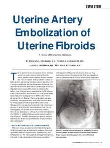
Figure 1. Digital subtraction angiogram (right anterior oblique projection) obtained with selective injection via the left internal iliac artery shows the division of the artery into two main stems (anterior stem, A; and posterior stem, P) and three branches (inferior gluteal artery, 1; uterine artery, 2; and superior gluteal artery, 3).
Introduction Embolization has become a first-line treatment for symptomatic uterine fibroid tumors (1) and is currently offered as an alternative to hysterectomy and multiple myomectomy (2– 4). Although there is widespread acceptance of embolization for treatment of uterine fibroid tumors, this treatment method requires great familiarity with the pelvic arterial anatomy. Variations in the vascularization of uterine fibroid tumors may account for treatment failures and complications, including uterine necrosis and amenorrhea (1–5). The uterus and uterine fibroid tumors are mainly supplied by the uterine artery, but ovarian arteries and round ligament arteries also may play a role (6 – 8). Angiography is useful for comprehensive assessment of the anatomy of the internal iliac artery, its pattern of division, and its branches (6 – 8). Doppler ultrasonography (US), contrast material– enhanced magnetic resonance (MR) imaging, and MR angiography also may be useful for evaluation of the arterial supply to the fibroid tumor before embolization (7). The authors have performed more than 2000 embolization procedures for treatment of uterine fibroid tumors. The purpose of this article is to provide a comprehensive review of the arterial anatomy of the female pelvis for diagnostic radiologists interested in women’s imaging and for interventional radiolo-
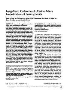
Figure 2. Digital subtraction angiogram obtained with selective injection via the right common iliac artery shows the absence of the right internal iliac artery and the origin of all pelvic branches, including the uterine artery (UA), in the common iliac artery (arrow). (Courtesy of JeanLouis Bertrand, MD, Centre Hospitalier de Perpignan, Perpignan, France.)
gists who wish to learn more about uterine fibroid embolization. The normal arterial anatomy is reviewed because it is the key to the procedure. Anatomic variants that may result from uterine fibroid tumor vascularization also are presented because such variants help to determine the outcome of embolization. The clinical relevance of the arterial anatomy for the selection of patients and of the technique to be used in catheterization and embolization, as well as for the possibility of complications, also is discussed.
Normal and Variant Arterial Supply to the Uterus Internal Iliac Artery (Hypogastric Artery) The internal iliac artery (also called the hypogastric artery) supplies the pelvic walls and the pelvic viscera (6 – 8). The internal iliac artery terminates in two main stems (bifurcation), one anterior and one posterior, in 57%–77% of the general population (Fig 1) (6,8). The anterior branches (visceral and parietal) of the internal iliac artery include the inferior gluteal, obturator, internal pudendal, vesical, middle hemorrhoidal, and genital (uterine and vaginal) arteries (6,8). The posterior branches (parietal only) include the superior gluteal, iliolumbar, and lateral sacral arteries (6,8). In individuals with bifurcation of the inter-
Teaching Point
●
Special Issue
Pelage et al
S101
RadioGraphics
RG f Volume 25
Figure 3. (a, b) Digital subtraction angiograms obtained with selective injection via the right internal iliac artery show, in left anterior oblique projection (a) and right (ipsilateral) oblique projection (b), the origin of the uterine artery (UA), which is best depicted in b (arrow) because of its location closer to the origin of the hypogastric artery. (c) Right anterior projection obtained with digital subtraction angiography shows superselective catheterization achieved by using a microcatheter and a guidewire with 90° angulation.
nal iliac artery, the contralateral anterior oblique (25°– 40° angle) projection is best for identification of the uterine artery (Fig 1) (6). Absence of the internal iliac artery is rarely observed (Fig 2).
Uterine Artery
Figure 4. Digital subtraction angiogram obtained with selective uterine artery injection shows the characteristic arterial course, with an initial descending segment (D), a transverse segment (T) from which the cervicovaginal artery (CV) originates, and an ascending segment (A). Numerous intramural arteries (arrows) also are visible.
Uterine fibroid tumors derive their main peripheral blood supply almost exclusively from the uterine arteries (9). The origin of the uterine artery is well identified on the contralateral oblique projection in most patients (Fig 1) (6). The uterine artery is the first or the second branch of the anterior division of the internal iliac artery (from the inferior gluteal artery) in 51% of cases (Fig 1) (6,7). In 6% of cases, the uterine artery is the first branch of the internal iliac artery above the level of the inferior gluteal and superior gluteal arteries (Fig 3) (6). In the presence of this anatomic variant, or when the internal iliac artery terminates in trifurcation (15%– 40% of the general population), the ipsilateral anterior oblique projection is used (Fig 3) (6). The uterine artery has a characteristic U shape, with a descending segment that parallels the lateral pelvic wall, a transverse segment that crosses the distal ureter at the level of the cervix, and an ascending segment that courses along the uterine margin at the medial edge of the broad ligament (Figs 1, 3, 4) (6 – 8). The uterine artery may have a common trunk with the vesical or vaginal artery (Fig 5) (6). The uterine artery
Teaching Point
RadioGraphics
S102
October 2005
RG f Volume 25
●
Special Issue
has several branches: the cervicovaginal artery, which arises from the transverse segment and which should be spared during embolization, and the intramural (arcuate) arteries, which course through the outer third of the myometrium (Fig 4) (6,8). The uterine artery may be replaced by small arterial branches (Fig 6) or may be absent; it is often replaced by the ipsilateral ovarian artery. The congenital absence of both uterine arteries is encountered in less than 1% of cases (Fig 7). The presence of aberrant uterine vessels that originate in the abdominal aorta also has been reported (10).
Ovarian Artery
Teaching Point
The ovarian artery arises anteromedially from the abdominal aorta a few centimeters below the renal arteries in 80%–90% of cases and has a characteristic corkscrew appearance (6,8,11–13). Rarely, the ovarian artery arises from the renal, lumbar, adrenal, or iliac artery (Figs 8, 9) (11– 13). Identification of the normal ovarian artery is not usually possible with angiography because of the small diameter of the artery (usually, less than 1 mm) (6,11,12). The ovaries are supplied by ovarian arteries in 40% of cases, by both uterine and ovarian arteries in 56% of cases, and by uterine arteries alone in 4% of cases (12,13).
Figure 5. Digital subtraction angiogram obtained with selective injection via the right genitourinary artery trunk shows right cervicovaginal (CV), vesical (V), and uterine (UA) arteries that arise from the anterior division of the internal iliac artery via a common genitourinary artery trunk (GU).
Round Ligament Artery Teaching Point
The round ligament artery, which arises from the external iliac artery or from the inferior epigastric artery, plays a minor role in uterine vascularization in normal physiologic conditions (Fig 10) (6,14).
Figure 6. Flush pelvic aortogram shows replacement of both uterine arteries by multiple small arteries (arrows).
●
Special Issue
Pelage et al
RadioGraphics
RG f Volume 25
Figure 7. (a) Flush pelvic aortogram shows no flow through the uterine arteries from the internal iliac artery, while both ovarian arteries, which arise from the infrarenal aorta, are visible (arrows). (b, c) Digital subtraction angiograms obtained with selective injection via the right (b) and left (c) ovarian arteries confirm ovarian arterial supply to the uterus.
Figures 8, 9. Anatomic variants. (8) Flush pelvic aortogram depicts two moderately enlarged ovarian arteries (arrows): a left ovarian artery that arises from the aorta, and a right ovarian artery that arises from the right renal artery (*). (9) Flush pelvic aortogram shows a right ovarian artery that arises from the right common iliac artery and supplies a large fundal fibroid tumor (F). (Fig 9 courtesy of Gerald Zemel, MD, Miami Cardiac and Vascular Institute, Miami, Fla.)
S103
October 2005
RG f Volume 25
●
Special Issue
RadioGraphics
S104
Figure 10. Digital subtraction angiogram obtained with selective injection via the left external iliac artery (*) shows a left round ligament artery (arrow) that arises from the inferior epigastric artery and supplies the left side of the uterus. (Reprinted, with permission, from reference 13.)
Figure 11. Digital subtraction angiogram obtained with selective injection via the left uterine artery (LUA) shows transverse anastomosis (arrow) between that artery and the right uterine artery (RUA) and retrograde opacification of the left artery via a utero-ovarian anastomosis (OA).
Figures 12, 13. (12) Digital subtraction angiogram obtained with selective injection via the left uterine artery (UA) depicts reflux into the ovarian artery (OA), a sign of anastomosis between the uterine and ovarian arteries. (13) Digital subtraction angiogram obtained with selective injection via the right uterine artery shows reflux into the ovarian artery, with resultant opacification of the ovary (arrow). Ut ⫽ uterus.
●
Special Issue
RadioGraphics
RG f Volume 25
Pelage et al
S105
Figure 14. Anatomic drawing shows the arterial blood supply (short arrows) to the uterus and three fibroid tumors (F). The perifibroid plexus is composed of arteries with diameters of 500 –1000 m in most cases. The diameter of the utero-ovarian anastomosis (long arrow) is usually less than 500 m. (Reprinted, with permission, from reference 7.)
Figure 15. Photomicrograph (hematoxylin-safran-eosin stain; magnification, ⫻200) of a specimen from the periphery of an intramural fibroid tumor (F), which was resected immediately after embolization, shows targeted occlusion of the perifibroid arterial plexus with 500 –700m-diameter calibrated microspheres (arrows).
tomosis may not be present at the beginning of the procedure and may become obvious only with flow redistribution after embolization of the main arterial supply to the fibroid tumor (11,12).
Arterial Anastomoses The interventional radiologist should be aware of two different types of arterial anastomosis between the uterine arteries and the ovarian arteries: left-to-right anastomosis (between the left and the right uterine arteries) and utero-ovarian anastomosis (between the uterine artery and the ovarian artery) (6,8). Left-to-right anastomosis is visible in about 10% of cases (Fig 11) (6,8), and uteroovarian anastomosis is identified in 10%–30% of Teaching cases (Figs 11, 12) (6,8,15). Retrograde filling of Point the ovarian artery is sometimes observed when contrast material is selectively injected via the ipsilateral uterine artery (6). The ovary itself is occasionally depicted (Fig 13). The usual diameter of the utero-ovarian anastomosis has been estimated to be less than 500 m (Fig 14) (9). Anas-
Arterial Supply to Uterine Fibroid Tumors The presence of uterine fibroids usually results in distortion and enlargement of the uterine arteries (Figs 1, 4) (9,16,17), which supply both normal myometrium and the fibroids (Fig 14) (9,16 –18). Usually, the flow to the fibroid tumor is substantially increased; perifibroid arteries are larger in diameter than are those that supply normal myometrium (Figs 14, 15) (9,17,18). The tumor itself is relatively hypovascular, and its interior is supplied by small centripetal arteries that originate in a rich perifibroid arterial plexus (16 –19). In the presence of multiple fibroid tumors, diffuse uterine hypervascularity is usually visible at
October 2005
RG f Volume 25
●
Special Issue
RadioGraphics
S106
Figures 16, 17. (16) Flush pelvic aortogram shows diffuse uterine hypervascularity in a woman with a uterus enlarged (volume, 800 mL) by multiple fibroid tumors. (17) Late arterial phase flush pelvic aortogram in a woman with three intramural fibroid tumors shows three separate areas of localized hypervascularity with dimensions that correspond to those of the fibroid tumors.
Figures 18, 19. (18) Flush pelvic aortogram in a patient with multiple fibroids depicts two enlarged ovarian arteries (arrows) that originate from the aorta. (19) Digital subtraction angiogram obtained with selective injection via a left ovarian artery with origin in the aorta shows enlargement of the artery and a large hypervascular fundal fibroid tumor (F).
●
Special Issue
Pelage et al
S107
RadioGraphics
RG f Volume 25
Figure 20. (a) Sagittal T2-weighted MR image shows a large fundal fibroid tumor (F) in a patient who previously underwent myomectomy. (b) Flush pelvic aortogram in the same patient depicts an enlarged right ovarian artery (OA) that originates from the aorta and supplies the fundal fibroid. Both uterine arteries (arrows) are also visible.
Figure 21. Flush pelvic aortogram in a patient with a pedunculated subserosal fibroid tumor shows a large left lumbar artery that supplies the fibroid (*). (Courtesy of Woodruff Walker, MBBS, FRCR, Royal Surrey Hospital, Guildford, England.)
angiography (Fig 16). In a few cases, a separate arterial supply to the fibroid may be observed (Fig 17). Even in the presence of two apparently normal uterine arteries, an additional supply to the fibroid may come from another source, such as the ovarian artery. This occurs in about 5%–10% of cases (Figs 9, 18, 19) (6 – 8,11,12,20,21). Ovarian artery supply to uterine fibroids is more fre-
quently found in women who have undergone pelvic surgery and those with previously diagnosed tubal or ovarian disease and/or large fundal fibroids (Figs 19, 20) (12). In women with one or more of these predisposing factors, the extent of ovarian artery supply to the fibroid tumor should be assessed with flush pelvic aortography (11). This may be done before or after uterine artery embolization (10,11). Since only residual blood flow to the fibroid tumor may be of clinical relevance, aortography is generally advocated after uterine artery embolization (10,11). Other sources of supply may be the round ligament artery and the lumbar artery (Figs 10, 21) (6,15). Unlike other types of fibroid tumor, pedunculated susbserosal fibroid tumors may have a specific feeding artery that corresponds to the arterial pedicle (Fig 22). A fibroid tumor located in a bicornate uterus may be supplied by one uterine artery only (Fig 23).
October 2005
RG f Volume 25
●
Special Issue
RadioGraphics
S108
Figure 22. (a) Contrast-enhanced sagittal MR image in a patient with a left-sided pedunculated subserosal fibroid tumor (F) shows the arterial pedicle (arrow). (b) Digital subtraction angiogram obtained with selective injection via the left uterine artery in the same patient depicts the same arterial pedicle (arrow) and tumor (F).
Figure 23. (a) Axial T2-weighted image shows a bicornate uterus with a single intramural fibroid tumor (F) on the left side. (b) Early arterial phase flush pelvic aortogram in the same patient shows exclusive supply to the fibroid tumor from the left uterine artery (arrow) and a small right uterine artery. (c) Late phase digital subtraction angiogram depicts hypervascularity of the fibroid (F). (Courtesy of Woodruff Walker, MBBS, FRCR, Royal Surrey Hospital, Guildford, England.)
●
Special Issue
Pelage et al
S109
RadioGraphics
RG f Volume 25
Figure 24. (a) Power Doppler US image in a patient with a 5-cm-diameter intramural fibroid tumor shows characteristic peripheral arterial flow that corresponds to the perifibroid plexus (arrows). (b) Pulsed Doppler US image in the same patient shows a low resistance index (0.50) in the periphery of the fibroid tumor.
Figure 25. (a) Color Doppler US image in a woman with diffuse adenomyosis shows dense intramural vascularity. (b) Flush pelvic aortogram shows multiple small abnormal intramural arteries and patchy uterine vascularization, findings that are consistent with adenomyosis.
Preembolization Imaging Imaging is very important in preprocedural planning for uterine fibroid embolization because it allows assessment of uterine fibroids and of concurrent conditions that may imitate or exacerbate the symptom complex. Deciding who is an appropriate candidate for the procedure involves careful evaluation of uterine fibroid tumors, particularly their number, size, and location and the degree of their vascularization (22–27).
Gray-scale and Color Doppler US US is the primary imaging modality used to evaluate patients in whom the presence of uterine fibroid tumors is suspected (22,23,26,27). Transabdominal and transvaginal US are used in conjunction with color and pulsed Doppler US (22,23,26). Doppler US can be used to assess fibroid and uterine vascularity and flow patterns (22,23). Typically, uterine fibroid tumors have a
marked peripheral blood flow (perifibroid plexus) and decreased central flow (Fig 24) (22,23). The resistance index is usually decreased in the perifibroid plexus, compared with that in the surrounding normal myometrium (Fig 24) (23). Doppler US also may be helpful for distinguishing an endometrial polyp with a single feeding vessel from an intracavitary submucosal fibroid tumor (26). The diagnosis of adenomyosis is usually made with gray-scale US, but Doppler US may be helpful for differentiating adenomyosis from fibroid tumors (Figs 24, 25) (24). In adenomyosis, multiple scattered vessels or intratumoral vascularity are depicted, whereas peripheral vessels and outer feeding vessels are visible in uterine fibroid tumors (Fig 25) (24).
RG f Volume 25
October 2005
●
Special Issue
RadioGraphics
S110
Figure 26. (a) MR angiogram before uterine fibroid embolization nicely depicts trifurcation of the right internal iliac artery and a kink at the origin of the uterine artery (UA). (b) Digital subtraction angiogram obtained with selective injection via the right internal iliac artery in the same patient demonstrates good correlation between MR angiography and digital subtraction angiography.
Figure 27. MR angiogram in a patient with multiple uterine fibroid tumors shows two uterine arteries and two enlarged ovarian arteries.
US plays a vital role also in the exclusion of associated pathologic conditions in the pelvic region, such as adnexal masses and endometrial carcinoma (26). Adnexal masses may mimic sub-
Figure 28. Sagittal contrast-enhanced MR image in a patient with multiple fibroid tumors shows intense enhancement of intramural fibroid tumors (F).
serosal fibroid tumors, but the two entities usually have very different vascular features (26). Because a pedunculated subserosal fibroid tumor has only one feeding artery, it may separate from the uterus and drift into the peritoneal cavity after embolization (Fig 22) (3,5). Pedunculated subse-
●
Special Issue
Pelage et al
S111
RadioGraphics
RG f Volume 25
Figure 29. (a, b) Digital subtraction angiograms obtained with injections in the right (a) and left (b) uterine arteries after embolization with 355–500-m-diameter nonspherical polyvinyl alcohol particles show an area of stasis and myometrial devascularization (arrow). (c) Postembolization contrast-enhanced MR image shows the same area of devascularization (arrows) and helps confirm the infarction of all fibroids (F).
rosal fibroid tumor is therefore considered a contraindication to embolization (2– 4). Preprocedural identification of the pedicle is crucial to separate broad-based fibroid tumors from pedunculated ones (3,27).
Contrast-enhanced MR Imaging and MR Angiography MR angiography may help the physician to obtain a better understanding of the arterial anatomy before embolization (Figs 26, 27) and may enable detection of an ovarian artery supply to the tumor (26,27). The majority of uterine fibroid tumors are markedly enhanced after the administration of a gadolinium-based contrast material (Fig 28) (26,27). Hypervascular uterine fibroid tumors may decrease more in size after embolization than do iso- or hypovascular fibroid tumors (27).
Anatomy and Angiographic End Points of Embolization Complete occlusion of the uterine arteries with stasis of contrast material is the usual angiographic end point when embolization is performed with nonspherical polyvinyl alcohol par-
ticles (1– 4). Embolization is stopped when a standing column of contrast material is observed in the uterine artery or when reflux of contrast material toward the uterine artery origin or into the hypogastric artery is observed (Fig 29) (1,3). Complete embolization of both uterine arteries (to stasis) may lead to myometrial and/or endometrial ischemia, with potentially disastrous consequences, such as endometrial atrophy or extensive uterine necrosis (3,28 –30). Recently, the use of a different angiographic end point was investigated at microsphere embolization (18). Since the target of embolization is the perifibroid arterial plexus, the main uterine artery should be spared (18). With calibrated microspheres, it is appropriate to stop embolization when the fibroid branch arteries have become occluded, even if antegrade flow is still present in the main uterine artery (the so-called “prunedtree” appearance). Additional angiographic criteria to identify the proper end point also include identification of uterine artery–to– ovarian artery
October 2005
RG f Volume 25
●
Special Issue
RadioGraphics
S112
Figure 30. (a, b) Digital subtraction angiograms obtained with selective injection via the left uterine artery before (a) and after (b) limited embolization with 500 –700-m-diameter tris-acryl microspheres. The postembolization image shows patency of the main uterine artery (arrow) and cervicovaginal branches (CV). (c, d) Contrast-enhanced sagittal MR images obtained before (c) and 24 hours after (d) embolization. The postembolization image shows normal perfusion of the myometrium and infarction of the three fibroid tumors (F).
or left-to-right anastomoses, which are usually not present initially (18). Limited embolization of the uterine arteries should be performed to reduce ischemic injury to normal myometrium
and/or endometrium as a result of complete occlusion of collateral vessels (Figs 14, 29). When calibrated microspheres with diameters of more than 500 m are used, a limited embolization, with targeted devascularization of the perifibroid plexus, is recommended (Figs 14, 29) (18,31).
RadioGraphics
RG f Volume 25
●
Special Issue
With the gradual shift over time both in technique and in end point, from the achievement of complete stasis of uterine arterial flow to the maintenance of persistent flow in the main uterine artery, the risk to normal tissue has decreased; sufficient blood flow is left to sustain portions of the myometrium (Figs 29, 30) (26,31).
Normal Postembolization Imaging Findings In the initial studies about uterine fibroid embolization, US was used to evaluate the uterus and the fibroids after treatment (1,3,27). A marked 35%– 60% reduction in uterine volume and 40%– 80% reduction in fibroid volume have been observed at 3– 6 months after embolization (2– 4). The disappearance of fibroid vascularization is usually observed at Doppler US after successful embolization (25–27). Given the limitations of US, most investigators currently use MR imaging for follow-up of patients who have undergone embolization for uterine fibroid tumors (3,4,25,26). After embolization, fibroid tumors usually show signal intensity changes that are consistent with hemorrhagic infarction, including increased signal intensity on T1-weighted images and homogeneous decreased signal intensity on T2-weighted images (25,26). In addition, the immediate reduction in perfusion after embolization in a fibroid tumor correlates well with the clinical response (25).
Common Causes of Treatment Failure and Recurrence The goal of embolization is the ischemic infarction of the fibroid tumor, a result that is verifiable with follow-up imaging after embolization (25,26). From a technical point of view, failure of embolization occurs when the fibroid tumor is not infarcted (32). In about 10% of treated patients, no improvement of symptoms, which corresponds to treatment failure, is observed (2– 4). In others, despite an initial short-term clinical improvement, clinical recurrence is observed (32). The durability of the clinical response depends on the completeness of treatment of all the fibroid tumors that are present at the time of embolization. Recurrence may be observed when fibroid tumors
Pelage et al
S113
are not infarcted after unilateral uterine artery embolization either because of an additional supply from other sources or because of associated adenomyosis (32–36).
Unilateral Uterine Artery Embolization The most common reason for treatment failure is difficulty in catheterization of one or the other uterine artery because of a faulty determination of the arterial origin (32). The appropriate oblique projection must be selected (Figs 1, 3) (6). A kink or angulation in the artery at or near its origin may make catheterization difficult (Fig 3). The use of a microcatheter is recommended to achieve a safe catheterization (18,32). Clinical failure is expected in most patients after unilateral arterial embolization (3,32). However, in rare cases (eg, in patients with a bicornate uterus), unilateral embolization may be successful (Fig 23) (33,36).
Extrauterine Arterial Supply to the Tumor A potential predictor of treatment failure or recurrence is the presence of ovarian arterial supply not only to the uterus but also to a fibroid tumor or a portion of the tumor that is not supplied by the uterine arteries (11,12,20,21). The extent of ovarian artery supply to the fibroid tumor should be assessed with aortography, which, ideally, is performed after embolization of both uterine arteries (11,12,32). It has been shown that women with viable fibroid tumors after embolization are at a higher risk for clinical recurrence than are those with infarcted fibroid tumors (Fig 31) (37). The use of contrast-enhanced MR imaging may allow early detection of failure by depicting fibroid tumor viability after embolization (37). In women with an additional supply to the fibroid tumor from the ovarian arteries, a delay of a few months is recommended before repeat embolization is considered (11). Additional embolization of the ovarian artery is considered if no clinical improvement has been observed and the fibroids are still viable, and the procedure is performed only with specific informed consent (11,32).
October 2005
RG f Volume 25
●
Special Issue
RadioGraphics
S114
Figure 31. (a) Flush pelvic aortogram obtained after bilateral uterine artery embolization in a woman with a large fundal fibroid shows additional supply to the uterus via an ovarian artery (arrow). Embolization of the ovarian artery was not performed because specific informed consent had not been obtained from the patient. (b) Postembolization contrast-enhanced MR image shows persistent perfusion (arrow) of the right inferior portion of the fibroid tumor. (c) Digital subtraction angiogram obtained with selective injection via the ovarian artery because of clinical recurrence 1 year later shows additional arterial supply to the fibroid. Embolization was performed by using 500 –700-m-diameter tris-acryl microspheres. (d) Postembolization contrast-enhanced MR image shows complete infarction of the fibroid and normal perfusion of the surrounding myometrium.
●
Special Issue
RadioGraphics
RG f Volume 25
Figure 32. Photomicrograph (hematoxylin-safraneosin stain; magnification, ⫻4) of a resected fallopian tube 6 months after embolization of a uterine fibroid tumor with 300 –500-m-diameter tris-acryl microspheres shows nontarget embolization of a tubal branch by a microsphere (arrow).
Pelage et al
S115
pain experienced by most such patients after embolization (2–5). It is encouraging that MR imaging of the uterus after embolization depicts rapid revascularization of the normal myometrium, particularly when a limited embolization of the uterine arteries has been performed (18,27,31). The rapid revascularization may be due to the rich collateral supply in the pelvis, which compensates for the complete occlusion of the uterine vessels at embolization (31). In rare cases, however, transient ischemia worsens and the uterus becomes completely infarcted (28 –30). Uterine necrosis that leads to hysterectomy because of persistent pain or infection has been reported (28 –30). Excessive embolization of the uterine arteries, particularly after the use of a secondary embolization agent (a supplemental embolization agent such as metal coils or gelatin sponge pledgets injected after the particles) or distal embolization with small embolization particles, may account for extensive uterine ischemia (28 –30).
Adenomyosis Any potential benefits of uterine artery embolization for controlling the symptoms of adenomyosis are still unclear (33–35). Although short-term results are encouraging, there have been cases in which the presence of adenomyosis has been implicated as the cause of failure of embolization of uterine fibroid tumors (34,35). In addition, the recurrence rate associated with uterine artery embolization for isolated adenomyosis is higher than that for treatment of uterine fibroid tumors with uterine artery embolization (35). The reason is that vascularization in diffuse adenomyosis (in which smaller arteries extend throughout the myometrium) is different from that in fibroid tumors (24,26). It has been hypothesized that embolization with the use of large particles may not be effective for treating adenomyosis (35). However, the use of small particles, while it may be more effective, may also cause injury to normal tissue (35). Therefore, appropriate diagnosis of this condition is mandatory at the time of the initial evaluation. The depiction of vascularity with US may help to distinguish adenomyosis from fibroid tumors when MR imaging is not available (Fig 25) (25).
Complications Uterine Ischemia and Necrosis Transient uterine ischemia occurs in most patients in whom fibroid tumors are treated with complete occlusion of both uterine arteries (3). Ischemia likely contributes to the postprocedural
Embolization of Nontargeted Cervicovaginal Branches Embolization of nontargeted cervicovaginal branches may cause ischemic vaginal damage and sexual dysfunction (38). To reduce the risk of cervical or vaginal injury, the tip of the catheter should be placed distally to these branches, or these branches should be spared by using large embolization particles (Fig 4) (38). Nontarget embolization in the internal pudendal artery may cause vulvar pain and labial necrosis (39).
Amenorrhea and Ovarian Failure Definitive amenorrhea related to ovarian failure may be caused by nontarget embolization through a patent utero-ovarian anastomosis or by embolization of a uterine artery that provides the only blood supply to the ovary (Figs 11–14, 32) (3,5,15,40 – 43). Ovarian Doppler flow measurements indicate a decrease in ovarian arterial flow in approximately 50% of patients after bilateral embolization is performed to the point of stasis (44). To reduce the risk of nontarget ovarian or tubal embolization through a patent anastomosis, the size of the embolic particles should be carefully chosen (18,38). Currently, calibrated microspheres with a diameter of more than 500 m are recommended (18). In the presence of a largediameter anastomosis, ovarian protection can be
RG f Volume 25
October 2005
●
Special Issue
RadioGraphics
S116
Figure 33. (a) Digital subtraction angiogram obtained with selective injection via the left uterine artery during embolization shows a large collateral artery that supplies the ovary (arrow) and patency of some arterial branches in the fibroid tumor. (b) After embolization of the utero-ovarian anastomosis with a 2 ⫻ 20-mm coil (arrow) to protect the ovary, uterine artery embolization was safely completed.
achieved by using larger particles or coils to occlude the anastomosis (Fig 33) (45– 47). In rare cases, a third mechanism may account for amenorrhea (3,48). Amenorrhea related to endometrial injury is usually caused by ischemic endometrial damage from uterine artery embolization to stasis (48).
Conclusions Embolization of uterine fibroid tumors requires a thorough knowledge of the pelvic arterial anatomy. Identification of the normal arterial anatomy and main variations in uterine fibroid tumor vascularization are key to the safety and success of the procedure. All interventional radiologists who plan to perform uterine fibroid tumor embolization should receive specific training in pelvic anatomy and procedural technique.
5. 6.
7.
8. 9.
10.
References 1. Ravina JH, Herbreteau D, Ciraru-Vigneron N, et al. Arterial embolisation to treat myomata. Lancet 1995;346:671– 672. 2. Spies JB, Ascher SA, Roth AR, Kim J, Levy EB, Gomez-Jorge J. Uterine artery embolization for leiomyomata. Obstet Gynecol 2001;98:29 –34. 3. Walker WJ, Pelage JP. Uterine artery embolisation for symptomatic fibroids: clinical results in 400 women with imaging follow-up. BJOG 2002;109: 1262–1272. 4. Pron G, Bennett J, Common A, et al. The Ontario uterine fibroid embolization trial: uterine fibroid
11.
12. 13. 14.
reduction and symptom relief after uterine artery embolization for fibroids. Fertil Steril 2003;79: 120 –127. Spies JB, Spector A, Roth AR, et al. Complications of uterine artery embolization for leiomyomata. Obstet Gynecol 2002;100:873– 880. Pelage JP, Le Dref O, Soyer P, et al. Arterial anatomy of the female genital tract: variations and relevance to transcatheter embolization of the uterus. AJR Am J Roentgenol 1999;172:989 –994. Gomez-Jorge J, Keyoung A, Levy EB, Spies JB. Uterine artery anatomy relevant to uterine leiomyomata embolization. Cardiovasc Intervent Radiol 2003;26:522–527. Merland JJ, Chiras J. Vascular territories. In Merland JJ, Chiras J, eds. Arteriography of the pelvis. Berlin, Germany: Springer-Verlag, 1981; 69 –72. Pelage JP, Le Dref O, Beregi JP, et al. Limited uterine artery embolization with tris-acryl gelatin microspheres for uterine fibroids. J Vasc Interv Radiol 2003;14:15–20. Binkert CA, Andrews RT, Kauffman JA. Utility of nonselective abdominal aortography in demonstrating ovarian artery collaterals in patients undergoing uterine artery embolization for fibroids. J Vasc Interv Radiol 2001;12:841– 845. Pelage JP, Walker WJ, Le Dref O, Rymer R. Ovarian artery: angiographic appearance, embolization and relevance to uterine fibroid embolization. Cardiovasc Intervent Radiol 2003;26:227–233. Borell U, Fernstro¨m I. The ovarian artery. Acta Radiol 1954;42:253–265. Saraiya P, Chang T, Pelage JP, Spies JB. Uterine artery replacement by the round ligament artery. J Vasc Interv Radiol 2002;13:939 –941. Ambekar A, Vogelzang RL. Aberrant uterine artery as a cause of uterine artery embolization treatment failure. Int J Gynaecol Obstet 2001;74:59 – 60.
RadioGraphics
RG f Volume 25
●
Special Issue
15. Razavi MK, Wolanske KA, Hwang GL, Sze DY, Kee ST, Dake MD. Angiographic classification of ovarian artery–to– uterine artery anastomoses. Radiology 2002;224:707–712. 16. Sampson JA. The blood supply of uterine myomata. Surg Gynecol Obstet 1912;14:215–234. 17. Holmgren B. Some observations on the blood vessels of the uterus under normal conditions and in myoma. Acta Obstet Gynecol Scand 1938;18: 192–213. 18. Farrer-Brown G, Beilby JO, Tarbit MH. The vascular patterns of myomatous uteri. J Obstet Gynaecol Br Commonw 1970;77:967–975. 19. McCluggage WG, Ellis PK, McClure N, Walker WJ, Jackson PA, Manek S. Pathologic features of uterine leiomyomas following uterine artery embolization. Int J Gynecol Pathol 2000;19:342–347. 20. Andrews RT, Bromley PJ, Pfister ME. Successful embolization of collaterals from the ovarian artery during uterine artery embolization for fibroids. J Vasc Interv Radiol 2000;11:607– 610. 21. Barth MM, Spies JB. Ovarian artery embolization supplementing uterine embolization for leiomyomata. J Vasc Interv Radiol 2003;14:1177–1182. 22. Weintraub JL, Romano WJ, Kirsch MJ, et al. Uterine artery embolization: sonographic imaging findings. J Ultrasound Med 2002;21:633– 637. 23. Tranquart F, Brunereau L, Cottier JP, et al. Prospective sonographic assessment of uterine artery embolization for the treatment of fibroids. Ultrasound Obstet Gynecol 2002;19:81– 87. 24. Chiang CH, Chang MY, Hsu JJ, et al. Tumor vascular pattern and blood flow impedance in the differential diagnosis of leiomyoma and adenomyosis by color Doppler sonography. J Assist Reprod Genet 1999;16:268 –275. 25. deSouza NM, Williams AD. Uterine arterial embolization for leiomyomas: perfusion and volume changes at MR imaging and relation to clinical outcome. Radiology 2002;222:367–374. 26. Jha RC, Allison SJ, Ascher SM. Imaging of leiomyomata, the uterus and the pelvis. In Spies JB, Pelage JP, eds. Uterine artery embolization and gynecologic embolotherapy. Baltimore, Md: Lippincott Williams & Wilkins, 2005; 37–51. 27. Fleischer AC, Donnelly EF, Campbell MG, et al. Three-dimensional color Doppler sonography before and after fibroid embolization. J Ultrasound Med 2000;19:701–705. 28. Spies JB. Uterine artery embolization for fibroids: understanding the technical causes of failure. J Vasc Interv Radiol 2003;14:11–14. 29. Smith SJ, Sewall LE, Handelsman A. A clinical failure of uterine fibroid embolization due to adenomyosis. J Vasc Interv Radiol 1999;10:1171–1174. 30. Jha RC, Takahama J, Imaoka I, et al. Adenomyosis: MRI of the uterus treated with uterine artery embolization. AJR Am J Roentgenol 2003;181: 851– 856. 31. Pelage JP, Jacob D, Fazel A, et al. Mid-term results of uterine artery embolization for symptomatic adenomyosis: initial experience. Radiology 2005;234:948 –953. 32. Nicholson T. Outcome in patients undergoing unilateral uterine artery embolization for symptomatic fibroids. Clin Radiol 2004;59:186 –191. 33. Pelage JP, Guaou-Guaou N, Jha RC, Ascher SM, Spies JB. Uterine fibroid tumors: long-term MR imaging outcome after embolization. Radiology 2004;230:803– 809.
Pelage et al
S117
34. de Blok S, de Vries C, Prinssen HM, Blaauwgeers HL, Jorna-Meijer LB. Fatal sepsis after uterine artery embolization with microspheres. J Vasc Interv Radiol 2003;14:779 –783. 35. Gabriel H, Pinto CM, Nikolaidis P, Miller FH, Weinrach DM, Vogelzang RL. MRI detection of uterine necrosis after uterine artery embolization for fibroids. AJR Am J Roentgenol 2004;183:733– 736. 36. Godfrey CD, Zbella EA. Uterine necrosis after uterine artery embolization for leiomyoma. Obstet Gynecol 2001;98:950 –952. 37. Banovac F, Ascher SM, Jones DA, Black MD, Smith JC, Spies JB. Magnetic resonance imaging outcome after uterine artery embolization for leiomyomata with use of tris-acryl gelatin microspheres. J Vasc Interv Radiol 2002;13:681– 688. 38. Yeagley TJ, Goldberg J, Klein TA, Bonn J. Labial necrosis after uterine artery embolization for leiomyomata. Obstet Gynecol 2002;100:881– 882. 39. Lai AC, Goodwin SC, Bonilla SM, et al. Sexual dysfunction after uterine artery embolization. J Vasc Interv Radiol 2000;11:755–758. 40. Walker WJ, Carpenter TT, Kent AS. Persistent vaginal discharge after uterine artery embolization for fibroid tumors: cause of the condition, magnetic resonance imaging appearance and surgical treatment. Am J Obstet Gynecol 2004;190:1230 – 1233. 41. Chrisman HB, Saker MB, Ryu RK, et al. The impact of uterine fibroid embolization on resumption of menses and ovarian function. J Vasc Interv Radiol 2000;11:699 –703. 42. Ahmad A, Qadan L, Hassan N, Najarian K. Uterine artery embolization treatment of uterine fibroids: effect on ovarian function in younger women. J Vasc Interv Radiol 2002;13:1017–1020. 43. Tropeano G, Di Stasi C, Litwicka K, Romano D, Draisci G, Mancuso S. Uterine artery embolization for fibroids does not have adverse effects on ovarian reserve in regularly cycling women younger than 40 years. Fertil Steril 2004;81:1055–1061. 44. Ryu RK, Chrisman HB, Omary RA, et al. The vascular impact of uterine artery embolization: prospective sonographic assessment of ovarian arterial circulation. J Vasc Interv Radiol 2001; 12(9):1071–1074. 45. Payne JF, Robboy SJ, Haney AF. Embolic microspheres within ovarian arterial vasculature after uterine artery embolization. Obstet Gynecol 2002; 100:883– 886. 46. Nikolic B, Nguyen K, Martin LG, Redd DC, Best I, Silverstein MI. Pyosalpinx developing from a preexisting hydrosalpinx after uterine artery embolization. J Vasc Interv Radiol 2004;15:297–301. 47. Wolanske KA, Gordon RL, Wilson MW, Kerlan RK Jr, LaBerge JM, Jacoby AF. Coil embolization of a tuboovarian anastomosis before uterine artery embolization to prevent nontarget particle embolization of the ovary. J Vasc Interv Radiol 2003;14:1333– 1338. 48. Marx M, Wack JP, Baker EL, Stevens SK, Barakos JA. Ovarian protection by occlusion of uteroovarian collateral vessels before uterine fibroid embolization. J Vasc Interv Radiol 2003;14:1329 – 1332.
RG
Volume 25 • Special Issue • October 2005
Pelage et al
Teaching Points for Uterine Fibroid Vascularization and Clinical Relevance to Uterine Fibroid Embolization Jean-Pierre Pelage, MD, et al RadioGraphics 2005; 25:S99 –S117 ● Published online 10.1148/rg.25si055510 ● Content Codes:
Page S100 The internal iliac artery terminates in two main stems (bifurcation), one anterior and one posterior, in 57%– 77% of the general population (Fig 1) (6,8). The anterior branches (visceral and parietal) of the internal iliac artery include the inferior gluteal, obturator, internal pudendal, vesical, middle hemorrhoidal, and genital (uterine and vaginal) arteries (6,8). The posterior branches (parietal only) include the superior gluteal, iliolumbar, and lateral sacral arteries (6,8). Page S101 The uterine artery is the first or the second branch of the anterior division of the internal iliac artery (from the inferior gluteal artery) in 51% of cases (Fig 1) (6,7). In 6% of cases, the uterine artery is the first branch of the internal iliac artery above the level of the inferior gluteal and superior gluteal arteries (Fig 3) (6). Page S102 The ovarian artery arises anteromedially from the abdominal aorta a few centimeters below the renal arteries in 80%–90% of cases and has a characteristic corkscrew appearance (6,8,11–13). Rarely, the ovarian artery arises from the renal, lumbar, adrenal, or iliac artery (Figs 8, 9) (11–13). Page S102 The round ligament artery, which arises from the external iliac artery or from the inferior epigastric artery, plays a minor role in uterine vascularization in normal physiologic conditions (Fig 10) (6,14). Page S105 Left-to-right anastomosis is visible in about 10% of cases (Fig 11) (6,8), and utero-ovarian anastomosis is identified in 10%–30% of cases (Figs 11, 12) (6,8,15). Retrograde filling of the ovarian artery is sometimes observed when contrast material is selectively injected via the ipsilateral uterine artery (6). The ovary itself is occasionally depicted (Fig 13). The usual diameter of the utero-ovarian anastomosis has been estimated to be less than 500 μm (Fig 14) (9).