TRAUMA
Total knee arthroplasty following tibial plateau fracture A MATCHED COHORT STUDY C. E. H. Scott, E. Davidson, D. J. MacDonald, T. O. White, J. F. Keating From Royal Infirmary of Edinburgh, Edinburgh, United Kingdom
Radiological evidence of post-traumatic osteoarthritis (PTOA) after fracture of the tibial plateau is common but end-stage arthritis which requires total knee arthroplasty is much rarer. The aim of this study was to examine the indications for, and outcomes of, total knee arthroplasty after fracture of the tibial plateau and to compare this with an age and gendermatched cohort of TKAs carried out for primary osteoarthritis. Between 1997 and 2011, 31 consecutive patients (23 women, eight men) with a mean age of 65 years (40 to 89) underwent TKA at a mean of 24 months (2 to 124) after a fracture of the tibial plateau. Of these, 24 had undergone ORIF and seven had been treated nonoperatively. Patients were assessed pre-operatively and at 6, 12 and > 60 months using the Short Form-12, Oxford Knee Score and a patient satisfaction score. Patients with instability or nonunion needed total knee arthroplasty earlier (14 and 13.3 months post-injury) than those with intra-articular malunion (50 months, p < 0.001). Primary cruciate-retaining implants were used in 27 (87%) patients. Complication rates were higher in the PTOA cohort and included wound complications (13% vs 1% p = 0.014) and persistent stiffness (10% vs 0%, p = 0.014). Two (6%) PTOA patients required revision total knee arthroplasty at 57 and 114 months. The mean Oxford knee score was worse preoperatively in the cohort with primary osteoarthritis (18 vs 30, p < 0.001) but there were no significant differences in post-operative Oxford knee score or patient satisfaction (primary osteoarthritis 86%, PTOA 78%, p = 0.437). Total knee arthroplasty undertaken after fracture of the tibial plateau has a higher rate of complications than that undertaken for primary osteoarthritis, but patient-reported outcomes and satisfaction are comparable. Cite this article: Bone Joint J 2015;97-B:532–8.
C. E. H. Scott, BSc, MSc, FRCS (Tr&Orth), Orthopaedic specialty registrar E. Davidson, BSc, MRCS, Orthopaedic specialty registrar D. J. MacDonald, BA (Hons), Research Associate T. O. White, MD, FRCS (Orth), Consultant Orthopaedic Surgeon J. F. Keating, FRCS (Orth), Consultant Orthopaedic Surgeon Royal Infirmary of Edinburgh, Little France Crescent, Edinburgh EH16 4SA, UK. Correspondence should be sent to C. E. H. Scott; e-mail:
[email protected] hs.uk ©2015 The British Editorial Society of Bone & Joint Surgery doi:10.1302/0301-620X.97B4. 34789 $2.00 Bone Joint J 2015;97-B:532–8. Received 11 July 2014; Accepted 27 November 2014
532
Between 25% and 45% of patients who have had a fracture of the tibial plateau have radiological evidence of post-traumatic osteoarthritis (PTOA) at long-term follow-up.1-4 This incidence increases with the severity of the fracture, any loss of reduction and malalignment of more than 5° (Fig. 1).1-5 Patients are also at greater risk of needing total knee arthroplasty (TKA) than matched individuals without a fracture: this risk is even higher in those with a bicondylar fracture, those who are older and those with more substantial comorbidities.6 Despite this, the proportion of patients who need TKA after the development of post-traumatic OA is low at 3% to 7% at ten years.4,6-8 However, when needed, it has been associated with a poorer outcome and a rate of complications of between 26% and 60%.9-11 There is little literature about this: many studies combine TKA for PTOA secondary to a fracture of the tibial plateau with TKA for PTOA secondary to a fracture of the distal femur in order to have a
sample of sufficient size.9,10,12,13 Though many papers quote a worse outcome from TKA for post-traumatic arthritis than for primary OA,9,13,14 we are unaware of any studies which have directly compared the outcomes of these two groups of patients. The aim of this study was to examine the outcome of TKA after fracture of the tibial plateau and to compare it with that of a cohort of age and gender matched patients who had undergone TKA for primary OA. The primary outcome measure was the Oxford Knee Score (OKS):15 secondary outcome measures included complications, patient satisfaction and survivorship.
Patients and Methods From our prospectively collected trauma database, we identified 888 fractures of the tibial plateau (875 patients) that had been treated at our hospital between 1995 and 2008. We included patients who had been managed THE BONE & JOINT JOURNAL
TOTAL KNEE ARTHROPLASTY FOLLOWING TIBIAL PLATEAU FRACTURE
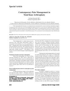
Fig. 1a
Fig. 1b
Hip-knee-ankle radiographs of a 41-year-old man who sustained a right bicondylar Schatzker VI tibial plateau fracture, taken a) at 14 months after ORIF and removal of metalwork and b) after TKR with a standard cruciate retaining implant.
operatively and non-operatively. Five elderly (> 75 years) patients (0.5%) who had undergone TKA as the primary management of an unreconstructable tibial plateau fracture were excluded from the study. Of the patients, twenty five (25 knees; 2.8%) went on to develop PTOA and needed TKA between 1997 and 2011.8 Six others underwent TKA for PTOA following a tibial plateau fracture which had initially been treated elsewhere during the period of study. These patients were identified from our prospectively collected arthroplasty database. This gave a study population of 31 TKAs in 31 patients. Each procedure was carried out by one of nine senior consultant orthopaedic surgeons. A postal questionnaire which included the SF-12 general health questionnaire16 and OKS was sent to patients before they underwent TKA. The SF-12 is a validated health questionnaire with both physical (PCS) and mental (MCS) health components. The OKS is a reliable, validated, kneespecific outcome measure designed to minimise the influence of comorbidities. It consists of 12 questions, each with five possible answers giving a score from 0 to 48, a higher score representing better function. The completed questionnaires were collected at a pre-assessment clinic. Similar post-operative questionnaires were sent to patients before they attended their six and 12 month follow-up appointments. In February 2014, patients who had died were identified and their date of death confirmed from the Scottish register of deaths. All remaining patients were contacted by telephone and an OKS recorded in addition to a measure of VOL. 97-B, No. 4, APRIL 2015
533
patient satisfaction,17 and details of any re-operations, including revision TKA. The collection of data was independent of the routine clinical care of the patient. The medical records and short-leg, weight-bearing radiographs of all patients were reviewed: we retrieved data on patient demographics; fracture date; Schatzker classification;18 fracture management; mechanism and symptoms of failure; re-operations other than TKA and femorotibial alignment (FTA) prior to TKA. Operation notes were reviewed to determine the surgical approach used, including the use of extensile exposures, the classification of any bone defect and its management, intra-operative complications and the type of implant used. Early and late post-operative complications and re-operations were recorded up to the time of final follow-up. Post-operative short-leg weight-bearing radiographs taken at most recent orthopaedic review were examined by one author (CEHS) who had no clinical contact with the patients. The coronal and sagittal plane alignment of the implants was measured19 and peri-prosthetic radiolucencies reported by femoral and tibial region in accordance with the Knee Society Score.20 If radiolucencies were present, all radiographs pertaining to that TKA were examined for progression. The outcomes (PROMs and complication rates) of this PTOA cohort were compared with those of an age and gender matched cohort of patients undergoing TKA for primary OA. The exposure under investigation in this cohort study was therefore tibial plateau fracture: matching was carried out to limit the effect of age and gender as confounding factors. A 3:1 matched cohort of patients undergoing TKA for primary osteoarthritis was identified from our arthroplasty database. This group was followed-up for five years with patient-reported outcomes pre-operatively and at six months, one and five years. The same complication, re-operation and revision data were collected for both cohorts in addition to radiological data. Statistical analysis. Statistical analysis was undertaken using Statistical Package for Social Sciences version 19.0 (SPSS Inc., Chicago, Illinois). One-way ANOVA was used to compare continuous variables with multiple groups such as age at fracture and time to TKA for different modes of failure of fracture fixation. Parametric (unpaired t-tests) and non-parametric (Mann–Whitney U test) tests were used as necessary to assess continuous variables for significant differences between the two TKA cohorts. Nominal categorical variables were assessed using a chi square or Fisher’s exact test. Relative risk was calculated for binary outcomes (i.e. complications) where possible. ANOVA was used to examine changes in parametric variables over the five-year period of study. A p-value of < 0.05 was considered to be statistically significant. Post-hoc analysis of OKS, PCS and MCS changes over time was undertaken using paired ttests. The significance level for this was set at p < 0.0125 incorporating a Bonferroni correction to correct for multiple testing. Survival analysis was undertaken using KaplanMeier analysis with failure for any reason as the endpoint.
534
C. E. H. SCOTT, E. DAVIDSON, D. J. MACDONALD, T. O. WHITE, J. F. KEATING
10%
6%
I
13% Nonsurgical
22%
II
10%
ORIF screws
13% III 48%
16%
ORIF screws and CaP04
IV 13% V
10%
ORIF plate
16%
ORIF plate and CaP04 23%
VI
Fig. 2a
External fixator
Fig. 2b
Pie charts showing fracture by a) Schatzker classification and b) management in the study group (CaPO4, calcium phosphate cement; ORIF, open reduction and internal fixation).
Table I. Modes of failure necessitating total knee arthroplasty (TKA) Mode of failure
N (%)
Mean age at fracture (yrs) (SD)
Valgus collapse Varus collapse Joint incongruity Articular surface subsidence Nonunion
13 (41.9) 1 (3.2) 5 (16.1) 5 (16.1) 7 (22.6)
67.5 (11.7) 41 51.6 (4.7) 53.3 (15.7) 75.3 (14.2)
p-value
Mean time to TKA (mths) (SD)
p-value
0.003*
13.9 (6.2) 14.0 38 (12.3) 61 (42.8) 13.3 (13.3)
0.001*
* ANOVA SD, standard deviation
Results In total, 31 patients underwent 31 TKAs for PTOA after a fracture of the tibial plateau over the period of study: 23 (74%) were female. The mean age at fracture was 64 years (30 to 89). Of the 31 patients, 24 had undergone ORIF and seven had been treated non-surgically or had presented late with a neglected fracture. Fig. 2a shows the distribution of fracture types and Fig. 2b their initial management. One fracture was open (Gustilo-Anderson type IIIA, Schatzker IV), the rest were closed. In the 24 patients who underwent ORIF, the operation was carried out through a longitudinal (midline or lateral parapatellar) incision in 14 (58%), an incision with both longitudinal and transverse elements (e.g. a hockey-stick incision) in five (21%), percutaneous incisions in three (13%) and a longitudinal posteromedial incision in one patient (4%). The approach for one patient was not recorded. Knee arthroplasty was performed at a mean of 24 months (2 to 124) after fracture at mean age of 65 years (40 to 89). Ten patients died of unrelated causes at a mean of 65 months (15 to 120) after TKA. The mean follow-up of those who survived was 85 months (26 to 169). Modes of failure. The modes of failure which resulted in the need for TKA are shown in Table I. These presented as pain in ten patients (32.2%), pain and stiffness in ten (32.3%) and valgus instability in 11 (35.5%). The time to TKA differed significantly between different modes of failure: collapse, malalignment and nonunion required earlier
conversion to TKA (Table I). Patients with instability were revised to TKA earlier (mean 13.4 months: 5 to 28) than those without (mean 30.5 months: 2 to 124). This difference was significant (p = 0.024 unpaired t-test, 95% CI -31.7 to -2.5). Those with nonunion (7 of 31) were significantly older (mean 75.3 years, SD 8.7) than those whose fractures had united (mean 60.5, SD 13.9, t-test p = 0.013). All nonunions occurred in women. Of the 24 patients, ten had the metalwork used for ORIF removed before undergoing TKA. Surgical data. All patients with PTOA underwent TKA through a medial parapatellar approach. One required an osteotomy of the tibial tuberosity for access and this healed without complication. There were 19 patients with no tibial defect and 12 patients with a defect after initial resection of the tibia. The tibial defects were graded using the Anderson Orthopaedic Research Institute classification:21 Grade I contained metaphyseal defect < 5 mm manageable with cement or cancellous bone graft; Grade II - uncontained metaphyseal defect, or larger contained defect, affecting one plateau only (Grade IIa) or both plateaus (Grade IIb), requiring cement, bone graft or augmentation; and Grade III deficient metaphyseal bone with bone loss of major portion of plateau/condyle with or without collateral instability. Three patients (10%) had grade I defects with grade IIA in nine (29%). These were managed with autologous bone graft or cement in ten patients: two needed tibial augments. In those in THE BONE & JOINT JOURNAL
TOTAL KNEE ARTHROPLASTY FOLLOWING TIBIAL PLATEAU FRACTURE
190
535
1
0.8 Cumulative survival
(°)
180
170
Survival function Censored
0.6
0.4
0.2
160
0 150
0 Pre-op FTA
Post-op FTA
2
4
6
8
10
12
14
Survived (yrs)
Fig. 3
Fig. 4
Boxplot showing the femorotibial angle (FTA) before and after total knee arthroplasty in patients with a previous tibial plateau fracture. Whiskers indicate 95% confidence intervals.
Kaplan-Meier survival graphs for total knee arthroplasty after fracture of the tibial plateau with the endpoint as failure for any reason: 96% survival at five years (95% CI 88.4 to 100) and 82.3% at ten years (95% CI 56.5 to 100).
whom calcium phosphate cement had been used at ORIF (11/31), there was a non-significant trend towards grade IIA defects at TKA (5/11 compared with 4/20 p = 0.186 Fisher’s exact test) and the need for augments (2/11 compared with 0/20, p = 0.118 Fisher’s exact test). The use of calcium phosphate cement had no significant effect on time to TKA or mode of failure of ORIF. Cruciate retaining (CR) primary implants were used in 27/31 (87%) patients: augmented tibial stems were needed in 11/31 (35.5%). Those who needed a stem were significantly older (mean age 71.7 years, SD 8.7) than those who did not (59.5 years, SD 15.1, p = 0.021 unpaired t-test, 95% CI 2 to 22) and underwent TKA earlier after fracture (mean 12.9 months, SD 10.2) than those who did not (mean 31.7 months, SD 29.5, p = 0.018 unpaired t-test, 95% CI -3.5 to -34). The median polyethylene insert was 12 mm (8 to 16). The mean pre-operative FTA of 170.1° (SD 8.0: 158 to 186) was corrected to a mean 174.7° (SD 2.83: 170° to 179°) after TKA: this difference was significant (p = 0.014, Paired t-test) (Fig. 3). Complications. Intra-operative complications occurred in three (10%) patients: these were partial division of the patella tendon in one, MCL division in one (both of which were managed with bracing) and an intra-operative fracture of the femoral condyle (managed with an augment) (Table II). Early complications occurred in five patients (16%): four superficial wound infections and one persistent common peroneal nerve palsy (Table II). Of those who developed problems with early wound healing, all had undergone ORIF: one through a midline longitudinal incision, one through percutaneous incisions and two through
an approach with a transverse element. The wound complication rate after TKA for each type of ORIF incision was therefore 7% for longitudinal incisions, 40% for incisions with a transverse element and 33% for percutaneous incisions. This was not significant (p = 0.27, chi-squared). All initially settled with antibiotics and did not need surgical debridement. However, one went on to develop a deep infection and required revision (see below). Late complications occurred in seven patients (22%) (Table II). Stiffness was the most prevalent late complication (4, 13%): one patient needed an MUA. Over the period of study, two of the 31 TKAs for PTOA were revised: one for septic loosening at 57 months (a CR implant with stem and medial augment) and the other for aseptic loosening at 114 months (a CR implant with no stem or augments). All-cause survival was 96% at five years (95% CI 88.4 to 100) and 82.3% (95% CI 56.5 to 100) at ten years (Fig. 4). A further four patients developed progressive lucent lines beneath the tibial component at a mean of 97 months (69 to 122). None were symptomatic. Three of these patients had standard CR implants, one with a stem. There were no failures amongst those managed with more constrained implants (4/31). Outcomes and matched cohort study. The age and gender matched primary OA cohort consisted of 93 patients with a mean age of 66 (37 to 89). Table II compares the demographics, complications and outcomes between groups. The rate of intra-operative complications was significantly higher in the PTOA cohort (p = 0.010, Fisher’s exact test). Though differences in the overall incidence of early and late complications failed to reach significance (p = 0.054,
VOL. 97-B, No. 4, APRIL 2015
536
C. E. H. SCOTT, E. DAVIDSON, D. J. MACDONALD, T. O. WHITE, J. F. KEATING
Table II. Comparative outcome of TKA after tibial plateau fracture (PTOA) and that of primary osteoarthritis (OA) in an age and gender matched cohort at a ratio of 1:3
Mean age at TKA (SD) Female (n,%) Complications (n,%) Intra-operative Early
Late
Mean PROMs (SD) PCS MCS OKS
Fracture Ligament injury Superficial infection/dehiscence CPN palsy Venous thromboembolism Cardiovascular Stiffness Instability Ongoing pain Heterotopic ossification Periprosthetic Deep infection Pre-op 1 year Pre-op 1 year Pre-op 6/12 1 year 5 years
PTOA (n = 31)
Primary OA (n = 93)
p-value
65.7 (13.1) 23 (74.2)
66.3 (12.3) 69 (74.2)
0.809*
1 (3.2) 2 (6.4) 4 (12.8) 1 (3.2) 0 0 3 (9.2) 0 1 (3.2) 1 (3.2) 0 1 (3.2) 31.2 (7.3) 37.9 (9.3) 42.3 (10.8) 50.6 (7.9) 29.9 (15.1) 33.2 (9.0) 34.4 (9.1) 32.4 (10.3)
0 0 1 (1) 0 3 (3) 2 (2) 0 1 (1) 2 (2) 0 1 (1) 1 (1) 30.7 (7.1) 42.9 (12.6) 50.1 (11.5) 52.3 (13.5) 18.2 (7.2) 33.5 (10.9) 34.8 (9.1) 35.5 (10.7)
0.010*
0.054*
0.054* 0.789* 0.14* 0.014† 0.240† < 0.001* 0.607† 0.561† 0.297*
* unpaired t-test † Mann–Whitney U test CPN (common peroneal nerve); CRPS (chronic regional pain syndrome); PROMs (Patient Reported Outcome Measures); PCS (Physical Component Score of the SF-12); MCS (Mental Component Score of the SF-12); OKS (Oxford Knee Score); SD (Standard Deviation)
Fisher’s exact test), the incidence of specific complications did differ significantly between groups: wound infection was significantly higher in the PTOA cohort (4/31, 13%) than in the OA cohort (1/93, 1%) (p = 0.014 Fisher’s exact test), as was persistent stiffness which occurred in three PTOA patients (9%) but none of the OA group (p = 0.014 Fisher’s exact test). The risk of orthopaedic complications was higher in the PTOA cohort throughout follow-up: the relative risk of an early wound complication in the PTOA cohort was 3.53 (95% CI 2.04 to 6.11); of an intra-operative or early orthopaedic complication (iatrogenic fracture or ligament injury, wound complication or CPN palsy) was 6.35 (95% CI 1.01 to 39.8); and of late mechanical problems (stiffness or instability) was 3.21 (95% CI 1.67 to 6.17). The post-operative FTA was significantly more valgus in the PTOA cohort (mean 174.7, SD 2.6) than in the primary OA cohort (mean 178.3, SD 2.7, p < 0.001 unpaired t-test, 95% CI 2.4 to 4.7). The mean pre-operative OKS was significantly better for the PTOA cohort than the OA cohort (29.9 compared with 18.2, p < 0.001, Table II, Fig. 5). The mean OKS in the PTOA cohort showed no significant improvement over time (p = 0.543, repeated measure ANOVA, Fig. 5). In the OA group, the OKS improved significantly postoperatively (p < 0.001, repeated measure ANOVA) pre-
dominantly in the first post-operative year (p < 0.001, paired t-test) with no significant improvement thereafter (p = 0.777, paired t-tests). There were no significant differences between the two cohorts in absolute post-operative OKS at any time point (Table II, Fig. 5). The pre-operative MCS was significantly worse in the PTOA cohort than in the OA cohort, but again with no significant differences post-operatively. There was no significant difference in satisfaction at more than five years: 86% of the OA cohort were satisfied, as were 78% of the PTOA cohort (p = 0.437, Fisher’s exact test, Fig. 6). Within the PTOA cohort, the need for stems or augments had no significant effect on OKS at any time point, nor did the original fracture type, mode of failure, pre-operative instability or removal of metalwork prior to TKA. The five-year OKS was significantly better (p = 0.042 unpaired t-test, 95%CI -4 to -19) in those initially managed non-operatively for their tibial plateau fracture (mean five year OKS 39.5, SD 6.0) than in those who had undergone ORIF (mean 29.4, SD 10.5).
Discussion We studied 31 patients who had undergone TKA after sustaining a fracture of the tibial plateau and found a higher rate of intra-operative complications, problems with wound healing and late stiffness than in TKA undertaken for primary OA. However, the post-operative patientTHE BONE & JOINT JOURNAL
45 40 35 OA
30 25
TKA indication
20 15 45 40 35
PTOA
Oxford Knee Score (95% confidence intervals)
TOTAL KNEE ARTHROPLASTY FOLLOWING TIBIAL PLATEAU FRACTURE
30 25 20 15 Pre-operative 6 mths
12 mths > 60 mths
Time Fig. 5 Functional outcome of total knee arthroplasty (TKA) after fracture of the tibial plateau (PTOA) and primary osteoarthritis measured using the Oxford knee score (OKS). Significant differences exist in pre-operative, but not post-operative OKSs. Whiskers indicate 95% confidence intervals
80 70 OA
Patients (%)
60
PTOA
50 40 30 20 10 0 Very Satified
Satisfied
Uncertain
Dissatisfied
Fig. 6 Histogram showing patient satisfaction at > five years for total knee arthroplasty (TKA) after fracture of the tibial plateau (PTOA) and TKA for primary osteoarthritis.
reported outcomes were no different to those seen after TKA for primary osteoarthritis. This is at odds with previous studies which report worse outcome scores for TKA after a fracture of the tibial plateau.3,10,11 Lonner et al10 described 31 TKAs for post-traumatic arthrosis, which included both distal femoral and tibial plateau fractures, Saleh et al11 reported on 15 TKAs after a fracture of the tibial plateau and Weiss et al3 reported on 62 TKAs after a fracture of the tibial plateau. In these studies, outcome was measured using the Knee Society Score (KSS) and the Hospital for Special Surgery Knee Score (HSSKS). These knee scores are very similar and are completed by a clinician. Most elements relate to objective clinical measures such as range of movement and alignment. They VOL. 97-B, No. 4, APRIL 2015
537
are not patient-reported outcomes. By contrast, the OKS is entirely reported by the patient on the basis of their day-today experience of their knee. The poor outcomes previously reported by the KSS and HSS-KS may therefore reflect residual malalignment and stiffness, also found in this study, rather than a poor patient-reported functional outcome. To the best of our knowledge, this is the first report on the outcome of TKA after a fracture of the tibial plateau which uses the OKS, the official patient-reported outcome measure of the National Joint Registry of England and Wales and the most widely used in the United Kingdom. Five of the 12 OKS questions specifically relate to pain and seven to function. In one-third of the patients with a fracture of the tibial plateau the main indication for surgery was instability rather than pain: the OKS may have underestimated the level of disability as a consequence. By contrast, patients undergoing TKA for primary OA will, by definition, be restricted by their painful knee and this may account for their significantly worse pre-operative Oxford Knee Scores. Patients who undergo TKA relatively soon after their fracture may also skew the pre-operative scores by reporting their pre-fracture level of symptoms. Patient satisfaction after TKA was higher in the primary OA cohort (86% compared with 78% following tibial plateau fracture) but this difference was not significant. Though the absolute OKS did not differ between the two groups, the primary OA group had a worse pre-operative OKS and as such experienced greater improvement in individual scores than did the fracture group. The greater improvement could help explain the higher satisfaction rate in the group with primary OA. The most common mode of failure of fracture management necessitating TKA was valgus/varus collapse. This is in keeping with what we know about the management of plateau fractures in which the outcome is more closely related to restoration of the mechanical axis than to accurate reduction of the joint surface. Patients with instability needed TKA sooner than those who had malunion of the joint. Though associated malalignment was significantly improved, the FTA of the PTOA group remained in more valgus than the primary OA group. This is likely to reflect the predominance of lateral compartment involvement in the PTOA group and medial compartment involvement in the group with primary OA. The rate of nonunion for these metaphyseal fractures is low, in the region of 1.6%,4 but appears to be another significant factor in the need for revision TKA (7/31, 22%) because of pain and collapse. Though the mean age of the PTOA (65 years) and the OA (66 years) groups were comparable, it should be noted that this is not coincident with the mean age of our tibial plateau fracture population, which is 56 years.8 The rate of intra-operative complication was higher in the PTOA group. Access to the knee after a fracture and previous surgery can be difficult and the techniques used in revision arthroplasty, such as osteotomy of the tibial tuberosity, are sometimes needed. Such difficulties predispose to
538
C. E. H. SCOTT, E. DAVIDSON, D. J. MACDONALD, T. O. WHITE, J. F. KEATING
iatrogenic avulsion of the patella tendon and division of the medial collateral ligament3,10 both of which occurred in this series and were successfully managed with a period of bracing without the need for more constrained implants or formal repair. Our intra-operative complication rate of 9.7% is comparable to that of Weiss et al3 who reported a rate of 10%. The rate of post-operative complications was also similar to that previously recorded, affecting 11/31 (35%) patients in this study and 16/62 (26%) in Weiss’s study.3 TKA after fracture of the tibial plateau has previously been associated with early wound complications, which sometimes require flap cover.10,11 While the overall rate of early complications did not differ between our PTOA and OA groups, the type of complication did. There were significantly more wound complications in the PTOA group, all following ORIF, and more medical complications in the OA group. Surgical scars and retained metalwork may increase the risk of wound breakdown and superficial infection after TKA and must be managed appropriately. We found a lower rate of wound complications in patients who had a longitudinal scar from ORIF: this could be fully incorporated into the TKA approach, unlike ORIF incisions with a transverse element, which could not. This did not reach statistical significance but may have done so in a larger series. None of our patients needed further surgery for wound problems, but one went on to develop a deep infection. The only late complication which differed between the two groups was ongoing knee stiffness which occurred in 9% of patients with PTOA, again similar to the rates reported in previous studies (e.g. 8%).3 The use of stems and augments and the management of tibial defects were similar to those described in previous studies. 3,10,11 Though Kaplan–Meier survivorship is difficult to interpret with a small sample size, no worrying early failure rate was encountered. The limitations of this study include its small sample size. This is a consequence of the rarity of TKA after fractures of the tibial plateau: our sample is still the second largest reported. The pre-operative FTA of the primary OA group was not available for comparison with that of the PTOA group. No consideration has been given to comorbidities in either group. It is however, the first study to record patientreported outcomes of TKA after fracture of the tibial plateau, the first to use the OKS and the first to compare outcomes with an age and gender matched control group with primary OA. Matching is important because tibial plateau fractures follow a bimodal age distribution: consequently, if TKA is needed, it often takes place at the extremes of the age of those typically undergoing TKA for primary OA. In conclusion, after a fracture of the tibial plateau, patients with instability or nonunion are likely to need TKA earlier than those with malunion of the joint. While revision techniques may be needed to gain access to knee joint in these patients, primary cruciate-retaining implants can normally be used, with an augmented stem if required. The rate of complications of TKA for PTOA was higher at each
time point than that of TKA for primary OA: this was more marked when previous surgical fixation had been undertaken. However, the implants survived well and patientreported outcome and satisfaction were no different postoperatively than those of the matched group with TKA for primary OA. Author contributions C. E. H. Scott: Data collection, Analysis, Write-up and revisions E. Davidson: Data collection, Analysis, Write-up and revisions D. J. MacDonald: Data collection and analysis T. O. White: Performed surgery, Co-authored paper J. F. Keating: Performed surgery, Co-authored paper Thanks go to all of the orthopaedic surgeons whose patients were included in this study, and to the Scottish Orthopaedic Research Trust into Trauma (SORT-IT). No benefits in any form have been received or will be received from a commercial party related directly or indirectly to the subject of this article. This article was primary edited by A. Simpson and first proof edited by A. C. Ross.
References
1. Rasmussen PS. Tibial condylar fractures. Impairment of knee joint stability as an indication for surgical treatment. J Bone Joint Surg [Am] 1973;55-A:1331–1350. 2. DeCoster TA, Nepola JV, el-Khoury GY. Cast brace treatment of proximal tibia fractures. A ten-year follow-up study. Clin Orthop Relat Res 1988;231:196–204. 3. Weiss NG, Parvizi J, Trousdale RT, Bryce RD, Lewallen DG. Total knee arthroplasty in patients with a prior fracture of the tibial plateau. J Bone Joint Surg [Am] 2003;85-A:218–221. 4. Manidakis N, Dosani A, Dimitriou R, et al. Tibial plateau fractures: functional outcome and incidence of osteoarthritis in 125 cases. Int Orthop 2010;34:565–570. 5. Rademakers MV, Kerkhoffs GM, Sierevelt IN, Raaymakers EL, Marti RK. Operative treatment of 109 tibial plateau fractures. Five- to 27-year follow-up results. J Orthop Trauma 2007;21:5–10. 6. Wasserstein D, Henry P, Patterson JM, Kreder HJ, Jenkinson R. Risk of total knee arthroplasty after operatively treated tibial plateau fracture. A matched-population-based cohort study. J Bone Joint Surg [Am] 2014;96-A:144–150. 7. Mehin R, O’Brien P, Broekhuyse H, Blachut P, Guy P. Endstage arthritis following tibia plateau fractures. Average 10-year follow-up. Can J Surg 2012;55:87–94. 8. Davidson E, Oliver W, White T, Keating J. Tibial plateau fractures. When will I need a knee replacement? Bone Joint J 2014;96-B:7. 9. Parratte S, Bonnevialle P, Pietu G, et al. Primary total knee arthroplasty in the management of epiphyseal fracture around the knee. Orthop Traumatol Surg Res 2011;97:S87–S94. 10. Lonner JH, Pedlow FX, Siliski JM. Total knee arthroplasty for post-traumatic arthrosis. J Arthroplasty 1999;14:969–975. 11. Saleh KJ, Sherman PJ, Katkin P, et al. Total knee arthroplasty after open reduction and internal fixation of fractures of the tibial plateau: a minimum five-year follow-up study. J Bone Joint Surg [Am] 2001;83-A:1144–1148. 12. Wu LD, Xiong Y, Yan SG, Yang QS. Total knee replacement for post-traumatic degenerative arthritis of the knee. Chin J Traumatol 2005;8:195–199. 13. Weiss NG, Parvizj J, Hanssen AD, Trousdale RT, Lewallen DG. Total knee arthroplasty in post-traumatic arthrosis of the knee. J Arthroplasty 2003;18:23–26. 14. Civinini R, Carulli C, Matassi F, Villano M, Innocenti M. Total knee arthroplasty after complex tibial plateau fractures. Chir Organi Mov 2009;93:143–147. 15. Dawson J, Fitzpatrick R, Murray D, Carr A. Questionnaire on the perceptions of patients about total knee replacement. J Bone Joint Surg [Br] 1998 80-B63–69. 16. Dunbar MJ, Robertsson O, Ryd L, Lidgren L. Appropriate questionnaires for knee arthroplasty. Results of a survey of 3600 patients from the Swedish Knee Arthroplasty Register. J Bone Joint Surg [Br] 2001;83-B:339–344. 17. Scott CE, Howie CR, MacDonald D, Biant LC. Predicting dissatisfaction following total knee replacement. A prospective study of 1217 patients. J Bone Joint Surg [Br] 2010;92-B:1253–1258. 18. Schatzker J, McBroom R, Bruce D. The tibial plateau fracture. The Toronto experience 1968-1975. Clin Orthop Relat Res. 1979;138:94–104. 19. Sarmah SS, Patel S, Hossain FS, Haddad FS. The radiological assessment of total and unicompartmental knee replacements. J Bone Joint Surg [Br] 2012;94-B:1321– 1329. 20. Ewald FC. The Knee Society total arthroplasty roentgenographic evaluation and scoring system. Clin Orthop Relat Res 1989;248:9–12. 21. Mulhall KJ, Ghomrawi HM, Engh GA et al. Radiographic prediction of intraoperative bone loss in knee arthroplasty revision. Clin Orthop Relat Res. 2006;446:51–58. THE BONE & JOINT JOURNAL