Proceedings of the Indiana Academy of Science (1988) Volume 98 p. 109-116.
THIN LAYER CHROMATOGRAPHIC IDENTIFICATION OF PHENOL IN THE GLANDULAR SECRETORY SYSTEM OF CANNABIS SATIVA L. (CANNABACEAE) CHARLES T. HAMMOND Department of Biology St. Meinrad College St. Meinrad, Indiana 47577 and PAUL G. MAHLBERG Department of Biology Indiana University Bloomington, Indiana 47405
ABSTRACT: A phenolic glycoside was isolated from vege tative stem exudates from a Mexican strain of Cannabis and tentatively identified by thin layer chromatography (TLC) as phloroglucinol-glucoside. Incubation at 100°C for 30 min in 2N HCl liberated a free phenol which was identified by mass spec troscopy and 1 H-nuclear magnetic resonance as phloroglucinol. Glandular heads were isolated in large numbers with high purity from floral regions by blending, microfiltration and cen trigugation in cold. Gland samples extracted for phenols as above revealed by TLC the presence of phloroglucinol. Phlo roglucinol may play an important role in the in vivo biosyn thesis of cannabinoids. INTRODUCTION Although there are over 300 cannabinoid or cannabinoid-related compounds reported as natural constituents of Cannabis (Turner, et al., 1980) most strains of marihuana are characterized by the production of a biogenetically related series of compounds (Figure 1). Cannabinoids are terpenophenolic compounds believed to originate in the plant from condensation of a terpene derivative (such as geranyl pyrophosphate) with the phenol olivetol (as olivetolic acid) to form the cannabinoid series which includes cannabigerolic acid (CBGA), cannabichromenic acid (CECA), cannabidiolic acid (CBDA), and 69-tetrahydrocannabinolic acid (69THCA) (Mechoulam, 1970; Shoyama, et al., 1975). These cannabinoids occur nat urally predominantly in their water-soluble acid forms but are readily decarbox ylated to their water-insoluble neutral forms by mild heating or drying of plant tissues or extracts. Glandular hairs, which are most abundant on the surface of pistillate floral bracts and bracteoles (Figure 2), have been studied developmentally (Hammond and Mahlberg, 1977) and ultrastructurally (Hammond and Mahlberg, 1978) and are considered to be a major site of synthesis and localization of these cannabinoids (Lanyon, et al., 1971; Malingre et al., 1975; Turner, et al., 1977, 1978). In an effort to further characterize the glandular site and mechanism of cannabinoid biogen esis, thin layer chromatography (TLC) was used to screen, isolate, and identify 109
110
INDIANA ACADEMY OF SCIENCE
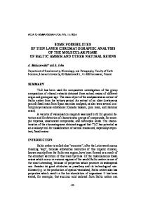
Geranyl Pyrophosphate
Vol. 98 (1988)
Olivetol
CBC
FIGURE 1. Biogenetic pathway for cannabinoid biosynthesis. Cannabinoids shown as neutral forms. Acid forms possess carboxyl group at R position. major phenolics characteristic to Cannabis tissues and also common to glandular sites of synthesis. MATERIALS AND METHODS Roots, stems, leaves, petioles, bracts, and stem exudates of a Mexican strain of Cannabis were screened for phenolic profiles using a phenolic extraction and TLC separation procedure modified from Harborne (Harborne and Williams, 1969). Fresh tissues were typically blended in water in cold, filtered, adjusted to 2N with HCl, heated for 30-60 min at 105°C, extracted in peroxide-free ethyl
Vol. 98 (1988)
BOTANY
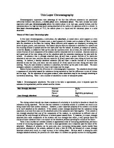
FIGURE
2. Glandular hairs on floral bract of Cannabis.
FIGURE
3. Purified glandular hairs isolated on Nitex by filtration. Bar
µm.
111
100
112
INDIANA ACADEMY OF SCIENCE
Vol. 98 (1988)
ether, and concentrated to near dryness under nitrogen gas. Extracts were spottted at 1 to 3 µl on 5x10 cm strips of Eastman Kodak Chromogram Sheet 13179 Silica Gel and developed in a non-equilibrated capped jar using either a polar (ethyl acetate-methanol-aqueous 2% acetic acid; 67:7:1) or more non-polar (toluene-ethyl acetate-formic acid; 17:10:1) solvent. Visualization of phenolics was by long and short wave UV and spraying with a freshly prepared 0. 1% aqueous-methanol (1:3) solution of fast blue salt B (FBSB). Glandular heads were isolated in large numbers with high purity from floral regions by blending in cold sodium acetate buffer (pH 5.0) and filtration through cheese cloth followed by filtration through 20 mesh Nitex in a Buchner funnel (Figure 3). Glands were rinsed off the Nitex with cold buffer and concentrated by low speed centrifugation. Glands were then disrupted with an amalgamator, using 0.5 mm zirconium beads, and the supernatant was subjected to phenol extraction using the acid-heat treatment described above. RESULTS The most promising results from the initial screening were afforded by stem exudates which produced abundant phenol in simple profiles. Young stem regions of large vegetative shoots, when cut, produce abundant exudate presumably from phloem and associated laticifer elements. Thin layer separation of extracts of stem exudates are shown in Figure 4. These chromatograms are reconstructions for illustrative purposes of several chromatograms run at different times. The two chromatograms in Figure 4 are identical in setup except that "A" is run in the polar system and "B" in the more non-polar system. Reading from left to right on each chromatogram, Sample 1 is a mixture of cannabinoid standards (0.5 µl of 1 mg/ml) which run together near the solvent front at the top of the chromatogram and which stains red with FBSB. Sample 2 is an olivetol standard (0.5 µl of 10 mg/ml) which stains purple with FBSB. In the polar system, it runs close to the cannabinoid standards, but in the non-polar system, it is clearly separated from the cannabinoid standards running in the middle of the chromatogram. Our oliv etol standard produced a faint second spot in the non-polar system at a slightly higher Rf position which was an artifact of undetermined origin. Sample 6 is the acid-heat treated extract of stem exudate. In the more polar system "A", three phenolic sports are resolved along with streaks of unresolved phenolics. In the non-polar system "B", nine phenolic spots are resolved. In both solvent systems, one purple staining spot appeared in greatest abundance and was targeted for further study. Stem exudate (1.28 ml) from 64 large vegetative plants was collected, acid heat treated, and extracted in ethyl ether as before, but separated by preparative thin layer chromatography in the non-polar solvent system. The abundant, purple staining phenolic of interest from the initial study was then isolated from the preparative chromatogram and taken to dryness. The extract yielded 1.4 mg of phenol per ml of stem exudate. This purified phenol sample was identified by proton nuclear magnetic resonance and mass spectroscopy as trihydroxybenzene, or, more commonly, phloroglucinol. These spectroscopy data will be published separately (Hammond and Mahlberg, in press). When a phloroglucinol standard (Figure 4, Sample 3; upper Rf spot) was run against stem exudate extract (Figure 4, Sample 6), it had a similar purple color reaction with FBSB and ran at an identical Rf position as the abundant unknown
Vol. 98 (1988)
BOTANY
113
FIGURE 4. Demonstration of phloroglucinol glucoside in stem exudate of Can nabis. Chromatogram A developed in Ethyl acetate:methanol: 2% aqueous acetic acid. Chromatogram B developed in Toluene: Ethyl acetate: formic acid. See text for sample discussion. phenol in both polar and non-polar solvent systems. This provided TLC confir mation that the unknown phenol was phloroglucinol. Fresh stem exudate collected in 80% ethanol but not acid-heat treated produced a phenolic profile shown in Figure 4, Sample 4. In the polar system "A", a single, pink staining, low Rf spot was formed. Considering the polar nature of this spot, its association with phloroglucinol, its location in the phloem, the hydrolytic function of acid-heat treatment in phenol extraction, and the common existence of phenolics as glycosides, we postulated that the phloroglucinol in stem exudates may occur as a phloroglucinol glycoside. This was confirmed by preparation of a phloroglucinol j3-d glucoside standard using a Vicia seed synthesizing system after Pridham (Pridham and Saltmarsh, 1963). This is shown in Figure 4, Sample 3, where the phloroglucinol glucoside standard (lower Rf spot) has a similar pink staining reaction with FBSB and an identical Rf position as the fresh stem exudate extract in chromatogram "A". Further confirmation was provided by incubation of the fresh stem exudate extract with 13-d glucosidase (Sigma G-8625) as shown in Figure 4, Sample 5. In both chromatograms "A" and "B", phloroglucinol is liberated after enzyme hydrolysis.
114
INDIANA ACADEMY OF SCIENCE
Vol. 98 (1988)
FIGURE 5. Demonstration of phloroglucinol in glandular hairs of Cannabis. Chro matograms A and B developed in Toluene: Ethyl acetate: formic acid. See text for sample discussion. With the identification of phloroglucinol as a characteristic and abundant phenolic of stem exudate, it was important to ascertain if phloroglucinol was a characteristic phenolic of the glandular secretory system. Glandular heads were isolated in large numbers with high purity and subjected to phenol extraction using the acid-heat treatment as described previously. TLC of the glandular phe nolic extract using the non-polar toluene-ethyl acetate-formic acid solvent system is shown in Figure 5, chromatogram "A". In Figure 5, reading from left to right, Sample 1 is a phloroglucinol standard (0.5 µl of 1 mg/ml), and Sample 4 is a mixture of cannabinoid standards (2 µl of 1 mg/ml). Figure 5, Samples 2 and 3 are 1 and 3 µl respectively of the gland phenolic extract. Note the presence of phloroglucinol as well as cannabinoids in gland heads. Figure 5, chromatogram "B" demonstrates that phloroglucinol is a natural constituent of gland heads and not an artifact of acid-heat treatment breakdown of cannabinoids or even olivetol. In Figure 5, reading again from left to right, Sample 1 is a mixture of phloroglucinol (0.5 µl of 1 mg/ml) and olivetol standards (0.25 ml of 10 mg/ml). Figure 5, Samples 3 and 4 and 1 are 2 µl respectively of
Vol. 98 0988)
BOTANY
115
acid-heat treated olivetol standard. Figure 5, Samples 5 and 6 are 1 and 2 µ.l respectively of acid-heat treated cannabinoid standard mixture. Note that in nei ther the treated olivetol nor the treated cannabinoid mixture is phloroglucinol generated by the phenol extraction procedure. Acid-heat treatment of the can nabinoid mixture did generate "new" phenolic spots, which are clearly artifacts of the procedure. DISCUSSION The chemical composition of marihuana is characterized by the production of a series of terpenophenolic cannabinoids (Mechoulam, 1970). The presence of cannabinoids and their interactions with man, while generally known for millenia, have only recently received serious study. While man has either wanted to enhance or inhibit cannabinoid production in the plant, little has become known about natural cannabinoid biogenesis. The currently accepted biosynthetic pathway (Figure 1) deduced from structural and synthetic studies postulates that CBGA is the first cannabinoid to be formed in the plant by a condensation of an activated monoterpene such as neryl or geranyl pyrophosphate with the phenolic olivetol. CBGA is then a precursor to CBCA as well as separately to L'::.9-THCA through CBDA (Mechoulam, 1970; Mechoulam and Gaoni, 1967; Shoyama, et al., 1975). Recent work with an in vitro system using labeled isotope incorporation with developing seedlings found the first cannabinoid to appear to be CBCA not CBGA (Turner and Mahlberg, 1988). Similarly, Mexican strains of Cannabis produce abundant THCA but lack CBDA. Clearly, either the postulated pathway is in error, pathways undergo change during ontogeny, or more than one pathway is involved in cannabinoid biosynthesis. The biogenetic scheme reported above is largely the result of a chemical systhetic approach rather than a botanical plant-related approach. Although THCA and other cannabinoids can now be readily synthesized from condensation of a variety of terpenes with olivetol (Churchill, 1983; Mechoulam and Gaoni, 1967), it is not clear that this is the mechanism of synthesis in an enzyme-mediated plant system. With this problem in mind, we thought it would be useful to intially approach the biosynthetic pathway by looking at phenolics in Cannabis. It was clear from our initial results that olivetol was not present in stem exudates, nor could it be identified in our screening of all major organs of Cannabis. Instead, the major phenolic of stem exudates and glandular heads was phloroglu cinol. Olivetol was tentatively identified, by TLC alone, as a decomposition product of acid-heat treatment of cannabinoids. There are reports of olivetol artifacts generated in marihuana smoke (Novotny, et al., 1982) or in the high temperature injection port of the gas-liquid chromatograph (Bailey, 1978). The presence of phloroglucinol, as a glucoside or glucoside derivative, as an abundant and characteristic phenolic of Cannabis glands and the absence of oliv etol raises important questions about the in vivo mechanism of biogenesis of the marihuana cannabinoids. This study suggests that phloroglucinol may play an important role in the natural biogenesis of cannabinoids. It is of biological interest to note that Humulus, the only other genus in the family Cannabaceae, is widely reported to utilize phloroglucinol derivatives in the synthesis of its specialized terpenophenolic compounds, humulone and lupulone (Reidl, 1962).
116
INDIANA ACADEMY OF SCIENCE
Vol. 98 (1988)
ACKNOWLEDGMENT This research was supported in part with a grant from the Indiana Academy of Science to C.T.H. DEA registration No. 011704. Indiana Board of Pharmacy No. PH0106852.
LITERATURE CITED Bailey, K. 1978. Formation of olivetol during gas chromatography of cannabinoids. J. Chromatog. 160: 288-290. Churchill, K.T. 1983. Synthetic tetrahydrocannabinol. J. Forensic Sci. 28: 762772. Hammond, C.T. and P.G. Mahlberg. 1977. Morphogenesis of capitate glandular hairs of Cannabis sativa L. (Cannabaceae). Amer. J. Bot. 64: 1023-1031. -----· 1978. Ultrastructural development of capitate glandular hairs of Cannabis sativa L. (Cannabaceae). Amer. J. Bot. 65: 140-151. -----· in press. Phloroglucinol glycoside as a natural constituent of Can nabis sativa (Cannabaceae). J. Natur. Prod. Harborne, J.B. and C.A. Williams. 1969. The identification of orcinol in higher plants in the family Ericaceae. Phytochem. 8: 2223-2226. Lanyon, V.S., J.C. Turner, and P.G. Mahlberg. 1981. Quantitative analysis of cannabinoids in the secretory product from capitate-stalked glands of Can nabis sativa L. (Cannabaceae). Bot. Gaz. 142: 316-319. Malingre, T., H. Hendriks, S. Batterman, R. Bos, and J. Visser. 1975. The essential oil of Cannabis sativa. Planta Med. 28: 56-61. Mechoulam, R. 1970. Marihuana chemistry. Science 168: 1159-1166. _____ and Y. Gaoni. 1967. Recent advances in the chemistry of hashish. Fortsch. Chem. Org. Natur. 25: 175-213. Novotny, M., F. Merli, D. Wiesler, M. Fencl, and T. Saeed. 1982. Fractionation and capillary gas chromatographic-mass spectrometic characterization of the neutral components in marijuana and tobacco smoke condensates. J. Chro matography 238: 141-150. Pridham, J.B. and M.J. Saltmarsh. 1963. The biosynthesis of phenolic glucosides in plants. Biochem. J. 87: 218-224. Reid!, W. 1962. Naturally occuring phloroacylophenones. Jn: T.S. Gore, B.S. Joshi, S.V. Sunthankar, and B.D. Tilak (Eds.), Recent Progress in the Chemistry of Natural and Synthetic Colouring Matters, Academic Press, New York. Shoyama, Y., M. Yagi, I. Nishioka, and T. Yamauchi. 1975. Biosynthesis of can nabinoid acids. Phytochem. 14: 2189-2192. Turner, C., M. Elsohly, and E. Boeren. 1980. Constituents of Cannabis sativa L. XVII. A review of the natural constituents. J. Natur. Prod. 43: 169-234. Turner, J., J. Hemphill, and P.G. Mahlberg. 1977. Gland distribution and can nabinoid distribution and cannabinoid content in clones of Cannabis sativa L. Amer. J. Bot. 64: 687-693. -----· 1978. Quantitative determination of cannabinoids in individual glandular trichomes of Cannabis sativa L. (Cannabaceae). Amer. J. Bot. 65: 1103-1106. ----- and P.G. Mahlberg. 1988. Jn vivo incorporation oflabeled percursors into cannabinoids in seedlings of Cannabis sativa L. (Cannabaceae). In: G. Chesher, P. Consroe, and R. Musty (Eds.), Marihuana, pp. 263-270, Australian Gov't, Canberra.