The surgical treatment of bony metastases of the spine and limbs P. Böhm, J. Huber From the Eberhard-Karls-Universität, Tübingen, Germany
he skeleton is the most common site to be affected by metastatic cancer. The place of surgical treatment and of different techniques of reconstruction has not been clearly defined. We have studied the rate of survival of 94 patients and the results of the surgical treatment of 91 metastases of the limbs and pelvis, and 18 of the spine. Variables included the different primary tumours, the metastatic load at the time of operation, the surgical margin, and the different techniques of reconstruction. The survival rate was 0.54 at one year and 0.27 at three years. Absence of visceral metastases and of a pathological fracture, a time interval of more than three years between the diagnosis of cancer and that of the first skeletal metastasis, thyroid carcinoma, prostate carcinoma, renal-cell carcinoma, breast cancer, and plasmacytoma were positive variables with regard to survival. The metastatic load of the skeleton and the surgical margin were not of significant influence. In tumours of the limbs and pelvis, the local failure rate was 0% after biological reconstruction (10), 3.6% after cemented or uncemented osteosynthesis (28) and 1.8% after prosthetic replacement (53). The local failure rate after stabilisation of the spine (18) was 16.6%. There was local recurrence in seven patients (6.4%), and in four of these the primary tumour was a renal-cell carcinoma. The local recurrence rate was 0% after extralesional (24) and 8.2% after intralesional resection (85). Improvements in the oncological management of patients with primary and metastatic disease have resulted in an increased survival rate. In order to avoid additional surgery, it is essential to consider the expected time of survival of the reconstruction and, in
T
P. Böhm, MD, Professor Orthopädische Universitätsklinik, Eberhard-Karls-Universität Tübingen, Hoppe-Seyler-Strasse 3, 72076 Tübingen, Germany. J. Huber, MD Medizinische Klinik Kreiskrankenhaus Reutlingen, Steinenbergstrasse 31, 72764 Reutlingen, Germany.
bony metastases with a potentially poor response to radiotherapy, the surgical margin. J Bone Joint Surg [Br] 2002;84-B:521-9. Received 5 June 2001; Accepted after revision 21 August 2001
The skeleton is the most common site to be affected by 1 metastatic cancer and metastatic deposits are the most 2 common malignant tumours affecting the skeleton. Solid cancers metastasise to bone by a complex multistep process which involves interactions between tumour cells and nor3-5 mal host cells. Despite an incidence of bony metastases of between 25% and 100% in advanced myeloma and 1 carcinoma of the breast, prostate, thyroid, lung and kidney, the incidence of complications is much lower. The incidence of pathological fracture of long bones in patients with metastatic bone disease is uncertain, but may be about 1 10%. Spinal instability is the cause of back pain in about 6 10% of patients with cancer. In those in whom metastatic disease is confined to the skeleton, decline in the quality of life and eventual death are almost entirely a result of complications such as bone pain, hypercalcaemia, pathological fracture, and dural compression. Because of improvements in oncological management, the long-term survival of patients after the first pathological fracture of a 7,8 long bone has increased during the past 30 years. The aims of treatment are to relieve pain, prevent pathological fractures, improve mobility and function, and pro9,10 There remains a lack of consensus about long survival. the optimum management of bony metastases. The role of surgical treatment and of different techniques of reconstruc9,10 tion has not been clearly defined. Since the treatment of bony metastases is primarily palliative, the possible benefit of treatment must be judged against the possible complications and the expected life span of the patient. Overtreatment of terminally ill patients must be avoided, but many patients may survive for some years and benefit greatly from surgical treatment. We have therefore analysed the survival of patients and the results of the surgical treatment of metastases of the spine and limbs.
Correspondence should be sent to Professor P. Böhm.
Patients and Methods
©2002 British Editorial Society of Bone and Joint Surgery 0301-620X/02/412495 $2.00
We studied retrospectively a consecutive series of 94
VOL. 84-B, NO. 4, MAY 2002
521
522
P. BÖHM, J. HUBER
2 1
14 2
8
5 4
9 4 30 19 8 3
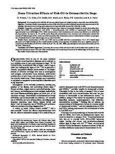
Fig. 1 Location of 109 bony metastases treated surgically in 94 patients.
patients who had undergone surgical treatment for 109 metastatic lesions in the skeleton (Fig. 1) between January 1988 and December 1997. There were 56 women and 38 men with a mean age of 58.5 years (15.2 to 86.7) at the time of the first operation for a metastasis. The breast was the most common site of the primary tumour (Table I) which explains the predominance of women. No patient was excluded. In 68 patients, the nature of the primary malignancy was known before the presentation of the first
skeletal metastasis, and in two a carcinoma of unknown origin had been diagnosed before the first bony metastasis was surgically treated. In the other 24 patients, the malignant disease presented with a painful bony metastasis. In 17 of these, a primary tumour was identified; non-small-cell bronchial carcinoma in five, small-cell bronchial carcinoma in one, renal-cell carcinoma in four, plasmacytoma in two, highly malignant non-Hodgkin's lymphoma in two, breast cancer in one, prostate cancer in one, and thyroid cancer in one. In seven patients, the nature of the primary tumour was not identified. The mean age of the patients at the time of diagnosis of malignancy was 54.7 years (13.8 to 86.7). The mean time between the diagnosis of malignancy and that of the first bony metastasis was 36.1 months (0 to 273.5) (Table I). At the time of the first operation in 25 patients (27%) there was only a solitary bony metastasis; only one of these had visceral metastases (lung). In 54 patients, there were multiple bony metastases without visceral metastases, and in 15 multiple bony and visceral metastases. A total of 81 patients had one bony metastasis requiring operation. Two bony metastases were treated surgically, in four patients synchronously and in seven sequentially with a mean interval of 17.2 months (0.25 to 79). Three bony metastases were treated surgically in two patients sequentially with an interval of 15 and 20 months between the first and the latest operation, respectively. The indication for operation was impending fracture in 42, pathological fracture in 54, dural compression in six, lack of another available mode of treatment (e.g. poor response to radiotherapy) in five and uncontrollable pain in two. The surgical margin of the procedures was classified as intralesional or extralesional (marginal, wide or radical) 11 according to the histological findings. There were 24 extralesional and 85 intralesional resections. Reconstruction of the pelvis or limbs was carried out using a standard or tumour prosthesis, osteosynthesis, often aug-
Table I. The site of the primary tumour and mean (range) survival times after diagnosis and first operation of a bony metastasis of the spine and limbs
Primary tumour Breast cancer Renal-cell carcinoma Plasmacytoma Carcinoma of unknown primary Non-small-cell lung cancer Thyroid carcinoma Non-Hodgkin's lymphoma Prostate carcinoma Small-cell lung cancer Melanoma Oesophageal cancer Endometrial carcinoma Scar tissue carcinoma Rectal carcinoma Cervical carcinoma Rhabdomyosarcoma All
Number of patients (%) 29 15 10 9 9 5 4 3 2 2 1 1 1 1 1 1 94
(31) (16) (11) (10) (10) (5) (4) (3) (2) (2) (1) (1) (1) (1) (1) (1) (100)
Age at first operation of bony metastases (yrs) 57.1 59.7 60.3 55.1 64.1 60.8 53.9 66.8 53.2 58.4 58.4 69.7 65.5 76.0 41.2 15.2 58.5
(31.0 (39.6 (46.3 (39.0 (54.0 (38.2 (39.8 (58.6 (53.0 (52.6
to to to to to to to to to to
81.0) 84.2) 73.8) 86.7) 78.4) 78.2) 65.1) 74.7) 53.4) 64.3)
(15.2 to 86.7)
Time between diagnosis of cancer and diagnosis of bony metastasis (mths)
Survival time after diagnosis of cancer (mths)
70.8 22.6 0.2 1.0 2.7 48.4 68.4 18.5 0.0 91.9 36.0 0.9 133.0 20.0 19.0 0.0 36.1
114.9 53.2 47.8 14.8 17.6 103.0 79.1 58.5 8.1 102.8 43.0 17.4 137.6 23.8 35.0 19.1 68.1
(0 (0 (0 (0 (0 (0 (0 (0
to to to to to to to to
246) 177.7) 2.0) 9.7) 17.0) 122.0) 273.5) 55.5)
(61.9 to 121.9)
(0 to 273.5)
(3.7 to 321.5) (6.2 to 238.7) (6.5 to 128.7) (1.4 to 39.2) (2.9 to 51.0) (56.5 to 145.8) (5.9 to 288.5) (15.4 to 119.0) (5.6 to 10.6) (67.0 to 138.7)
(1.5 to 321.5)
Survival time after first operation of a bony metastasis (mths) 24.4 27.3 25.4 13.3 12.0 53.0 8.6 35.1 7.0 10.9 3.8 11.2 4.2 3.1 12.8 1.9 22.1
(0.4 to 80.8) (6.2 to 60.9) (0.2 to 80.8) (0.6 to 38.8) (2.4 to 50.6) (15.9 to 89.1) (2.6 to 14.2) (5.4 to 61.5) (4.1 to 9.9) (5.1 to 16.8)
(0.2 to 89.1)
THE JOURNAL OF BONE AND JOINT SURGERY
THE SURGICAL TREATMENT OF BONY METASTASES OF THE SPINE AND LIMBS 12-14
mented by polymethylmethacrylate (PMMA), or by techniques of biological reconstruction such as bone transplantation. In 17 of 27 operations (25 patients) undertaken on the upper limb, there was a pathological fracture. In 11, a tumour prosthesis was used. In seven, osteosynthesis with, and in three without PMMA augmentation was carried out. In six, a biological reconstruction such as a fibular graft was used. Resection of the affected segment of bone with an extralesional margin was undertaken in 14. Preoperative or postoperative radiotherapy was given to ten sites. In 34 of 60 operations (56 patients) involving the lower limb, there was a pathological fracture. In 40 operations, a prosthesis was used. A standard total hip (25) or hinged total knee (1) replacement was used in 26. In 14, the affected segment of bone was resected and replaced by a tumour prosthesis (12 proximal femur, 1 distal femur, 1 total femur). In two, a biological reconstruction and in 18, cemented osteosynthesis were undertaken. There were nine extralesional resections. Preoperative or postoperative radiotherapy was given to 39 sites. In two of the four metastases of the pelvis, the indication for surgery was a pathological fracture after radiotherapy. Stabilisation of the hip was achieved with a reinforcement ring augmented by PMMA and a total hip replacement. In the third patient, the indication for surgery was progressive destruction of the right hemipelvis and protrusion of the femoral head 13 years after mastectomy and three years after irradiation of the hemipelvis for localised metastatic disease. Although a total internal hemipelvectomy was 15 carried out, she died 25 months later from progressive disease without local recurrence. A wide pelvic resection 16 type I was undertaken in the fourth patient who had a solitary metastasis from a renal-cell carcinoma; no radiotherapy was given. For operations on the spine posterior decompression and instrumentation was used in four, anterior resection of the vertebral body, implantation of a spacer and instrumentation in one, and anterior resection of the vertebral body and implantation of a spacer with anterior and posterior instrumentation in 13. In only one operation was there a marginal surgical margin; in the other 17, the procedure was intralesional. In 13 metatases, preoperative or postoperative radiotherapy was given. 11 The surgical margin, as defined by Enneking, the technique of reconstruction, the complications, and the functional result were evaluated for the different sites. The scoring of the International Symposium on Limb Salvage 17 (ISOLS) and the Musculoskeletal Tumor Society (MSTS) was used to assess the functional result after surgery for metastases of the pelvis or long bones. The neurological 18 grading of Frankel et al was used for spinal metastases. The evaluation of the oncological, functional, and radiological results was undertaken by an independent investigator (JH). Survival curves were calculated for both the patients and for the different reconstruction techniques. We VOL. 84-B, NO. 4, MAY 2002
523
used the Kaplan-Meier method (JMP software, version 3.1.6.2, SAS Institute Inc, Cary, North Carolina).
Results All patients were followed until December 2000, or until death except for two who were lost to follow-up. By December 2000, 88 patients had died and four were alive after a mean follow-up of 54.9 months (39.1 to 80.8). The mean follow-up from the first operation for a bony metastasis or the time of diagnosis of malignancy until the latest follow-up or death was 22.1 months (0.2 to 89.1) and 68.1 months (1.5 to 321.5), respectively. Chemotherapy. A total of 49 patients (52%) received chemotherapy. In tumours with a good response to chemotherapy such as plasmacytoma and non-Hodgkin's lymphoma, this was always given whereas in those with a poor response such as renal-cell carcinoma, it was only used on the basis of a decision by the patient and the institution which was responsible for the treatment of the primary tumour. Chemotherapy was given to 35 of 79 patients with primary epithelial tumours. Most patients with breast cancer (20 of 29) received chemotherapy. Upper limb. The mean functional score after the 27 operations on the upper limb improved from 36.0% (0 to 100) preoperatively to 64.3% (17 to 100) after operation. In the 11 using megaprosthetic replacement, the score improved from 30.6% (4 to 60) to 56.1% (17 to 97). The mean score with 16 operations using cemented or uncemented osteosynthesis improved from 42.9% (20 to 100) to 73.0% (33 to 100). There were no postoperative surgical complications, but in one patient with multiple bony metastases from a renal-cell carcinoma, a local recurrence after PMMA-augmented osteosynthesis following pathological fracture of the distal humerus was diagnosed radiologically nine months after operation. There had been no pre- or postoperative radiotherapy. Because of the progressive disease, no local treatment was necessary. Lower limb. The mean functional score after the 60 operations on the lower limb improved from 21.5% (0 to 100) preoperatively to 55.9% (3 to 100) after operation. Differentiating between endoprosthetic (40) and osteosynthetic (20) reconstruction, the mean score improved from 17.4% (0 to 100) to 57.1% (3 to 100), and from 29.9% (0 to 100) to 53.6% (7 to 100), respectively. There were three local recurrences. In one patient with multiple bony metastases from a non-small-cell bronchial carcinoma, an intertrochanteric osteolytic lesion had been stabilised by a PMMA-augmented osteosynthesis but, despite postoperative radiotherapy, progressive local growth of tumour led to a pathological fracture and a total hip replacement was required. Retrospectively, in this patient with the only failure after osteosynthesis of a metastasis of the limbs, fixation was not indicated since the metastasis was located too proximally. In osteolytic lesions extending proximally to the lower margin of the lesser trochanter, primary endo-
524
P. BÖHM, J. HUBER 19
prosthetic reconstruction is recommended. Another two patients with metastases in the proximal femur, from breast and from non-small-cell bronchial lesions, respectively, developed a local recurrence after total hip replacement. The first had received no radiotherapy but the proximal femur of the second patient had been irradiated preoperatively; the implants remained stable until death at 52.5 and 12 months, respectively. Of the 38 hip prostheses which were used for reconstruction of the proximal femur, two dislocated. Although dislocation occurred in two of six megaprostheses which included an acetabular component, there was no dislocation in seven megaprostheses with bipolar components. In both cases of dislocation, open reduction was necessary. In one patient, a 74-year-old woman with breast cancer, deep infection occurred after open reduction and, after two revisions, 23 months after operation the loosened megaprosthesis was removed and a disarticulation carried out. Pelvis. The mean functional score improved from 42% (28 to 80) before to 73.3% (30 to 97) after operation. There was no local recurrence. In the patient with an internal hemipelvectomy three years after irradiation there was delayed wound healing. At the latest follow-up, one patient was still alive 80.8 months after operation and three had died at 11.2, 25.0 and 45.9 months, respectively, after operation. Spine. Whereas before operation only seven patients were mobile without needing a wheelchair, this increased to 16 after operation. The mean pain score (0 to 5 points) improved from 3.2 points before to 4.7 points after operation. With regard to neurological function preoperatively, all 18 patients had been in Frankel grade D (4) or E (14). In two patients after posterior instrumentation and irradiation of a pathological fracture of a metastasis of the thoracic spine from carcinoma of the breast and as a result of recurrent growth of tumour from a renal-cell carcinoma, there was deterioration of the neurological function at the latest follow-up; grade E to grade A and grade D to grade B, respectively. In two patients, revision after deep infection of a posterior wound was required. In another two patients with renal-cell carcinoma and anterior resection of the vertebral body of T7 and L4, respectively, in whom implantation of a spacer with combined anterior and posterior instrumentation had been performed, revision was required because of progressive deterioration of neurological function. Despite radiotherapy in both patients, recurrent tumour developed in an adjacent vertebra. In another patient with metastatic breast cancer and anterior and posterior instrumentation at T5, progressive painful (pain score, 0 points) kyphosis developed two years after operation because of osteolytic destruction of the T4 and T6 vertebrae in which the instrumentation had been anchored. After resection of the vertebral bodies of T4 and T6, implantation of a spacer (T3 to T7), and anterior and posterior instrumentation, the patient was free from pain (pain score, 5 points; Frankel grade E). A total of 20 months later and 50 months after proximal femoral replace-
ment which had been done in another institution, a total femoral replacement was required because of progressive destruction and fracture of the distal femur. Survival. Details about the influence of the site of the primary tumour, various clinical factors, and chemotherapy on the survival of the patients are given in Tables I to III. Figures 2 and 3 show the survival curves for the various groups of patients. Using a reoperation as the definition of failure, the cumulative survival of all 109 procedures was 0.87 (95% CI 0.75 to 0.99) after 89.1 months. When differentiating between the different procedures for biological reconstruction (10) the cumulative survival was 1.0 after 89.1 months, for cemented or uncemented osteosynthesis (28) it was 0.96 (95% CI 0.88 to 1.0) after 80.8 months, for endoprosthetic replacement (53) 0.9 (95% CI 0.71 to 1.0) after 61.5 months and for spinal procedures (18) 0.61 (95% CI 0.21 to 1.0) after 68.2 months (Fig. 4). The cumulative survival of all 91 procedures of the limbs and pelvis was 0.94 (95% CI 0.85 to 1.0) after 89.1 months. When testing between the groups, procedures of the spine had a significantly worse outcome than procedures of the limbs or pelvis (log-rank test, p = 0.0195). A woman with multiple bony metastases and pulmonary metastases from a follicular thyroid carcinoma had a biological reconstruction after wide resection of a lytic metastasis of the femoral shaft. The metastases showed good response to radioiodine therapy and the patient had, despite her high metastatic load, the longest postoperative survival (89.1 months) of the series.
Discussion The development of a bony metastasis is a catastrophic complication for most patients with cancer. Metastatic destruction of bone reduces its load-bearing capabilities, resulting initially in trabecular disruption and microfractures and subsequently in loss of bony integrity. Bony metastases thus cause considerable morbidity, including pain, impaired mobility, hypercalcaemia, compression of the spinal cord or nerve roots and, particularly with osteolytic lesions, pathological fracture. Fracture of a long bone or the epidural extension of a tumour cause the most disability. Apart from these local complications, metastatic bone disease indicates that the malignant process is incurable and that only palliation is available. In our patients, we saw no cures after extralesional resection of a solitary metastasis from a renal-cell carcinoma as described by 20 2 Francis and Dahlin. Nevertheless, there were some patients with a long interval between operation and death. The long-term survival of patients after their first pathological fracture of a long bone has more than tripled for the 8,21 and most common cancers during the past 25 years, survival in our study was high compared with those under14,19,22-29 taken in the 1970s and 1980s. In two Swedish studies which described patients treated surgically between 1986 and 1995, the one-year survival rate (0.3) was in the THE JOURNAL OF BONE AND JOINT SURGERY
THE SURGICAL TREATMENT OF BONY METASTASES OF THE SPINE AND LIMBS
525
Table II. Mean survival time after first operation of a bony metastasis based on clinical factors Factor Gender Male Female All Age at surgery (yrs)