Boston University OpenBU
http://open.bu.edu
Theses & Dissertations
Boston University Theses & Dissertations
2014
The development of intratumoral heterogeneity in ovarian tumors: role of cancer stem cells in disease progression Lunsford, Elaine Patricia http://hdl.handle.net/2144/14678 Boston University
BOSTON UNIVERSITY SCHOOL OF MEDICINE
Thesis
THE DEVELOPMENT OF INTRATUMORAL HETEROGENEITY IN OVARIAN TUMORS: ROLE OF CANCER STEM CELLS IN DISEASE PROGRESSION
by ELAINE P. LUNSFORD B.S., Rensselaer Polytechnic Institute, 2008
Submitted in partial fulfillment of the requirements for the degree of Master of Science 2014
© 2014 by ELAINE P. LUNSFORD All rights reserved
Approved by
First Reader Theresa A. Davies, Ph.D. Director, M.S. in Oral Health Sciences Program Adjunct Assistant Professor of Biochemistry
Second Reader Joan S. Brugge, Ph.D. Professor and Chair of the Department of Cell Biology Harvard Medical School
ACKNOWLEDGEMENTS
I would like to thank Drs. Joan S. Brugge, Suha Abu Amara, Ioannis Zervantonakis and the wonderful members of the Brugge Lab for their instrumental guidance and critique. It has been a joy and a privilege to work amongst such a passionate and collaborative group of scientists and physicians.
In addition, Drs. Theresa Davies and Gwynneth Offner, for granting me a “second chance” to prove my readiness for medical school. I appreciate deeply the help I’ve received from the MAMS program in reaching for my dreams.
iv
THE DEVELOPMENT OF INTRATUMORAL HETEROGENEITY IN OVARIAN TUMORS: ROLE OF CANCER STEM CELLS IN DISEASE PROGRESSION
ELAINE P. LUNSFORD
ABSTRACT Like with many cancers, a single ovarian tumor can display remarkable diversity in genetics, epigenetics, expression profiles, microenvironment and cell differentiation and plasticity. This so-called intratumoral heterogeneity (ITH) is thought to greatly increase mortality by enabling tumors to adapt quickly to therapy, metastasize, and recur, thus the study of ITH holds great clinical significance. Clonal evolution and cancer stem cell (CSC) theory are two models for the initiation and propagation of a tumor, which offer differing views on the way that ITH is developed and maintained. In the clonal evolution model, cancer arises from a single cell and, through genetic instability, proliferates into a diverse population of daughter cells, which develop additional mutations and undergo Darwinian selection under the influence of the tumor microenvironment. Each cell of the clonal evolution model may be capable of initiating a tumor independently. In CSC theory, cancer arises from the transformation of a stem cell that has the capacity to self-renew and differentiate into a diverse population of daughter cells. Each cell is NOT capable of tumorigenesis as most are terminally differentiated and do not harbor self-renewing capabilities. According to CSC v
theory, small, rare subpopulations of CSCs persist throughout chemotherapy and are responsible for repopulating the heterogeneous tumor post-treatment. The hypothesis that CSCs may play a role in ovarian cancer progression is the subject of this thesis. Many studies have detected the presence of stem cell markers and dysregulated stem cell signaling pathways in ovarian cancer, but doubts remain as to the existence of ovarian CSCs; critics have pointed out inherent flaws in experimental designs meant to identify and characterize CSCs. For example, the presence of cancer cells which express the stem cell marker CD133 has been correlated to both positive and negative impacts on prognosis. Further challenging the study of ovarian CSCs is the lack of consensus on the true cell of origin for ovarian cancer – whether it be from the fallopian tube epithelium or ovarian surface epithelium, or elsewhere in the peritoneal cavity – this will have important implications for the identification and characterization of tumorigenic ovarian CSCs. Advocates of clonal evolution theory have put forth incredible effort to reveal the extent of inter and intra-tumoral heterogeneity in ovarian cancer, and from these data there has arisen a general consensus that cancer cell populations do evolve in a step-wise fashion, accumulating additional mutations over time. The involvement of cancer stem cells in this progression and how exactly they fit in (as a cell of origin or arising from genetic mutations), as well as their significance for different cancer types, is a question worth answering. Despite the challenges facing the study of ovarian CSCs, the clinical impact of cells with stem-like properties has been repeatedly demonstrated, vi
especially with regard to metastatic processes and chemoresistance. Moreover, new drugs which target stem cell pathways have proven effective in the treatment of ovarian cancer. The existence of a rare subset of cells that have enhanced tumor-initiating properties is apparent in ovarian cancer, and more work is needed to characterize the unique identifiers and behavior of these cells in vivo. Future experiments involving lineage tracing promise to deepen our understanding of the nature of ovarian CSCs and address whether normal stem cells might serve as the cell of origin.
vii
TABLE OF CONTENTS TITLE PAGE …………………………………………………….……………………….i COPYRIGHT PAGE…………………………………………………………………….ii READER’S APPROVAL PAGE…………………………………………………….…iii ACKNOWLEDGEMENTS ....................................................................................iv ABSTRACT ...........................................................................................................v TABLE OF CONTENTS ..................................................................................... viii LIST OF TABLES ..................................................................................................x LIST OF FIGURES...............................................................................................xi ABBREVIATIONS ............................................................................................. xiii INTRODUCTION.................................................................................................. 1 The Importance of Intratumoral Heterogeneity.................................................. 1 Major Concepts and Discoveries in the Field of Intratumoral Heterogeneity..... 4 Models for Intratumoral Heterogeneity: Clonal Evolution Theory .................... 11 Models for Intratumoral Heterogeneity: Cancer Stem Cell Theory .................. 15 Specific Aims................................................................................................... 19 INTRATUMORAL HETEROGENEITY IN OVARIAN CANCER.......................... 20 Ovarian Cancer Subtypes and Pathogenesis.................................................. 20 Genetic, Epigenetic and Expression Profiles................................................... 29 Microenvironment............................................................................................ 39 Differentiation and Cell Plasticity..................................................................... 44 viii
THE ROLE OF CANCER STEM CELLS IN HETEROGENEOUS OVARIAN TUMORS............................................................................................................ 46 A Review of Stem Cell Biology........................................................................ 46 Stem Cells and Cancer ................................................................................... 55 Evidence for Ovarian Cancer Stem Cells ........................................................ 61 Validity of the cancer stem cell model in predicting intratumoral heterogeneity............................................................................................. 69 CONCLUSIONS AND FUTURE DIRECTIONS.................................................. 75 APPENDIX I ....................................................................................................... 78 APPENDIX II ...................................................................................................... 80 REFERENCES .................................................................................................. 81 CURRICULUM VITAE........................................................................................ 98
ix
LIST OF TABLES
Table 1
Title Timeline: Highlights in the Study of Intratumoral
Page 8
Heterogeneity 2
Ovarian Cancer Subtypes
23
3
Recurrent Genetic Mutations Found in HGSC
31
4
Regulators of Embryonic Stem Cell Differentiation
80
5
Qualities of a Cancer Stem Cell
61
6
Ovarian Cancer Stem Cell Markers
64
x
LIST OF FIGURES
Figure
Title
Page
1
Clonal Evolution Model
13
2
Phylogenetic Tree of Acute Myeloid Leukemia
14
3
Cell Division and Partitioning of DNA Strands
16
4
Cancer Stem Cell vs. Clonal Evolution Model
18
5
Ovarian Cancer Subtypes
22
6
Hypothesized Progression from Site of Origin in Serous
26
Ovarian Carcinoma 7
Pathogenesis of Ovarian Cancer Subtypes from
27
Hypothesized Sites of Origin 8
Ras-Raf and PI3K-AKT Signaling Pathways
34
9
Wnt Signaling Pathway
36
10
The SWF/SNF complex including BAF250A
37
11
The Cells of the Heterogeneous Tumor Microenvironment
40
12
Interaction pathways in the ovarian cancer
42
microenvironment 13
Key steps in tumor metastasis involve EMT and MET
45
programs 14
Regulatory Pathways in Embryonic Stem Cells xi
47
15
Hematopoeitic Stem Cells
48
16
The Notch Pathway
51
17
Wnt Signaling Pathway
52
18
The Hedgehog Pathway
53
19
TGF-beta Superfamily Signaling Pathways
54
20
Metastatic Teratocarcinoma
56
21
Normal Stem Cell Vs. Cancer Stem Cell
58
22
RT-PCR reveals connection between CD24 and stem cell
67
function
xii
ABBREVIATIONS
AML
acute myeloid leukaemia
BMPs
bone morphogenic protein
BRCA1/2
breast cancer type 1/2 susceptibility gene
CAF
cancer associated fibroblast
CAF
cancer associated fibroblasts
CCC
Clear Cell Carcinoma
CGL
Chronic Granulocytic Leukemia
CK1
casein kinase 1
CoR
corepressor
CSC
cancer stem cell
EC
Endometriod Carcinoma
EGFR
Epidermal growth factor receptor
EMT
epithelial-mesenchymal transition
ERK
extracellular signal-regulated kinases
EZH2
enhancer of zeste homolog 2’
GSIs
gamma-secretase Ibnhibitors
GSK
glycogen synthase kinase
HGSC
High Grade Serous Carcinoma
HSCs
hematopoeitic stem cells
ICN
intracellular fragment of Notch xiii
IL
Interleukin
ITH
Intratumoral heterogeneity
LGSC
Low Grade Serous Carcinoma
MAPK
mitogen-activated protein kinase
MC
Mucinous Carcinoma
MET
mesenchymal-epithelial transition
MSCs
Mesenchymal stem cells
Pi3K
phosphoinositide 3-kinase
SBT
serous borderline tumor
SCNAs
somatic copy number alterations
SMO
receptor Smoothened
STIC
serous tubal intraepithelial carcinoma
TAM
tumor associated macrophage
TAMs
Tumor-associated macrophages
TGF-β
transforming growth factor beta
TIC
tubal intraepithelial carcinoma
xiv
INTRODUCTION In the first part of this thesis, intratumoral heterogeneity (ITH) in ovarian cancer will be explored, including contributions of genetic mutations, epigenetic alterations, microenvironment and cell plasticity, and how each of these aspects are critical for disease progression. Next, the extent to which ITH in ovarian cancer can be explained by certain models of tumor progression, namely clonal evolution and cancer stem cell theory, will be assessed using data from the literature. In the following introduction, the clinical relevance and history of the field of ITH will be detailed, and background on the two models of tumor progression, clonal evolution and cancer stem cell theory, will be given. The Importance of Intratumoral Heterogeneity Ovarian cancer remains a highly lethal disease, with the 5-year survival rate for all types being only 44%, and even less for invasive epithelial types diagnosed at Stage III or IV (Cancer.org (n.d.)). Unfortunately, because women remain largely asymptomatic throughout the early stages of disease, 75% of ovarian cancer is diagnosed at stage III and IV (Marcus, Maxwell, Darcy, Hamilton, & McGuire, 2014). Efforts toward earlier detection have produced mixed results. On one hand, studies point out that screening of asymptomatic women has not proven beneficial and may lead to unnecessary surgery (Reade, Riva, Busse, Goldsmith, & Elit, 2013). Other studies show there is benefit, but limitations in study design render the results inconclusive. Current screening studies using new algorithms
1
to assess CA-125 levels are underway, with results expected in 2015 (Menon, Griffin, & Gentry-Maharaj, 2014). These new studies show increasing promise toward a recommendation for ovarian cancer screening in the general public which could possibly lead to earlier detection and decreased mortality. Once diagnosed, patients typically undergo a debulking surgery along with platinum/taxane chemotherapy (Collinson, Seligmann, & Perren, 2012). Despite many patients showing initial sensitivity to treatment, the mostly likely outcome is recurrence and death (Lopez, Banerjee, & Kaye, 2013; Marcus et al., 2014). Thus, as for many cancers, recurrence is a major challenge to the successful treatment of ovarian cancer. Although chemotherapy can successfully eliminate a large bulk of the tumor, certain cells, particularly at advanced stages of the disease, can develop or possess an inherent resistance to treatment (Collinson, Seligmann, & Perren, 2012). The fact that certain cells respond differently to treatment illustrates a widely-known and important aspect of cancer biology – namely that cells within tumors evolve to form a complex mix of subpopulations which differ widely in genetic and phenotypic traits. Some of these traits may confer survival advantages that are responsible for the persistence of cancerous subpopulations during and after treatment. Understanding the nature of these advantages, as well as the mechanisms underlying the development of resistant subpopulations will help in designing more effective treatment strategies.
2
Intratumoral heterogeneity has also been implicated in metastasis, as well as tumor expansion, relapse, and drug resistance (Brabletz, 2012; Elshamy & Duhé, 2013; Scheel & Weinberg, 2012). Throughout the evolution of a tumor, transformations occur such that cells develop certain “hallmarks of cancer,” including the ability to self-sustain growth factors, evade suppression of growth, escape apoptosis, self-renew, promote and maintain angiogenesis, and finally invade other host tissues and metastasize (Hanahan & Weinberg, 2011). At this stage, the invasive cells are said to be malignant. Many have proposed that these transformed subpopulations evolve in a Darwinian fashion, with cells undergoing a selection process whereby an aggressive, dominant population emerged (Greaves & Maley, 2012; Murugaesu, Chew, & Swanton, 2013). Current theory suggests, however, that subpopulations which are only partially transformed may work together, in commensal or mutual relationships. This synergy could allow for the hallmarks of cancer to be acquired much earlier, before one subpopulation has become fully transformed, so that metastasis occurs faster than a Darwinian model would predict (Axelrod, Axelrod, & Pienta, 2006). This theory of cooperation among subpopulations illustrates the importance of understanding intratumoral heterogeneity and the ways in which it can aid in tumor progression. Additional features of intratumoral heterogeneity, such as
3
contributions from the tumor microenvironment and cell plasticity, play crucial roles in the progression of disease and will be discussed in later sections. In summary, ITH has important clinical implications for the successful treatment of cancer, for example, while chemotherapy may be effective against a majority of the cancer cells, certain subpopulations can uniquely develop resistance and cause relapse. Further, ITH is theorized to expedite tumor progression toward aggressive metastases, and this process may involve the cooperation of various subpopulations. Understanding the nature of ITH will have major clinical implications, including the potential to design new drugs which may prevent relapse and metastases. Major Concepts and Discoveries in the Field of Intratumoral Heterogeneity Although the heterogeneous nature of bulk tumors had been recognized for some time, the field of intratumoral heterogeneity gained momentum in the late 1970s and early 1980s, as researchers considered the important implications for treatment and prognosis. Observations of phenotypic diversity were reported in various cancers, and included findings such as differences in proliferation rates (Rabes, Carl, Meister, & Rattenhuber, 1979), variety of cell surface markers (Davis, Zava, Locher, Goldhirsch, & Hartmann, 1984), and protein phosphorylation (Chakrabarty, Jan, Miller, & Brattain, 1985). In each case, the studies emphasized the significance of the findings for treatment response.
4
The clinical significance of intratumoral heterogeneity is the key behind the continued interest among researchers. As early as 1965, it was asserted that cancer treatment would not be effective without understanding the diversity found within the disease (Foulds, 1965). Toward this end, models of tumorigenesis were proposed, including a Darwinian evolutionary model, whereby genetic instability leads to accumulation of mutations and divergent clonal populations within the same tumor (Nowell, 1976). Others proposed mechanisms in which tumorigenesis bore striking similarity to embryonic development, such that a small population of initiating cells can give rise to a multitude of different cell phenotypes (Nicolson, 1987). Still others emphasized the “societal” aspect of tumors whereby various subpopulations work together to influence tumor growth and behavior (Heppner, 1993). Adding to the recognized importance of the field was the proposal that intratumoral heterogeneity promoted metastatic processes (Fidler, 1978) as well as drug resistance (Sirachý, 1979). In order to achieve malignancy, it was suggested that a tumor would need to diversify its population to manifest key alterations in cell physiology, termed ‘the hallmarks of cancer’ (Hanahan & Weinberg, 2000). These changes would allow a cancer cell to invade the surrounding stroma, enter the blood stream, and seed new tumor sites elsewhere in the body.
5
Thus, by the year 2000, the concept that intratumoral heterogeneity is considered a necessary component of cancer progression was emerging, and various models were being developed to explain the mechanisms behind creating this diversity. The key characteristics defining a malignant turning point, the ‘hallmarks of cancer,’ created a framework within which to study intratumoral heterogeneity. Other major concepts in the field of intratumoral heterogeneity include the contribution of epigenetic alterations – changes that affect gene transcription but do not involve alterations to the genome sequence – as an additional source of intratumoral heterogeneity (Frost & Kerbel, 1983). In parallel, the extracellular environment as well as the inflammatory response were shown to enhance and perpetuate intratumoral heterogeneity, cell proliferation, survival and invasion (Allavena, Sica, Solinas, Porta, & Mantovani, 2008; Gillies, Schornack, Secomb, & Raghunand, 1999; Liotta & Kohn, 2001; Talmadge, 2011). In the last 10 years, the debate over which model of tumorigenesis best describes the intratumoral heterogeneity found in various cancers has become a hot topic in cancer research. The idea of a ‘cancer stem cell’ has gained popularity and stands to challenge the existing theory of clonal evolution, as well as provide compelling explanations as to the mechanisms behind malignancy and drug resistance (Maugeri-Saccà, Vigneri, & De Maria, 2011; O’Brien, Kreso, & Jamieson, 2010; Soltysova, Altanerova, & Altaner, 2005; Tu, Lin, & Logothetis,
6
2002). Adding complexity to the cancer stem cell theory has been the most recent evidence for the epithelial-mesenchymal transition (EMT) as a necessary step for metastasis, and the plasticity of cellular differentiation which challenges the traditional hierarchy of ‘one-way’ differentiation originally proposed by the stem cell model (Kreso & Dick, 2014; Marjanovic, Weinberg, & Chaffer, 2013; Polyak & Weinberg, 2009). Cells which undergo an EMT bare striking resemblance to cancer stem cells with regard to cell surface markers and active signaling pathways which control cell behavior, and thus the theory of cancer stem cells has merged to some degree with the EMT, at least with regards to metastatic progression. The prevailing theories of tumorigenesis, clonal evolution and cancer stem cell theory, will be outlined in the following section. A timeline of the major events and influential papers in the study of intratumoral heterogeneity is given in Table 1.
7
Table 1. Timeline: Highlights in the Study of Intratumoral Heterogeneity
8
Table 1 continued. Timeline: Highlights in the Study of Intratumoral Heterogeneity
9
Table 1 continued. Timeline: Highlights in the Study of Intratumoral Heterogeneity
10
Table 1 continued. Timeline: Highlights in the Study of Intratumoral Heterogeneity
11
Models for Intratumoral Heterogeneity: Clonal Evolution Theory During the time that cancer was being redefined as a complex disease of multiple cell types, differing in surface protein expression, DNA ploidy, and genetic makeup, the question of how this progression occurs became of increasing interest. An early yet persistent theoretical model for the evolution of a tumor (Nowell, 1976) built upon the widely held notion that most neoplasms originated from a single cell or ‘stem line’, and emphasized the role of genetic instability and selection advantages throughout the evolution of ‘clones’ (daughter cells of the original neoplastic cell), suggesting a Darwinian progression (Figure 1). The effect of local tissue environment and drug treatment on tumor evolution, with particular reference toward fostering selection of subclones which persist in metastatic sites in contrast to the primary tumor, is also emphasized. Evidence for this model is supported by cytogenetic data, however at the time, limitations are noted in as much as genetic mapping was unavailable.
12
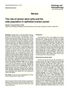
Figure 1. Clonal Evolution Model. Tumors evolve from a single normal cell (indicated by ‘N’) which undergoes a transformation that allows it to escape normal cell growth regulation. It rapidly divides, generating clones with increasing genetic instability and mutations. Here, Chronic Granulocytic Leukemia (CGL) is used as an example. Subpopulations which do not survive are indicated by the shaded circles, while those that acquire an additional mutation which confers a survival advantage are indicated by the numbers T1 through T6. Significant biological events and ploidy number are also indicated throughout the progression. Figure taken from (Nowell, 1976). Today, clonal evolution theory is generally accepted, and advanced versions of Nowell’s depiction of CGL progression are plotted in so-called phylogenetic trees (Figure 2), which contain step-wise progressive genetic mutations that lead to the eventual accumulation of a wide variety of subpopulations both within a single tumor and between patients with the same disease (Shlush et al., 2012).
13
Importantly, in the clonal evolution model, every cancer cell is capable of generating a tumor, regardless of its position in the evolutionary timeline.
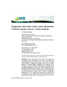
Figure 2. Phylogenetic Tree of Acute Myeloid Leukemia. Cells were sampled from two patients before and after treatment, and genetic sequencing revealed mutation progression. With each split in the tree, a new genetic identifier is found in a population of cells. The distance between each split in the tree indicates “cell depth,” and is related to the number of replications the cells underwent before gaining a unique genetic identity from the root. Large depths indicate frequent replication, while shallow depths indicate rare replication of a subclone. Figure taken from (Shlush et al., 2012).
14
The Swanton group has published extensively on the importance of genomic instability for generating intratumoral heterogeneity, including the selection of the ‘fittest’ clones within a given microenvironment or therapeutic context (Burrell, McGranahan, Bartek, & Swanton, 2013). This mechanism has essentially been proven as an influential force in tumorigenesis, and all other models have incorporated clonal evolution as part of the process alongside new theories of cancer development. Controversy has ensued, however, about the extent to which clonal evolution theory is responsible for intratumoral heterogeneity. Models for Intratumoral Heterogeneity: Cancer Stem Cell Theory A stem cell is an unspecialized progenitor which can give rise to a variety of more specialized cells, and is capable of self-renewal. In adults, stem cells make up rare populations that remain relatively dormant in tissues, waiting for queues from the extracellular environment to divide and differentiate. Around the same time that clonal evolution theory began to unfold, investigators were examining stem cells and how mutations in an immortal cell line may be involved in the development of a tumor. A model was proposed by John Cairns in 1975 in which “immortal” strands of DNA were passed on from one stem cell to its daughter stem cell, while its other daughter cell received a “mortal” strand and went on to terminally differentiate (Figure 3). In this way, genetic mutations could be immortalized and, as they accumulate throughout a person’s lifetime, dramatically increase the risk of
15
developing cancer, since several mutations are required for cancer to occur (Cairns, 1975; Knudson, Strong, & Anderson, 1973). One can imagine the accumulation of mutations would be many fold higher if a stem cell were to divide into two daughter stem cells, a process called ‘symmetric division’ for example (Kondo, 1977), creating two immortal DNA strands bearing oncogenic mutations instead of one.
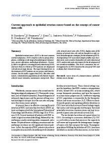
Figure 3. Cell Division and Partitioning of DNA Strands. a) normal semiconservative cell division where each parental DNA strand is randomly assigned to a daughter cell. A mutation is indicated by the double strike. b) stem-cell division where one parental strand (solid line) is preferentially passed on to stem cell progeny, in effect this “older” strand becomes immortalized. The other “younger” strand (dashed line) is passed on to daughter cells which go on to terminally differentiate. A mutation is indicated by the double strike. Taken from (Cairns, 1975).
16
Today, stem cell theory has taken hold and is a hot topic in the scientific community, although some would argue, little is understood about stem cell differentiation, including Cairns’ model of the immortalized DNA strand (Sell, 2004). In brief, cancer stem cell theory proposes that tumors are initiated when a normal stem cell undergoes a transformation which leads to dysregulation of its regulatory mechanisms. As a result, the stem cell divides abnormally, giving rise to either more stem cells, differentiated daughter cells, or both, and creating a diverse tumor environment of various phenotypes. Alternatively, differentiated cells which have adopted a mutator phenotype can de-differentiate and attain stem-like characteristics, which impart the capacity for self-renewal and thus tumorigenesis at metastatic sites as well (Figure 4A). At the heart of the cancer stem cell model is the hierarchy, whereby the tumorigenic stem cell is at the apex, and is capable of self-renewal and capacity to regenerate all phenotypic diversity found in a tumor (O’Brien et al., 2010).
17
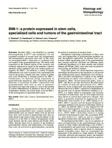
Figure 4. Cancer Stem Cell vs. Clonal Evolution Model. Two approaches for tumorigenesis. A) Classical stem cell theory whereby normal stem cells capable of self-renewal undergo a transformative genetic mutation, creating a cancer stem cell, which can differentiate to generate all other cells that comprise a tumor. B) Clonal evolution model whereby any cell can initiate a tumor, and through genetic mutations, establish intratumoral heterogeneity. These cells may also possess self-renewing capacity which was obtained through stochastic processes. Lightening bolts represent mutagenesis and asterisks represent mutations. Taken from (Campbell & Polyak, 2007).
In the following thesis, intratumoral heterogeneity within the context of ovarian cancer will be explored, as well as the applicability of the cancer stem cell model in describing this heterogeneity.
18
Specific Aims
Specific aims of the following thesis include: 1. Comprehensive review of literature to characterize the nature and extent of intratumoral heterogeneity found in ovarian cancer. 2. Investigation into the current evidence for the ovarian cancer stem cells. 3. Conclusion on the validity of the cancer stem cell model in predicting intratumoral heterogeneity in ovarian cancer.
19
INTRATUMORAL HETEROGENEITY IN OVARIAN CANCER Ovarian cancer is not a single disease, but includes several subtypes which range from relatively benign to aggressive, metastatic tumors. These subtypes vary greatly in morphology, genetic alterations, and interestingly, the proposed cell of origin, which in most cases is theorized to be from tissues outside of the ovary. In the following section will outline the ovarian cancer subtypes and discuss their proposed pathogenesis. Next, we will focus on intratumoral heterogeneity within ovarian cancer, specifically, how the genetic and epigenetic alterations, microenvironment, and cell plasticity contribute to the remarkable diversity of cancer cell populations typically found in this disease.
Ovarian Cancer Subtypes and Pathogenesis Ovarian cancer is not a single disease, but a grouping of cancers which have differing behaviors and morphologies, cells of origin and responses to treatment. Beyond the histological appearance of the cells (Figure 5), each subtype differs in its clinical course and genetic expression profile, although much overlap has been known to occur. The characteristics of each subtype of ovarian cancer are summarized in Table 2. The five most common forms of ovarian cancer are very diverse, yet high grade serous carcinoma research dominates the literature, presumably due to its aggressive nature and high frequency. An attempt was made to reclassify 20
ovarian cancer into only two types, ‘Type I and Type II’ (Kurman & Shih, 2010), however this approach has been criticized for lumping together the more rare subtypes of ovarian cancer and thus hindering our understanding and progress toward managing these important diseases (McCluggage, 2011; Prat, 2012). Although rare, the clear cell carcinoma subtype is actually more deadly when diagnosed in the late stages than the common HGSC, because of its poor response to chemotherapy (Prat, 2012). The study of this particular subtype of cancer is made difficult by its rarity, however recognition of it as a distinct form has advantages for a much needed focused study. For a detailed outline of each subtype see Appendix I.
21
Figure 5. Ovarian Cancer Subtypes. A) High-Grade Serous Carcinoma (HGSC); B) Low-Grade Serous Carcinoma (LGSC); C) Mucinous Carcinoma (MC); D) Endometriod Carcinoma (EC); E) Clear Cell Carcinoma (CCC)
22
Table 2. Ovarian Cancer Subtypes. Summary of distinguishing features of five ovarian cancer subtypes, which account for nearly 98% of all ovarian cancer manifestations. HGSC – High Grade Serous Carcinoma; LGSC – Low Grade Serous Carcinoma; MC – Mucinous Carcinoma; EC – Endometriod Carcinoma; CCC – Clear Cell Carcinoma. HGSC
LGSC
MC
EC
CCC
Frequency
68-71%