Achilles Tendon Injuries in Elite Athletes: Lessons in Pathophysiology from Their Equine Counterparts
Janet C. Patterson-Kane and Tina Rich
Abstract Superficial digital flexor tendon (SDFT) injury in equine athletes is one of the most well-accepted, scientifically supported companion animal models of human disease (i.e., exerciseinduced Achilles tendon [AT] injury). The SDFT and AT are functionally and clinically equivalent (and important) energy-storing structures for which no equally appropriate rodent, rabbit, or other analogues exist. Access to equine tissues has facilitated significant advances in knowledge of tendon maturation and aging, determination of specific exercise effects (including early life), and definition of some of the earliest stages of subclinical pathology. Access to human surgical biopsies has provided complementary information on more advanced phases of disease. Importantly, equine SDFT injuries are only a model for acute ruptures in athletes, not the entire spectrum of human tendonopathy (including chronic tendon pain). In both, pathology begins with a potentially prolonged phase of accumulation of (subclinical) microdamage. Recent work has revealed remarkably similar genetic risk factors, including further evidence that tenocyte dysfunction plays an active role. Mice are convenient but not necessarily accurate models for multiple diseases, particularly at the cellular level. Mechanistic studies, including tendon cell responses to combinations of exercise-associated stresses, require a more thorough investigation of cross-species conservation of key stress pathway auditors. Molecular evidence has provided some context for the poor performance of mouse models; equines may provide better systems at this level. The use of horses may be additionally justifiable based on comparable species longevity, lifestyle factors, and selection pressure by similar infectious agents (e.g., herpesviruses) on general cell stress pathway evolution. Key Words: achilles tendon; aging; athletes; horse; superficial digital flexor tendon; tendinopathy; tendon; tenocyte Janet C. Patterson-Kane, BVSc, PhD, is a professor and Tina Rich, BSc, PhD, is a lecturer at the Institute of Infection, Immunity and Inflammation at the University of Glasgow, Scotland. Address correspondence and reprint requests to Dr. Janet C. PattersonKane, Institute of Infection, Immunity and Inflammation, University of Glasgow, University Place, Glasgow G12 8TA, Scotland or email
[email protected].
T
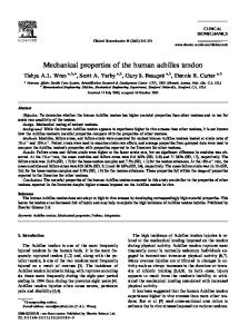
he Achilles tendon (AT) has been of pivotal evolutionary importance to the human species, facilitating bipedal and rapid locomotion (Malvankar and Khan 2011; Raichlen et al. 2011). However, AT rupture is a significant injury event in the most physically active of people. Up to 75% of ruptures are related to athletic activity ( particularly in men), and the incidence has been increasing in the Western world in particular, in association with greater sports participation (Maffulli et al. 1999; Nyyssönen et al. 2008). Incidences of 10–57% vary with the type of athletic activity (e.g., different types of running exercise and football, basketball, and gymnastics), and there are long recovery periods (up to 10 months), with reinjury rates of 2–31% (GajhedeKnudsen et al. 2013; Knobloch et al. 2008; Lopes et al. 2012; Raikin et al. 2013; van der Eng et al. 2013). In elite equine athletes, partial and complete clinical injuries of the forelimb superficial digital flexor tendon (SDFT; Figure 1) show significant epidemiological similarities. It has been estimated that up to 30% of racehorses are affected, with differences between types of activity (e.g., racing on the flat vs. over hurdles), high rates of retirement (19–70%) and re-rupture (23–67%), and long recovery periods (up to 18 months) (Goodship et al. 1994; reviewed in PattersonKane and Firth 2009). SDFT biology and pathology in the athletic horse is an excellent spontaneous (natural) model in many respects; this being particularly important in the absence of any clearly validated rodent or rabbit model for exercise-induced AT injury (Lui et al. 2011). There is no tendon in any other domesticated (and therefore accessible) species that has equivalent clinical similarity or importance. There is the added benefit that the equine injuries constitute an important clinical problem in their own right (i.e., horses and people are models for each other). Therapeutic interventions can often be tested more rapidly in horses in a high-pressure environment similar to that experienced by human athletes. For example, stem-cell repair in England was applied to hundreds of racehorse tendons (beginning more than 10 years ago; Smith et al. 2003) before being tested in treatment of AT injuries (http://bit.ly/1fT747Z), last accessed March 11, 2014. It is critical, however, to use the evolving therapeutic information in a cross-species manner only in appropriate circumstances and with adequate scientific rationale; as discussed herein, in most circumstances, equine athletes are not experimental
ILAR Journal, Volume 55, Number 1, doi: 10.1093/ilar/ilu004 © The Author 2014. Published by Oxford University Press on behalf of the Institute for Laboratory Animal Research. All rights reserved. For permissions, please e-mail:
[email protected].
86
importance. Much of the original work on elastic energy storage in tendons was performed using kangaroos (Dawson and Taylor 1973); however, this is of unknown clinical importance and, outside of Australia, of little use to the tendon researcher. This is an improvement on the situation in shoulder tendon research; most animals other than nonhuman primates (with their increased ethical concerns) are quadrupeds that use their forelimbs for weight-bearing to various degrees (i.e., their shoulder tendons have the same names but are remarkably different in structure and function) (Longo et al. 2011). The presence of a tendon in the horse that is functionally and clinically equivalent to the human AT is an enormous advantage for veterinary and medical researchers.
Importance of Energy Storage during High-Speed Exercise
Figure 1 Anatomy of tendons and ligaments of the distal limb of the horse. The superficial digital flexor tendon (SDFT) is the most clinically significant tendon in equine athletes and is functionally equivalent to the human Achilles tendon. CDET, common digital extensor tendon; DDFT, deep digital flexor tendon. Adapted from PattersonKane JC, Firth EC. 2014. Tendon, ligament, bone and cartilage anatomy, physiology, and adaptations to exercise and training. In: Hodgson DR, McKeever KH, McGowan CM, eds. The Athletic Horse. Principles and Practice of Equine Sports Medicine, 2nd ed. St. Lois MO: Elsevier Saunders. p 204.
animals, and their needs as patients are paramount (according to the principles of veterinary medical ethics). In this review we will explore the comparative aspects of human and equine tendon pathophysiology in these specific structures and the practical considerations in obtaining clinically relevant research data for both species.
Why a Functionally and Clinically Equivalent Tendon Is a Better Model Although the equine SDFT and human AT are not the same structure anatomically, they share a key function: both are energy-storing structures that significantly improve the efficiency of high-speed locomotion (Biewener 1998; Malvankar and Khan 2011). No other domestic animal has an energystoring tendon with this degree of specialization and Volume 55, Number 1, doi: 10.1093/ilar/ilu004
2014
Most tendons function simply to connect skeletal muscle to bone, allowing force transfer to move (and often stabilize) joints; they have been referred to as “positional” tendons (Birch 2007). The SDFT and AT serve those functions but additionally store potential and kinetic energy as elastic energy during weight bearing. The elastic recoil when the foot leaves the ground at the end of the weight-bearing phase of the stride results in significant recovery of mechanical work that reduces the need for muscular effort (i.e., these are “spring” mechanisms). In galloping horses, mechanical work savings have been estimated at 36%, and in humans they are greater than 50% (Biewener 1998; Farriss et al. 2011). During recoil, not all of the energy is returned, with some being lost as heat (hysteresis); comparable levels of core hyperthermia (43–45oC) have been measured/predicted in these tendons during high-speed locomotion (Farriss et al. 2011; Wilson and Goodship 1994). Further examples of energy-storing tendons include hind-limb tendons in hopping (saltatory) animals (kangaroos and wallabies) and ostriches, but these are species to which most researchers will not have access. However, this does raise the question of why rabbits would not be a preferable model, given their frequent (and more economically viable) use as laboratory animals.
Why Not Use Rodent and Rabbit Models? There is no energy-storing tendon of equivalent specialization and importance in rodents, despite the attraction of being able to use genetically engineered mice for mechanistic studies. The biomechanics of the rabbit hindlimb are less well understood, but studies of hares versus rabbits and of other small jumping mammals suggest that there are significant differences from larger athletes (Biewener and Blickhan 1988; Williams et al. 2007). Small animals must place greater reliance on the ability to accelerate or decelerate rapidly to avoid predation, and in those circumstances, the tendon energysaving function is not the primary concern (Biewener and Blickhan 1988). Larger animals are more likely to move at 87
steady speeds, these being conditions most suited to optimal energy storage and recovery. The theory of humans being adapted to long-distance running for “persistence hunting” by using their energy-storing ATs has been supported by recent data on calcaneus length (and hence AT moment arm length) relative to oxygen consumption during steady speed treadmill locomotion (Raichlen et al. 2011). Rabbits are a popular model species because of their availability, relatively large tendon size (this facilitating surgical manipulation), and low cost relative to horses. However, general disease problems and lack of knowledge regarding their behavior and physiology (including tendon physiology), in addition to the questions regarding direct comparability of the AT, should be taken into account (Lui et al. 2011).
How Studying Horses Has Increased Our Understanding of Tendon Physiology Access to fresh specimens of nondiseased human tendons (or other tissues) is often extremely difficult, particularly in the case of younger/immature individuals (Longo et al. 2011). The greater availability of specimens from horses of known (and widely varying) age and, in some cases, exercise history has significantly facilitated tendon research. Comparative Studies of Energy-Storing and Positional Tendons One area of advantage has been the opportunity to analyze large numbers of specimens from normal tendons that differ markedly in function (e.g., the SDFT vs. the anatomically opposing, positional, and non-injury-prone common digital extensor tendon [CDET]) (Figure 1). The SDFT is larger and
stiffer as a structure than the CDET, but its matrix is more elastic. Extension and recovery are facilitated at the fascicular subunit level (Figure 2) by greater amounts of sliding between them and their helical structure, respectively (Batson et al. 2003; Thorpe et al. 2012; Thorpe, Klemt, et al. 2013). Similar differences in mechanical properties have been found between the AT and the anatomically opposing anterior tibialis tendon (Birch 2007). The elasticity of energy-storing tendons allows greater storage of recoverable energy but is associated with lower mechanical safety margins (Batson et al. 2003). Comparisons of strains (elongation relative to original tendon length) in vitro and in vivo have confirmed that the SDFT and AT routinely function close to failure levels to store sufficient amounts of energy, although it should be noted that there is marked individual variation in both of these tendons (Birch 2007; Lichtwark and Wilson 2005; Stephens et al. 1989). There are also significant and corresponding tendon-specific differences in matrix composition: for example, the collagen content of the SDFT and CDET is the same but its organization is not, with the SDFT containing significant numbers of collagen fibrils (the longitudinally oriented units of tensile strength; Figure 2) that have a lower mass-average diameter. The mixture of small and large fibrils in the SDFT caters to elasticity and strength, respectively, whereas the more uniformly large fibrils of the CDET are appropriate for its role in force transmission (requiring stiffness only) (Edwards et al. 2005; Patterson-Kane, Wilson, et al. 1997). Analysis of Immature versus Adult Tendons Changes in the equine CDET and SDFT with maturation provide additional clues as to why the latter structure is so prone to injury. In horses of all ages, the SDFT contains
Figure 2 The hierarchical structure of the superficial digital flexor tendon (SDFT), the sectioned surface of which is shown in this diagram (following a transverse cut). (A) The planar, zig-zag waveform followed by in-register collagen fibrils can be seen at the fascicular (and whole tendon) level. (B) A fascicle with a roughly hexagonal outline (approximately 1 mm in diameter), that is comprised of collagen fibrils with intervening noncollagenous matrix and cells. Movement between the fascicles is essential for the high elasticity of this energy-storing tendon. (C) The circular collagen fibrils (in cross-section), measuring up to approximately 300 nm in diameter in this particular tendon. (D) The collagen fibrils in a longitudinal view showing the crimp waveform and their banding pattern; the latter is caused by the quarter-stagger arrangement (E) of their component collagen molecules. (F) Each collagen molecule is a triple helix of α-chains. Type I collagen, the predominant type in the adult tendon, has two α1 chains and one α2 chain. Adapted from Patterson-Kane JC, Firth EC. 2014. Tendon, ligament, bone and cartilage anatomy, physiology, and adaptations to exercise and training. In: Hodgson DR, McKeever KH, McGowan CM, eds. The Athletic Horse. Principles and Practice of Equine Sports Medicine. 2nd ed. St. Lois MO: Elsevier Saunders. p 205. 88
ILAR Journal
significantly more tenocytes (tendon fibroblasts; Figure 3) per unit area than the CDET (Stanley et al. 2007). Although both tendons reduce collagen turnover with age, the CDET consistently maintains the higher level (Birch, Worboys, et al. 2008); in adult tendons, the level of procollagen type I aminopropeptide (cleaved during collagen synthesis) is higher per square millimeter in the less cellular CDET (Young et al. 2009). This is, of course, relative because tendons in general have extremely slow collagenous matrix metabolism; the half-life of collagen in the SDFT and CDET has been estimated at 197.5 and 34.0 years, respectively (Thorpe et al. 2010). Reductions in gap junction expression and communication have also been measured with maturation in the SDFT, suggesting a reduced capacity of the tenocyte network to coordinate matrix control (Young et al. 2009). Measurement of retention of carbon-14 in people (those alive in the years 1955–1963 when nuclear bombs were tested) demonstrated almost no collagen turnover in the adult AT (Heinemeier et al. 2013). Noncollagenous matrix (NCM) turnover is relatively fast (2–4 years) (Thorpe et al. 2010); however, it is not known where this is occurring (i.e., inside fascicles where the tenocytes are situated or in the endotenon [the interfascicular connective tissue]) (Figure 3). It has been postulated that cells in energy-storing tendons essentially switch off to maintain the matrix within narrow optimal limits (for both strength and elasticity). However, the lack of evidence of turnover of extracellular collagen does not necessarily imply that tenocytes are completely inactive; it is advantageous to be continually synthesizing and degrading collagen to facilitate rapid responses to injury when/if it occurs, and a large proportion of that degradation can potentially be intracellular (Laurent 1987).
Figure 3 Photomicrograph of the superficial digital flexor tendon (SDFT) of a Thoroughbred racehorse. The tenocytes (tendon fibroblasts) are arranged in longitudinal rows, and only their nuclei are visible. There are large amounts of intervening matrix, which has a wavy appearance because the constituent collagen fibrils are crimped. Blood vessels are only located within the small amounts of endotenon tissue (arrow) that separate fascicles. Nuclear morphology is thought to relate to levels of activity. Hematoxylin and eosin (HE). Scale bar = 25 µm. Volume 55, Number 1, doi: 10.1093/ilar/ilu004
2014
Controlled Exercise Studies, Including Effects in Early Life The study of horses has also contributed significantly to our understanding of the positive and negative effects of exercise on energy-storing tendons. Responses of bone to exercise in both horses and people is relatively well understood, and significant data have been generated on possible positive effects of weight-bearing exercise during musculoskeletal development (defined as enhancement of bone mineral accretion and/or bone size) (Brama et al. 2009; Firth et al. 2011; Hind and Burrows 2007). Controlled studies of treadmillexercised foals and young adult horses have indicated that the situation in tendon tissue is significantly different, with little (if any) adaptation shown by the SDFT, even though the tenocytes are more synthetically active in immature tendon (Birch, Wilson, et al. 2008; Moffat et al. 2008; Young et al. 2009). Acceleration of SDFT growth has been noted in exercised foals, but this normalizes by the time they reach maturity (Cherdchutham, Meershoek, et al. 2001; Kasashima et al. 2002). Although there is no conclusive evidence that exercise during skeletal development induces greater resistance to SDFT injury (specifically) later in life, this training has not been associated with tendon injury when applied to young foals; this suggests that conditioning exercise to benefit later racing performance can be applied without ill effect (Kasashima et al. 2002; Moffat et al. 2008). Results from studies where foals were monitored at pasture and/or compared with animals effectively immobilized in stall boxes, have also indicated the importance of normal levels of activity for tendon development (Cherdchutham, Becker, et al. 2001; Kasashima et al. 2002). Studies of the human AT have (necessarily) been limited to in vivo measurements of structure, mechanical properties, and peri-tendonous biochemistry (Magnusson et al. 2008; Olesen et al. 2007). Some of the latter work indicating very rapid responses of collagen metabolism to single bouts of exercise was controversial (admittedly the measurements were not taken from within the tendon proper) (Olesen et al. 2007). However, in a recent study of horses in which ultrasound tissue characterization was used, there were subtle and temporary alterations in SDFT echo pattern immediately after racing. It was suggested that this occurs because of rapid and reversible alterations in NCM between the collagen fibrils (Docking et al. 2012). Locations and potential adaptive responses to exercise of various proteoglycans and glycoproteins at various levels of the structural hierarchy in the tendon are still poorly defined (Thorpe, Birch, et al. 2013). However, changes in both collagen and NCM in exercised horses have provided information regarding the development of pathology at injury-prone sites.
Pathophysiology of Athletic Tendon Injury: Complementary Information from Humans and Horses It is widely accepted that many tendon injuries occur during normal activity after an undefined period of accumulation of 89
tissue damage (microdamage) that precedes clinical symptoms and exceeds any capacity for repair. This does not preclude the ( probably less usual) rupture of a healthy tendon due to extreme mechanical circumstances occurring for some reason during exercise. It has been suggested that (in human tendons) there is a continuum of pathology, defined as tendonopathy/tendonosis, that ranges from early failure of adaptation to exercise, through progressive stages of proliferative reaction, disrepair (failed healing), and degeneration, to a potentially irreversible state (Cook and Purdam 2009). The term tendonitis is less frequently used because of the prevailing opinion that this is not primarily an inflammatory condition. However this microdamage occurs, there is an increased risk of partial or complete rupture once the tendon matrix is sufficiently weakened.
How Does the Term Tendonopathy Relate to Exercise-Induced Pathology in Horses? Increasingly it is becoming clear that human tendonopathy (often spelled tendinopathy) is a term better used to refer to a syndrome of chronic tendon disease rather than one specific situation, with significant variations in pathology, stages of presentation, and risk factors between various patient groups (Cook 2011). The horse is highly unlikely to be a useful model for all of those states and risk groups in the human species, including nonathletic individuals (e.g., postmenopausal women), patients taking fluoroquinolone antibiotics, and people with various metabolic disorders (Abate et al. 2013). Comparison of horses and humans is additionally complicated by the frequent definition of human tendonopathy as chronic tendon pain, this not being a known clinical feature of the equine disease. A horse experiencing SDFT injury is logically most equivalent to the athlete who experiences an acute rupture during exercise and has (usually) previously been asymptomatic. In both cases, pain is not a significant feature, other than for a few weeks after rupture (McIlwraith 2002). Differences in matrix gene expression (from normal) found between chronically painful and acutely ruptured ATs support this separation of human patient groups in studying pathogenesis (Corps et al. 2004; Jones et al. 2006).
Evidence of the Earliest Stages of Exercise-Induced Damage: The Value of the Horse In determining the very earliest steps in the pathogenesis of athletic (energy-storing) tendon injuries, the horse has been of value because of greater knowledge of normal SDFT biology/physiology and the fact that controlled high-speed exercise studies with subsequent tissue analysis have been possible. These studies have been correlated with findings in feral horses (with “normal” levels of activity) and in racehorses to confirm real-world relevance. Galloping exercise has been associated with multiple asymptomatic changes in matrix in the SDFT that would be expected to increase the 90
risk of overstrain. These include alterations in collagen crimp morphology (Figure 2), decreased collagen fibril diameter, accumulation of partially degraded collagen, and reductions in glycosaminoglycan content; some of these changes appear to represent accelerated aging (Birch, Wilson, et al. 2008; Patterson-Kane, Parry, et al. 1997; Patterson-Kane, Wilson, et al. 1997; Thorpe et al. 2010). Because surgical repair of the human AT is frequently performed, complementary information on later stages of pathology has come from analysis of biopsy specimens taken at the time of surgical repair. In one study, 97% of spontaneously ruptured tendons showed evidence of chronic degenerative change that must have preceded the clinical incident (Kannus and Józsa 1991). Surgical approaches to equine SDFT treatment have fallen from favor because the risks and costs involved cannot be justified by any consistent beneficial effect (Dowling et al. 2000). In both human and horse, changes in matrix metalloproteinase (MMP) gene expression have been measured in ruptured tendons, including similar reductions in MMP-3 (Clegg et al. 2007; Jones et al. 2006). These have also been found in the apparently normal contralateral SDFT, indicating a potential role for enzymatic perturbation in earlier stages of lesion development (Clegg et al. 2007).
Current Developments in Genomics That Favor Using Equine Populations It has been recognized for some time that in horses and humans there is significant individual variation in susceptibility to tendon injury due to interactions between extrinsic and intrinsic factors—the latter potentially including limb malalignment/conformation, susceptibility to fatigue, and the structure and mechanical properties of the tendon tissue itself (Birch 2007; Järvinen et al. 2005). Developments in genetic profiling techniques over the past 5 to 6 years have resulted in identification of a number of polymorphisms in matrix protein-encoding genes that have association with a higher risk of AT injury. These include genes encoding nonfibrillar collagens (e.g., COL5A1, COL11A2, and COL27A1), an associated microRNA (e.g., MIR608), a procollagen processing enzyme ( procollagen N-propeptidase; ADAMTS14), and tenascin C (TNC; a glycoprotein) (Abrahams et al. 2013; El Khoury et al. 2013; Hay et al. 2013; September et al. 2012). Collagen V regulates collagen fibril generation and organization, and tenascin C is involved in cell-matrix interactions. Interestingly, in recent work using samples from National Hunt Thoroughbred racehorses, (single nucleotide) polymorphisms in the COL5A1 and TNC genes were associated with an increased risk of SDFT injury (Tully et al. 2013). This suggestion of similar genetic risk factors further supports the use of the horse as a model for AT injury. How specific genetic alterations play a role in causing certain individuals to develop tendon lesions is not understood, but they are not confined to genes encoding extracellular matrix components. Certain polymorphisms in genes encoding effectors of the apoptosis pathway have also been associated ILAR Journal
with predisposition to human AT pathology; this includes polymorphisms that potentially reduce caspase-8 expression (i.e. should protect against apoptotic cell death) (Nell et al. 2012). These data raise the possibility that the fine balance between cell fate pathways is of significant importance. A failure to clear injured cells could result in the persistence of dysfunctional populations with multiple potential negative effects, including disruption of local/more extensive matrix control by tenocyte networks and inappropriate signaling to the immune system. The active participation of tenocytes in the pathogenesis of microdamage has been a rapidly increasing focus of research in recent years and is again an area in which comparative human–equine studies are likely to be synergistic.
The Tenocyte: Collateral Damage or Provocateur? Tenocytes are tendon fibroblasts (Figure 3) responsible for synthesis, degradation, and maintenance of the extracellular matrix, including repair of microdamage. Initially, tenocytes were seen as “victims” in the development and accumulation of damage (i.e., having their reparative capacities overwhelmed and/or being damaged or killed at the same time as a consequence of the injury to the surrounding matrix). Increasingly however, it has been recognized that cell death and, in particular, inappropriate cellular responses to either direct or indirect damage (or microenvironmental change) are likely to play major and active roles in microdamage accumulation (Leadbetter 1992).
Cellular Populations of Tendons Tenocytes are found within fascicles, arranged in widely spaced rows (oriented in the longitudinal axis) with large amounts of matrix between them (Figure 3). There is variation in nuclear morphology with larger, ovoid nuclei and thinner, elongate nuclei classified by some researchers as tenoblasts and tenocytes; the larger nuclei (the proportion of which reduces with age) have been associated with more active matrix remodeling activity (Chuen et al. 2004; Stanley et al. 2007). The tenocytes are linked into three-dimensional networks by cytoplasmic processes that extend both within and between rows and are joined by adherens and gap junctions. There are also fibroblastic cells within the endotenon (closely adjacent to the vascular supply; Figure 3), but differences or similarities between these and the tenocytes have not been defined. Cells with multilineage potential (i.e., stem or progenitor cells) have been isolated from tendons and termed tendon-derived stem cells (TDSCs) or tendon stem/progenitor cells (TSPCs). The relationship of TDSCs/TSPCs with bone marrow–derived mesenchymal stem cells, their anatomical locations, and roles played (if any) in response to microdamage or macroscopic tearing are not certain. Although there is some evidence for TDSCs in the paratendon as well as the tendon proper, various researchers claim to have identified Volume 55, Number 1, doi: 10.1093/ilar/ilu004
2014
them in perivascular locations and/or sites embedded in the extracellular matrix; there may, in fact, be multiple populations (reviewed in Lui 2013). Supporting histologic data to confirm the identity of these cells based on markers determined using ex vivo material would be helpful.
The Tendonosis Cycle and Why Energy-Storing Tendons Provide “the Perfect Storm” The “tendonosis cycle” refers to inappropriate repair of (or response to) microdamage by the tenocytes, with the resulting abnormal matrix being even more prone to exercise-induced damage (Leadbetter 1992). There has been complementary support for this theory from SDFT and AT research, respectively (e.g., evidence of abnormally high type III/ type I collagen ratios); type III collagen is associated with smaller, weaker fibrils and is usually a minor component in adult tendon (