Int J Clin Exp Med (2008) 1, 260-266 www.ijcem.com/IJCEM805001
Review Article Epithelial Ovarian Cancer Stem Cells---A Review Yueyin Pan1,2 and Xudong Huang2 1Department
of Medical Oncology, The First Affiliated Hospital of Anhui Medical University, Hefei, China; and Medicinal Chemistry Laboratory, Department of Radiology, Brigham and Women’s Hospital and Harvard Medical School, 75 Francis Street, Boston, MA 02115, USA 2Conjugate
Received May 2, 2008; accepted June 26, 2008; available online June 30, 2008 Abstract: Ovarian cancer is the fifth leading cause of cancer deaths among women and the most common type of gynecologic malignancy. Despite advances in surgery and chemotherapy, the survival rate of patients with epithelial ovarian cancer is still low. For a whole decade, that situation has not improved. Most ovarian cancer patients relapse and become drug-resistant. Increasing evidence has suggested that tumor growth capability is dependent upon cancer stem cells (CSCs) that represent a small subset of cells within a variety of human tumors. In immunocompromised mice, CSCs were assayed by their ability to initiate tumor growth. Although still being explored, the ovarian CSCs, which have been found in several ovarian cancer cell lines, are within a mixed population of cells derived from the ascites of a patient with advanced ovarian cancer. Hope may lie in killing of the ovarian CSCs. They are also likely to be the therapeutic target in the treatment of epithelial ovarian cancer. Thus, a thorough understanding of CSCs’ biology, particularly of how they differ from ordinary cancer cells and normal stem cells, might lead to a more effective ovarian cancer therapeutic strategy involving the selective targeting and elimination of ovarian CSCs. Key Words: Epithelial ovarian cancer, stem cell, cancer stem cell
Cancer stem cells in various tumors Two models explain tumor development. The first, termed the stochastic model, assumes that every cancerous cell has the capacity to extensively proliferate and regenerate a tumor. This model assumes that all cancer cells have an equal probability of regenerating a tumor. In contrast, the model of the cancer stem cell (CSC) assumes that only a very small subset of cells within the tumor population actually has the capacity to initiate and sustain tumor growth [1]. As we have known, human stem cells (HSCs), one of master cells in human body, are unspecialized cells with the same lineage. Unlike mature cells with permanently committed fate, HSCs are capable of dividing throughout life and creating highly specialized cells for cell renewal and tissue repair. On the other hand, CSCs are a subpopulation of tumorigenic cancer cells that possess properties of HSCs. Thus, they are sharply different from the bulk of cancer cells that are
believed to be non-tumorigenic. Indeed, increasing experimental evidence suggests that a distinct population of CSCs persist in tumors that may engender tumor relapse and metastasis [2-8]. Technical advancements in stem cell biology help to identify stem cells in tumors. These methods include: cell isolation using serial dilution and serial transplantation, side population (SP) sorting, and transplantation into immunodeficient mice. Side population cell sorting has been used to enrich stem cell compartments in diverse tissues and organs. SP cells are detected by their own ability to efflux Hoechst 33342 dye through an adenosine triphosphate (ATP)binding cassette (ABC) membrane transporter [9-11]. Through fluorescence-activated cell sorting (FACS) and with knowledge of the cell surface
Pan and Huang/Epithelial Ovarian Cancer Stem Cells
Table 1. Cell surface biomarkers associated with different cancer stem cells Tumor type Cell surface markers Acute myeloid leukemia
CD34+CD38-
(2)
Breast cancer
CD44+CD24-ESA+
(12)
Brain cancer
CD133+
(13)
Colon cancer
CD133+
(5,18)
Head and neck cancer
CD44+
(7)
Prostate cancer
CD44+
( 17)
Multiple myeloma
CD138-
(14)
Metastatic melanoma
CD20+
( 19)
Colorectal cancer
EpCAMhigh CD44+CD166+ CD24+CD44+ESA+
(20)
( 20)
Bone sarcoma
Sca1+CD45-PecamCD34+ Stro1+CD105+CD44+
( 21)
Hepatocellular carcinoma
Very heterogeneous
( 22)
Pancreatic cancer Lung adenocarcinoma
phenotypes for HSC and progenitor cells, John Dick and colleagues isolated and identified CD34+CD38- leukemic stem cells from human acute myeloid leukemia and, after comparing them with the CD34+CD38+and CD34fractions [2], demonstrated that the stem cells initiated leukemia in NOD-SCID mice. Using similar approaches and principles over the last 10 years, scientists have prospectively identified solid tumor stem cells from a variety of cancers, including isolating a minor distinct tumor cell population in breast cancer. This tumorigenic cell population was identified based on its cell surface phenotype, which was lineage-CD44+CD24-/low and able to form mammary tumors in NOD-SCID mice, whereas cells with alternative phenotypes, even when implanted at significantly higher cell numbers [12], were non-tumorigenic. Tumorigenic cells can be serially passaged, demonstrating selfrenewal capacity, generating tumor heterogeneity, and producing differentiated non-tumorigenic progeny. A similar finding was made in a human brain tumor as it was known that normal neuronal stem cells express the marker CD133. As few as 100 CD133+cells from human brain tumors initiated new tumors in the brains of immunocompromised mice, while CD133- cells did not contain any tumorinitiating activity [13].
261
References
(4)
This result was also obtained with cells isolated from multiple myeloma [14], head and neck squamous cell carcinoma (HNSCC) [7,15], pancreatic cancer [4,16], colon cancer [5, 8], and prostate cancer [3,17], etc. The specific cell surface biomarkers of CSC populations from these human solid tumors are summarized in Table 1. Epidemiology of ovarian cancer Epithelial ovarian cancer (EOC), originating from the surface epithelium of the ovary [23,24], consists of various histologic subtypes of Mullerian origin. It is the fifth leading cause of cancer deaths among women and the most common type of gynecologic malignancy. The poor ratio of survival to incidence in EOC results from the high percentage of cases diagnosed at an advanced stage. Despite advances in surgery and chemotherapy, the survival rate of patients with EOC stands at just 45% at 5 years, a statistic that has not changed over the last decade. Most patients relapse and become drug-resistant [25-28]. Complicating matters is that significant heterogeneity exists within the EOC group. For example, histologically defined subtypes such as serous, endometrioid, mucinous, low and high-grade malignancies all have variable clinical manifestations and underlying molecular
Int J Clin Exp Med (2008) 1, 260-266
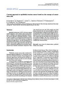
Pan and Huang/Epithelial Ovarian Cancer Stem Cells development into pluripotent mesenchyme and coelomic epithelium. Mullerian mesothelium is the precursor of oviductal, endometrial, and cervical epithelia. The ovarian surface epithelium differentiates after invagination of the coelomic mesothelium over the gonadal ridges [24,35,36]. As aforementioned, HSCs have the remarkable potential to develop into many different cell types in the body. Serving as a sort of repair system for the body, they can theoretically Figure 1. Three-dimensional view of mammalian ovarian architecture. divide without limit to replenish other cells as long as the person or animal is still alive. Quiescent somatic stem cells signatures. Chemotherapy alone can lead to reside in specific tissue niches and may be massive cytoreduction but seldom cures the activated by injury or other stimuli emanating disease. The majority of patients who respond from skin and hair follicles, mammary glands, to primary chemotherapy ultimately develop intestines, and other organs [37]. The evolving recurrent, usually drug-resistant, disease for evidence that somatic stem cells contribute to reasons that are still unknown [29]. CSCs, like normal tissue repair and regeneration somatic stem cells, are thought to be capable suggests the potential for multipotent somatic of unlimited self-renewal and proliferation. stem cells in the ovary to be responsible for Therefore, tumor stem cell escape from innate regulated surface epithelial repair after somatic niche regulators might occur due to a ovulatory rupture and possibly the generation variety of factors including: ovarian cancer of oocyte nurse cells for folliculogenesis [38]. progression, resistance to chemotherapy, Ovarian somatic stem cells would be expected recurrence and metastasis. Further, an to divide asymmetrically, yielding both a explanation for the histologic heterogeneity daughter cell that proceeds to terminal often found in tumors [30-33] might be that differentiation for epithelial repair and an CSCs are multipotent. undifferentiated self-copy. Repeated asymmetric self-renewal sets the stage for Architecture of the ovarian epithelium and its somatic stem cells or their immediate profile during development progenitors to accrue mutations over time, a phenomenon that might lead to their As shown in Figure 1 [34], ovarian surface transformation into CSCs and malignant epithelial cells vary in type from simple progression [39]. squamous to cuboidal to low pseudostratified columnar. The surface epithelium is supported Experimental evidence for ovarian epithelial over the ovarian cortical interstitium (tunica cancer stem cells albuginea) by a basement membrane and is held together laterally by desmosomes and As mentioned above, several human primary gap or tight junctional complexes. Surface tumors and human cancer cell lines were cells are continuous at the hilum with the shown to possess CSCs. Although it is very mesothelium of the ovarian ligament limited, work has been done by investigators (mesovarium) and peritoneum. Ovarian on CSCs in EOC. surface epithelial cells have a mesodermal derivation shared with the epithelia of the For example, Bapat and colleagues presented urogenital system and adrenal cortex. direct evidence that the aggressiveness of Mesoderm segregates during embryonic human ovarian cancer may result from
262
Int J Clin Exp Med (2008) 1, 260-266
Pan and Huang/Epithelial Ovarian Cancer Stem Cells transformation and dysfunction of stem cells in the ovary. They isolated a single tumorigenic clone among a mixed population of cells derived from the ascites of a patient with advanced ovarian cancer. During the course of this study, another clone underwent spontaneous transformation in culture. Both transformed clones possess stem cell-like characteristics and, although further maturation and tissue-specific differentiation was arrested, differentiate to grow in an anchorage-independent manner in vitro as spheroids. Tumors established from these clones in animal models are similar to those in the human disease in their histopathology and cell architecture and the tumorigenic clones. Even when involved in serial transplantation, they continue to establish tumors, thereby confirming their identity as tumor stem cells [40]. Szotek and colleagues identified and characterized SP cells from two distinct genetically engineered mouse ovarian cancer cell lines. They used Hoechst 33342 to isolate SP from these cell lines to define candidate CSCs. In vivo, when equal numbers of SP and non-SP cells were injected into the dorsal fat pad of nude mice, mouse SP cells formed measurable tumors sooner than non-SP (NSP) cells. They also found that Mullerian inhibiting substance (MIS) inhibited the proliferation of both SP and NSP cells, whereas the lipophilic chemotherapeutic agent doxorubicin more significantly inhibited the NSP cells [41]. Their findings suggest that CSCs are within EOC and that its underlying cause might be stem cell transformation. Further, the continuing stochastic events of stem and progenitor cell transformation may define ovarian cancer’s often increasing aggression. To investigate whether ovarian cancer cells express somatic and CSC surface markers as well as to identify differential expression between CSCs and non-CSCs, investigators analyzed mouse and human ovarian cancer cells. Stem cells isolated from the nervous system and prostate express CD133 antigen. As such, they are widely used to isolate hematopoietic stem and progenitor cells. Ferrandina G and colleagues investigated the expression of the CD133-1 and CD133-2 epitopes in primary ovarian tumors. The results demonstrated that CD133(+) cells gave rise to a larger number of colonies than those documented in a CD133(-) population.
263
Moreover, CD133(+) cells showed an enhanced proliferative potential compared to CD133(-) cells. The percentages of CD133-1and CD133-2-expressing cells were significantly lower in normal ovaries/benign tumors with respect to those in ovarian carcinoma. Both the percentages of CD133-1and CD133-2-expressing cells were significantly lower in omental metastases compared to those in primary ovarian cancer. CD133-1 and CD133-2 may be useful, therefore, to select and enrich the population of CD133(+) ovarian tumor cells that are characterized by a higher clonogenic efficiency and proliferative potential [42]. Bapat and colleagues found that the growth factor receptors c-met and epidermal growth factor receptor were upregulated in ovarian CSCs as was the surface adhesion molecule CD44. They also expressed E-cadherin. Correspondingly, Snail, a known mediator of epithelial-mesenchymal transition through transcriptional repression of E-cadherin in ovarian cancer, was expressed in some CSC clones and to a lesser extent in others. These expression patterns indicate the mesothelial nature of the cells [40]. Szotek and colleagues’ results showed, compared with NSP cells, the MOVCAR 7 SP cells were enriched in number of cells and intensity of expression of c-kit/CD117 (stem cell factor receptor), whereas 4306 and human SP and NSP cells did not express c-kit. MOVCAR7 SP and NSP cells strongly express the tumor metastasis marker CD 44 (hyaluronic acid receptor), whereas 4306 cells and most human ovarian cancer cells do not. MOVCAR 7 and 4306 SP and NSP cells did not express CD24, CD34, CD105, CD133, or Sca1. Human cell lines and ascites cells showed variable expression of the epithelial cell marker epithelial-specific antigen/EpCAM(epithelial specific antigen) and CD24. These markers, aside from c-kit in MOVCAR 7, did not add to the consistent SP phenotype and Bcrp1 immunostaining they have observed in identifying putative ovarian CSCs in both the mouse and human [41]. All these findings tell us that, up to now, no specific marker has been found from the surface of ovarian CSCs. Wani et al. described a strategy to explore genetic drift in the mitochondrial genome accompanying varying stem cell dynamics in
Int J Clin Exp Med (2008) 1, 260-266
Pan and Huang/Epithelial Ovarian Cancer Stem Cells EOC. A major and novel outcome is the identification of a specific mutant mitochondrial DNA profile associated with the CSC lineage that is drastically different from the germ line profile. Continuing mutagenesis in subsets with a mutant mitochondrial genome could result in transformation through a cooperative effect with nuclear genes. A representative example in their study is a tumor suppressor gene- cAMP responsive element binding protein. This specific profile could be a critical predisposing step undertaken by a normal stem cell to overcome a tightly regulated mutation rate and DNA repair in its evolution towards tumorigenesis [43]. These findings suggest that varying stem cell dynamics and mutagenesis define the CSC progression that may clinically translate into increasing tumor aggression with serious prognostic implications. Conclusion and future perspectives Despite advances in surgery and chemotherapy, most patients with ovarian cancer relapse and become drug-resistant. Therefore, the goal of existing therapies for ovarian cancer is to eliminate the bulk of cells within the ovarian tumor. Current treatment strategies cannot cure the disease. One reason may be that existing therapies fail to kill ovarian CSCs effectively. After all, normal stem cells from various tissues appear to be more resistant to chemotherapeutic reagents than do mature cell types and characteristically express drug-resistance proteins. If this were true of ovarian CSCs, therapies that are more specifically directed at the CSC might result in more durable responses for primary as well as metastatic disease. Further characterization of the tumorigenic populations will allow for the identification of molecules expressed in CSCs that could then serve as targets. With these targets known, this fraction of cancer cells that can rapidly develop the critical tumor cell mass can be eliminated. Consequently, defining the unique properties of ovarian CSCs remains a high priority for developing early diagnostic and effective therapeutic strategies against ovarian cancer. Acknowledgements The authors would like to thank Ms. Kimberly Lawson for her excellent manuscript editing. This work was supported by the funds from the
264
First Affiliated Hospital of Anhui Medical University and Radiology Department of Brigham and Women’s Hospital. Address correspondence to: Xudong Huang, Ph.D., Conjugate and Medicinal Chemistry Laboratory, Department of Radiology, Brigham and Women's Hospital and Harvard Medical School, 75 Francis Street, Boston, MA 02115, Fax: (617)582-0004, Email:
[email protected]
References [1] Al-Hajj M and Clarke MF. Self-renewal and solid tumor stem cells. Oncogene 2004; 23: 72747282. [2] Bonnet D and Dick JE. Human acute myeloid leukemia is organized as a hierarchy that originates from a primitive hematopoietic cell. Nat. Med. 1997; 3: 730–737. [3] Miki J, Furusato B, Li H, Gu Y, Takahashi H, Egawa S, Sesterhenn IA, McLeod DG, Srivastava S and Rhim JS. Identification of putative stem cell markers, CD133 and CXCR4, in hTERT-immortalized primary nonmalignant and malignant tumor-derived human prostate epithelial cell lines and in prostate cancer specimens. Cancer Res. 2007; 67: 31533161. [4] Li C, Heidt DG, Dalerba P, Burant CF, Zhang L, Adsay V, Wicha M, Clarke MF and Simeone DM. Identification of pancreatic cancer stem cells. Cancer Res. 2007; 67: 1030–1037. [5] O’Brien CA, Pollett A, Gallinger S and Dick JE. A human colon cancer cell capable of initiating tumour growth in immunodeficient mice. Nature 2007; 445: 106–110. [6] Cariati M and Purushotham AD. Stem cells and breast cancer. Histopathology 2008; 52: 99107. [7] Prince ME, Sivanandan R, Kaczorowski A, Wolf GT, Kaplan MJ, Dalerba P, Weissman IL, Clarke MF and Ailles LE. Identification of a subpopulation of cells with cancer stem cell properties in head and neck squamous cell carcinoma. Proc. Natl. Acad. Sci U S A 2007; 104: 973–978. [8] Ricci-Vitiani L, Lombardi DG, Pilozzi E, Biffoni M, Todaro M, Peschle C, and De Maria R. Identification and expansion of human coloncancer-initiating cells. Nature 2007; 445: 111– 115. [9] Montanaro F, Liadaki K, Schienda J, Flint A, Gussoni E and Kunkel LM. Demystifying SP cell purification: viability, yield, and phenotype are defined by isolation parameters. Experimental Cell Research 2004; 298: 144– 154. [10] Liadaki K, Kho AT, Sanoudou D, Schiendaa J, Flint A, Beggs AH, Kohane IS and Kunkel LM. Side Population cells isolated from different tissues share transcriptome signatures and express tissue-specific markers. Experimental Cell Research 2005; 303: 360– 374.
Int J Clin Exp Med (2008) 1, 260-266
Pan and Huang/Epithelial Ovarian Cancer Stem Cells [11] Hadnagy A, Gaboury L, Beaulieu R and Balicki D. SP analysis may be used to identify cancer stem cell populations. Experimental Cell Research 2006; 312: 3701-3710. [12] Al-Hajj M, Wicha MS, Benito-Hernandez A, Morrison SJ and Clarke MF. Prospective identification of tumorigenic breast cancer cells. Proc. Natl. Acad. Sci. U S A 2003; 100: 3983-3988. [13] Singh SK, Hawkins C, Clarke ID, Squire JA, Bayani J, Hide T, Henkelman RM, Cusimano MD and Dirks PB. Identification of human brain tumour initiating cells. Nature 2004; 432: 396–401. [14] Matsui W, Huff CA, Wang Q, Malehorn MT, Barber J, Tanhehco Y, Smith BD, Civin CI and Jones RJ. Characterization of clonogenic.multiple myeloma cells. Blood 2004; 103: 2332-2336. [15] Harper LJ, Piper K, Common J, Fortune F and Mackenzie IC. Stem cell patterns in cell lines derived from head and neck squamous cell carcinoma. J Oral Pathol. Med. 2007; 36: 594603. [16] Hermann PC, Huber SL, Herrler T, Aicher A, Ellwart JW, Guba M, Bruns CJ and Heeschen C. Distinct populations of cancer stem cells determine tumor growth and metastatic activity in human pancreatic cancer. Cell Stem Cell 2007; 1: 313-323. [17] Collins AT, Berry PA, Hyde C, Stower MJ and Maitland NJ. Prospective identification of tumorigenic prostate cancer stem cells. Cancer Res. 2005; 65: 10946-10951. [18] Dalerba P, Dylla SJ, Park IK, Liu R, Wang X, Cho RW, Hoey T, Gurney A, Huang EH and Simeone DM. Phenotypic characterization of human colorectal cancer stem cells. Proc. Natl. Acad. Sci. U S A 2007; 104: 10158-10163. [19] Fang D, Nguyen TK, Leishear K, Finko R, Kulp AN, Hotz S, Van Belle PA, Xu X, Elder DE and Herlyn M. A tumorigenic subpopulation with stem cell properties in melanomas. Cancer Res. 2005; 65: 9328-9337. [20] Kim CF, Jackson EL, Woolfenden AE, Lawrence S, Babar I, Vogel S, Crowley D, Bronson RT and Jacks T. Identification of bronchioalveolar stem cells in normal lung and lung cancer. Cell 2005; 121: 823–835. [21] Gibbs CP, Kukekov VG, Reith JD, Tchigrinova O, Suslov ON, Scott EW, Ghivizzani SC, Ignatova TN and Steindler DA. Stem-like cells in bone sarcomas: implications for tumorigenesis. Neoplasia 2005; 7: 967–976. [22] Chiba T, Kita K, Zheng Y-W, Yokosuka O, Saisho H, Iwama A, Nakauchi H and Taniguchi H. Side population purified from hepatocellular carcinoma cells harbors cancer stem cell–like properties. Hepatology 2006; 44: 240-251. [23] Young RH. A brief history of the pathology of the gonads. Mod. Pathol. 2005; 18 (suppl 2): S3–S17. [24] Murdoch WJ and McDonnel AC. Roles of the
265
ovarian surface epithelium in ovulation and carcinogenesis. Reproduction 2002; 123: 743–750. [25] Runnebaum IB and Stickeler E. Epidemiological and molecular aspects of ovarian cancer risk. Journal of Cancer Research and Clinical Oncology 2001; 127: 73–79. [26] Jemal A, Murray T, Ward E, Samuels A, Tiwari RC, Ghafoor A, Feuer EJ and Thun MJ. Cancer statistics, 2005. CA Cancer J. Clin. 2005; 55:10–30. [27] Jemal A, Siegel R and Ward E. Cancer statistics, 2007. CA Cancer J Clin 2007; 57: 43-66. [28] Parker SL, Tong T, Bolden S and Wingo PA. Cancer statistics, 1997. CA Cancer J. Clin. 1997; 47: 5–27. [29] Lage H and Denkert C. Resistance to chemotherapy in ovarian carcinoma. Recent Results Cancer Res. 2007; 176: 51-60. [30] Landen Jr CN, Birrer MJ and Sood AK. Early Events in the Pathogenesis of Epithelial Ovarian Cancer. J Clin. Oncol. 2008; 26: 9951005. [31] Florian S, Sonneck K, Hauswirth AW, Krauth MT, Schernthaner GH, Sperr WR and Valent P. Detection of molecular targets on the surface of CD34+/CD38- stem cells in various myeloid malignancies. Leukemia Lymphoma 2006; 47: 207–222. [32] Reya T, Morrison SJ, Clarke MF and Weissman IL. Stem cells, cancer, and cancer stem cells. Nature 2001; 414: 105–111. [33] Tu SM, Lin SH and Logothetis CJ. Stem-cell origin of metastasis and heterogeneity in solid tumours. Lancet Oncol. 2002; 3: 508–513. [34] http://www.answers.com/topic/oogenesis. [35] Walt ML, Stabenfeldt GH, Hughes JP, Neely DP and Bradbury R. Development of the equine ovary and ovulation fossa. Journal of Reproduction and Fertility 1979; Supplement 27: 471–477. [36] Byskov AG. Differentiation of mammalian embryonic gonad. Physiological Reviews 1986; 66: 71–117. [37] Li L and Xie T. Stem cell niche: structure and function. Annu. Rev. Cell. Dev. Biol. 2005; 21: 605–631. [38] Bukovsky A, Caudle MR, Svetlikova M and Upadhyaya NB. Origin of germ cells and formation of new primary follicles in adult human ovaries. Reprod. Bio. Endocrinol. 2004; 2: 20-26. [39] Soltysova A, Altanerova V and Altaner C. Cancer stem cells. Neoplasma 2005; 52: 435-40. [40] Bapat SA, Mali AM, Koppikar CB and Kurrey NK. Stem and progenitor-like cells contribute to the aggressive behavior of human epithelial ovarian cancer. Cancer Res 2005; 65: 30253029. [41] Szotek PP, Pieretti-Vanmarcke R, Masiakos PT, Dinulescu DM, Connolly D, Foster R, Dombkowski D, Preffer F, MacLaughlin DT and
Int J Clin Exp Med (2008) 1, 260-266
Pan and Huang/Epithelial Ovarian Cancer Stem Cells Donahoe PK. Ovarian cancer side population defines cells with stem cell-like characteristics and Mullerian Inhibiting Substance responsiveness. Proc. Natl. Acad. Sci. U S A 2006; 103: 11154-11159. [42] Ferrandina G, Bonanno G, Pierelli L, Perillo A, Procoli A, Mariotti A, Corallo M, Martinelli E, Rutella S, Paglia A, Zannoni G, Mancuso S and
266
Scambia G. Expression of CD133-1 and CD133-2 in ovarian cancer. Int. J. Gynecol. Cancer. 2008; 18: 506-514. [43] Wani AA, Sharma N, Shouche YS and Bapat SA. Nuclear–mitochondrial genomic profiling reveals a pattern of evolution.in epithelial ovarian tumor stem cells. Oncogene 2006; 25: 6336–6344.
Int J Clin Exp Med (2008) 1, 260-266