Oncogene (1997) 14, 171 ± 183 1997 Stockton Press All rights reserved 0950 ± 9232/97 $12.00
Nuclear accumulation of FGF-2 is associated with proliferation of human astrocytes and glioma cells A Joy1, J Moett1, K Neary1, E Mordechai1, EK Stachowiak1, S Coons1, J Rankin-Shapiro1, RZ Florkiewicz2 and MK Stachowiak1 1
Barrow Neurological Institute, 350 West Thomas Road, Phoenix, Arizona 85013; 2The Scripps Research Institute, La Jolla, California 92037, USA
FGF-2 has been implicated in the neoplastic transformation of glioma cells and in the transition of normal quiescent astrocytes to a proliferating, reactive state. In the present study we have observed that in human glial cells, levels and subcellular localization of FGF-2 are dierent in quiescent and proliferating cells. FGF-2 was detected in the cytoplasm of non-reactive astrocytes in human brain sections. In contrast FGF-2 was located within the cytoplasm and nuclei of reactive astrocytes in gliotic brain tissue and in neoplastic cells of glioma tumors. In vitro, FGF-2 was found predominantly in the nucleus of subcon¯uent proliferating astrocytes, but was detected only in the cytoplasm of density arrested quiescent astrocytes. Our results suggest that reduced cell contact stimulates nuclear accumulation of FGF-2, accompanying mitotic activation of reactive human astrocytes. FGF-2 was constitutively localized to the nucleus of continuously proliferating glioma cells independent of cell density. A role for intracellular FGF-2 was further suggested by the observation that glioma cells that are not stimulated to proliferate by extracellular FGF-2 proliferated faster when transfected with FGF-2 expressing vectors. This increased proliferation correlated with nuclear accumulation of FGF-2. Cell proliferation was attenuated by 5'-deoxy-5'methylthioadenosine, a FGF-2 receptor tyrosine kinase inhibitor that acts within the cell, but was unaected by myo-inositol hexakis [dihydrogen phosphate] that disrupts FGF-2 binding to plasma membrane receptors. Our results indicate that FGF-2 serves as a nuclear regulator of proliferation in astrocytic cells. In glioma cells, the constitutive presence of FGF-2 in the nucleus may promote proliferation that is insensitive to cell contact inhibition. Keywords: growth factor; nuclear localization; astrocytes; glioma; cell proliferation
Introduction In response to injury or neuronal degeneration quiescent astrocytes in the nature brain become hypertrophic and proliferate until the space left by the degenerated brain tissue is ®lled (Eng et al., 1992; Liu and Chen, 1994). In contrast to transient
Correspondence: M Stachowiak Received 22 May 1996; revised 11 September 1996; accepted 12 September 1996
astrocytic activation neoplastic glioma cells proliferate continuously showing a reduced response to cell contact inhibition (Rosenblum et al., 1978). The expression of growth factors, including the pleiotropic ®broblast growth factor-2 (FGF-2; bFGF), controls astrocytic development, as well as the transition of mature astrocytes from a quiescent to a reactive state. In vitro, FGF-2 stimulates proliferation of neonatal rat astrocytes (Kniss and Burry, 1988; Bogler et al., 1990; Arajuo and Cotman, 1992; Engele and Bohn, 1992; Vescovi et al., 1993). In normal adult rat brain astrocytes express low levels of FGF-2 protein. During gliosis, however, the number of astrocytes expressing FGF-2 increases dramatically along with the levels of FGF-2 protein and mRNA (Finkelstein et al., 1988; Frautschy et al., 1991; Liu and Chen, 1994; Gomez-Pinilla et al., 1992) and correlates with their proliferation (Liu and Chen, 1994). The autocrine action of FGF-2 has also been implicated in the abnormal growth of the transformed astrocytes in human brain neoplasms. Overexpression of FGF-2 in certain gliomas correlates with the grade of the tumor and the extent of anaplasia (Zagzag et al., 1990). Proliferation of glioma cells is inhibited by FGF-2speci®c antisense oligonucleotides (Liberman et al., 1987; Takahashi et al., 1990; Morrison, 1991; Morrison et al., 1994a). Unlike many well-characterized growth factors, FGF-2 does not contain a consensus secretory sequence (Abraham et al., 1986). Accordingly, only very low levels of FGF-2-like growth stimulatory activity have been detected in conditioned medium or associated with the extracellular matrix of cells that produce FGF-2 (Moscatelli et al., 1986; Vlodavsky et al., 1987). At least four amino terminus extended isoforms of FGF-2 arise from the same gene due to initiation of translation at dierent start codons with dierent cells expressing dierent isoforms (Florkiewicz and Sommer, 1989). All FGF-2 isoforms are detected in both the cytoplasm and the nucleus of expressing cells (Powell and Klagsbrun, 1991; Florkiewicz et al., 1991; Gualandris et al., 1993; Woodward et al., 1992; Vijayan et al., 1993; Stachowiak et al., 1994, 1996a,b). Nuclear presence of FGF-2 was detected in rat brain astrocytes both in vivo (Woodward et al., 1992) and in vitro (Vijayan et al., 1993) and in cultured human astrocytes and glioma cells (Moett et al., 1996; Stachowiak et al., 1996a). With the observation that FGF-2 remains cell associated and may not be secreted by a classical mechanism, a regulated targeting of endogenous FGF2 to the nucleus (Puchacz et al., 1993; Stachowiak et
Nuclear accumulation of FGF-2 and proliferation of glial cells A Joy et al
172
al., 1994, 1996b; Moett et al., 1994, 1996) suggests that this growth factor may serve as an intracrine signaling molecule. This study was undertaken to determine the signi®cance of nuclear localization of FGF-2 in human astrocytes and glioma cells (Moett et al., 1996; Stachowiak et al., 1996a) and its role in cell proliferation. In con¯uent, quiescent astrocytes FGF-2 is present exclusively in the cytoplasm. Release from cell-contact inhibition and mitotic activation are associated with nuclear accumulation of FGF-2. In contrast, continuously proliferating glioma cells express high levels of nuclear FGF-2 constitutively, independent of cell density. The mitogenic action of nuclear FGF-2 is further suggested by the observation that intracellularly expressed recombinant FGF-2 accumulates in the cell nucleus and stimulates proliferation of glioma cells. Thus, FGF-2 may serve as a nuclear regulator of astrocyte and glioma cell proliferation.
Results Expression of FGF-2 in normal and gliotic human brain tissues and in glioblastoma multiforme (GBM) To investigate the function of FGF-2 in reactive and neoplastic astrocytes we ®rst examined its expression in normal and gliotic brain tissues and in GBM. In normal lateral temporal lobe tissue, glial ®brillary acidic protein (GFAP) positive cells with typical processes were present in grey and white matter (Figure 1(I)). Medial temporal lobe (Ammon's horn) sclerosis associated with epilepsy, with marked neuronal loss and gliosis of the pyramidal cell layer, was present in the hippocampal region. Reactive astrocytes had enlarged oval or oblong nuclei. Their increased size and nonstellate morphology resembled reactive proliferating astrocytes in the gliotic tissue of the rat brain (Liu and Chen, 1994). GFAP staining was con®ned to the perinuclear cytoplasm; well-developed processes were rare (Figure 1(III)). Tumor tissues isolated from GBMs displayed hypercellularity, pleomorphism and necrosis. In normal tissue, stellate astrocytes demonstrated cytoplasmic FGF-2 immunoreactivity (FGF-2-IR), with the perinuclear region and cytoplasmic processes outlined (Figure 1(II)). Large neurons also stained diusely for FGF-2 in the perinuclear cytoplasm. Small round cells, consistent in appearance and distribution with oligodendrocytes, showed no detectable FGF-2 labeling (not shown). FGF-2-IR in less gliotic tissue was similar to normal lateral temporal lobe (not shown). However, in areas of severe neuronal loss, the majority of nonstellate cells with enlarged nuclei, identi®ed by GFAP staining as reactive astrocytes, demonstrated prominent nuclear FGF-2 immunostaining (Figure 1(IV)). Occasional nucleolar-like staining was also observed. Most neoplastic cells in GBM tissue were strongly positive for FGF-2 (Figure 1(V)). Immunoreactivity was more intense in the nuclei than in the cytoplasm. The intensity of nuclear staining was similar or stronger than that of reactive astrocytes from nonneoplastic tissue. We detected no FGF-2-IR in vascular cells or in the extracellular space.
Figure 1 FGF-2 expression in human brain tissues ± normal, gliotic and GBM. (I) ± GFAP (blue) immunoreactivity in typical stellate astrocytes in normal cortex. (II) ± FGF-2 immunoreactivity (red) in normal cortex. The section is counterstained with hematoxylin and eosin (dark blue) to visualize nuclei. In stellate astrocytes and in neurons (two large cells), FGF-2 is con®ned to the cytoplasm. (III) GFAP and (IV) FGF-2 immunoreactivity in reactive astrocytes with ill-de®ned cytoplasm and few processes in the gliotic hippocampus. Nuclear FGF-2 staining is present in the majority of reactive astrocytes. (V) GBM with diuse, predominantly nuclear, FGF-2 immunoreactivity. Vascular endothelial cells and pericytes/smooth muscle cells are unstained (*). (Original magni®cation 6250)
Localization of FGF-2 in cultured human astrocytes and glioma cells To identify what triggers nuclear accumulation of FGF-2 in gliotic tissue and in GBM and the function of nuclear growth factor, we have developed cultures of human astrocytes and utilized established glioma cell lines. We found that Waymouth 87/3 medium (MAB, Asch et al., 1988) supplemented with 10% to 20% serum supports proliferation of adult human astrocytes, while at the same time suppressing the growth of oligodendroglial and endothelial cells. Using this medium we obtained pure human astrocytic cultures as evidenced by the expression of GFAP by all cells (Figure 2a). In most cases (see example on Figure 2a), GFAP staining in glioma cells was less intense than in normal astrocytes. FGF-2-IR was localized predominantly in the nuclei and nucleoli of all astrocytic cultures obtained from dierent subjects and in U251MG and U343MG glioma cells (see examples on Figure 2b). Confocal
Nuclear accumulation of FGF-2 and proliferation of glial cells A Joy et al
microscopy was performed to distinguish between periand intra-nuclear localization of FGF-2. A composite of individual confocal sections taken at 1 mm intervals through the astrocytes is shown on Figure 2b(V). Inspection of individual sections con®rmed that FGF2-IR is localized inside the nucleus (not shown). Again, FGF-2-IR was strongest within the nucleus. Cell density-dependent regulation of nuclear FGF-2 in cultured human astrocytes and glioma cells In vitro, astrocytes transferred from con¯uent to subcon¯uent cultures underwent changes that mimicked reactive transformation in vivo. Con¯uent cultures of human astrocytes remained quiescent even in the presence of 20% serum. Following passage, subcon¯uent astrocytes re-entered a proliferative phase. Cells appeared enlarged, synthesized DNA, and showed karyokinesis. Incorporation of 5-bromo-2'deoxyuridine (BUdR) into nuclei of subcon¯uent, but not con¯uent, astrocytes within the same culture dish is shown in Figure 3. Before reaching a con¯uent state, proliferating astrocytes exhibited intense FGF-2-IR within nuclei and nucleoli (Figure 4a(I)). Gradually astrocytes began to extend processes and contact one another. Concomitantly, the intensity of the nuclear FGF-2 staining began to decrease. As shown in Figure 4a(II)), nuclear and nucleolar FGF-2-IR was reduced markedly between 1 and 3 weeks as the cells attained con¯uence. In con¯uent astrocytes antibody penetration was unimpaired since staining for human nucleolar antigen was unaected by cell density and time in culture (not shown). The depletion of nuclear FGF-2-IR was reversed after two additional weeks in culture when astrocytes detached leaving large empty surfaces. In those areas, astrocytes showed increased expression of nuclear and nucleolar FGF-2 (Figure 4a(III)). Some cells showed karyokinesis indicating renewed proliferation. The gradient of FGF-2 expression was observed even within the same culture dish (Figure 4b). Con¯uent astrocytes had no detectable nuclear FGF-2 expression, whereas subcon¯uent cells outside the dense core showed a gradual densitydependent increase in FGF-2-IR. These data suggest that loss of nuclear FGF-2-IR correlates with establishment of cell-cell contact rather than with the action of diusible factors. Nuclear FGF-2 was depleted in high-density astrocytic cultures prepared from dierent subjects and brain areas (frontal lobe cortex (Figure 4), frontal parietal area, or cerebellum (not shown)). In contrast to astrocytes, the two glioma lines examined, U251MG and SF-767, expressed high levels of nuclear FGF-2, even in cultures maintained in a con¯uent state for an extended period. Figure 4a(IV ± VI) shows expression of FGF-2 in U251MG cells. Three weeks after plating, cells that attained con¯uence showed a small reduction of nuclear and cytoplasmic FGF-2-IR(V). With an additional 2 weeks in culture, new cell layers formed with intense nuclear and nucleolar FGF-2IR(VI). Western analysis of biochemically fractionated astrocytes con®rmed the cell density-dependent expression of FGF-2. Cytoplasmic fractions of astrocytes
and glioma cells contained at least three FGF-2 proteins (Figure 5) with apparent molecular weights of 18, 22, and 24 kDa similar to human FGF-2 isoforms generated from alternate utilization of CUG and AUG translation initiation codons (Florkiewicz and Sommer, 1989; Powell and Klagsbrun, 1991). The 22 kDa band was previously shown to be a doublet of two closely migrating 22 and 23 kDa isoforms (Florkiewicz and Sommer, 1989). In nuclear and cytoplasmic extracts isolated from astrocytes (Figure 5a) or U251MG cells (Figure 5b) the 18 and 22 kDa FGF-2 isoforms were the most abundant. To investigate the eect of cell density on the nuclear content of FGF-2 independent of time in culture, astrocytes were plated at two densities. Three weeks post-plating the majority of astrocytes reached con¯uence in higher density cultures, while cells plated at lower density reached con¯uence after 4 weeks. At 3 weeks astrocytes attaining a con¯uent state in the higher density cultures demonstrated a partial depletion of nuclear FGF-2 and complete depletion of nuclear FGF-2 4 weeks post-plating. Reduction of nuclear FGF-2 content in astrocytes plated at lower density was delayed. Depletion of nuclear FGF-2 was accompanied by a less marked reduction of cytoplasmic FGF-2 content. These data provide further support for a correlation between nuclear accumulation of FGF-2 and cell proliferation. In addition, FGF-2 was not detected in isolated nuclei, while cytoplasmic FGF-2 remained high, indicating that the nuclear fraction was not significantly contaminated by cytoplasmic FGF-2. U251MG glioma cells showed little or no depletion of nuclear FGF-2 as the culture progressed from a subcon¯uent state (1 week in culture) to a con¯uent state (3 weeks in culture) (Figure 5b). During the next 2 weeks, multi layer cell foci formed and the nuclear content of FGF-2 further increased. Extra- and intracellular FGF-2 may serve as a mitogenic signal in glial cells Although a Western blot assay did not detect FGF-2 in the media conditioned by cultured glial cells (not shown), FGF-2 could reach the extracellular space in the event of cell injury or lysis (Schechter, 1992). Hence, to determine whether FGF-2 may control growth and proliferation of human glial cells, we ®rst treated astrocytic cultures with exogenous 18 kDa FGF-2. The cells appeared enlarged and responded with an increase in the rate of proliferation that was relative to the amount of FGF-2 added (Figure 6a). Astrocytes in FGF-2-treated cultures achieved higher saturation density than in control cultures. The mitogenic action of FGF-2 was con®rmed by measuring the fraction of astrocytes that incorporate BUdR. This fraction increased 3.2- to 5-fold in astrocytic cultures treated with FGF-2. Stimulation of proliferation by FGF-2 also was observed in 10% serum, where astrocytes had a higher basal proliferative rate and was accompanied by an accumulation of FGF-2 proteins in the nuclei (J Moett and MK Stachowiak, submitted for publication). To investigate the eects of FGF-2 on proliferation of glioma cells, the SF-767 glioma cells were treated with 18 kDa recombinant FGF-2. We found that
173
Nuclear accumulation of FGF-2 and proliferation of glial cells A Joy et al
174
proliferation of the SF-767 cells was not stimulated by exogenous FGF-2. Figure 6b shows that SF-767 cells maintained at 0.5%, 1% or 5% serum, exhibit dierent rates of basal proliferation. Recombinant FGF-2 added to the culture medium does not stimulate proliferation of SF-767 glioma cells under any of these conditions. The eect of intracellularly expressed FGF-2 on proliferation of glioma cells was examined by transfecting glioma cells with plasmids expressing the 18 kDa FGF-2 (CMV-18), 22, 23 and 24 kDa FGF-2 (CMV-HMW), or all four FGF-2 isoforms (CMVbFGF). Control cells were transfected with the plasmid CMV-Neo lacking the FGF-2 cDNA insert. In two independent experiments, populations of neomycinresistant, stably transfected SF-767 cells were selected. SF-767 cells transfected with CMV-bFGF displayed increased (1.5- to 3.1-fold) levels of all FGF-2 isoforms including a 20 kDa band that may result from proteolytic processing of the larger isoforms (Figure 7a, gel inserts). The 18 kDa FGF-2 content in cells transfected with CMV-18 increased 4.2-fold (Figure 7b). The levels of higher molecular weight isoforms (20, 22 and 24 kDa) increased also, probably due to increased expression of endogenous FGF-2 (J Moett and MK Stachowiak, submitted for publication). SF767 cultures expressing CMV-bFGF or CMV-18 showed a reproducible increase in the rate of exponential growth compared to CMV-Neo-transfected SF-767 cultures (Figure 7a and b). The rate of cell proliferation was also increased in CMV-HMWtransfected SF-767 cells (Figure 7b) that overexpressed only the high molecular weight FGF-2 isoforms. Those isoforms are known to be localized only intracellularly
(Florkiewicz et al., 1991; Bikfalvi et al., 1995). The proliferation of SF-767 cells transfected with CMVNeo or FGF-2-expressing plasmids was unaected by exogenous (1 ± 500 nM) 18 kDa FGF-2 (not shown). Like the SF-767 cells, U251MG glioma cells were also not stimulated with exogenous FGF-2 (Table 1). Similar to SF-767 cells, they increased proliferation when transfected with CMV-bFGF, CMV-18, or CMV-HMW (Table 1). After reaching a high density, the proliferation of CMV-Neo transfected cultures slowed. This inhibition was attenuated in U251MG cells transfected with FGF-2 expressing plasmids (not shown). These ®ndings demonstrate that intracellular expression of FGF-2 increases proliferations of SF-767 and U251MG glioma cell lines, even though the same cell lines are unresponsive to the addition of exogenous FGF-2. To con®rm that expression of intracellular FGF-2 stimulates glioma cells to enter the cell cycle, the proportion of cells residing in G0/G1, S and G2/M was evaluated by ¯ow cytometric analysis of DNA content in asychronous CMV-Neo- or CMV-bFGFtransfected glioma cells. The proportion of cells residing in G0/G1 was decreased by 10% in both U251MG and SF-767 cells transfected with FGF-2 relative to control transfected cells with a concomitant increase in the proportion of cells residing in S and G2/M. The proliferation rate of another glioma cell line, SF-763, was unaected by stable transfection of plasmids expressing 18 kD, only high molecular weight isoforms, or all isoforms of FGF-2 (Figure 7c and d). This is probably not due to insucient expression of FGF-2 protein since following transfec-
a
GLIOMA U251MG
ASTROCYTES FIBROBLASTS
Nuclear accumulation of FGF-2 and proliferation of glial cells A Joy et al
tion, levels of FGF-2 protein in SF-763 cells were similar to the responding SF-767 cell line (Figure 7a and b). Consistent with these results, CMV-bFGF and CMV-Neo-transfected SF-763 cells displayed no signi®cant dierence in the proportion of cells residing in G0/G1, S and G2/M as determined by ¯ow cytometric analysis. To identify parameters that may determine the functional response of glioma cells to transfected FGF-2 the subcellular localization of FGF-2 was investigated by immunohistochemical analysis. In agreement with the Western analysis, we found increased FGF-2-IR in glioma cells expressing CMVbFGF when compared to CMV-Neo transfected cells. In control SF-767 cells, FGF-2-IR was located predominantly outside the nucleus, but there was a
substantial increase in nuclear FGF-2-IR upon transfection of CMV-bFGF (Figure 8). The U251MG cells contain more endogenous nuclear FGF-2 than SF-767 cells, however a small increase in nuclear FGF2-IR was observed upon transfection with CMV-bFGF (Figure 8). In contrast, FGF-2-IR increased predominantly within the cytoplasm of SF-763 cells transfected with CMV-bFGF (Figure 8). These data demonstrate a further correlation between nuclear accumulation of FGF-2 and cell proliferation. Cells that accumulated FGF-2 within the nucleus (SF-767 and U251MG) increased their proliferative rate upon overexpression of FGF-2. Cells that showed no accumulation (SF-763) did not increase their proliferative rate. To further identify sites of FGF-2 action in CMVHMW transfected SF-767 glioma cells, we used agents
b
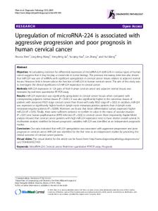
Figure 2 Expression of GFAP (a) and FGF-2 (b) in cultured human astrocytes and glioma cells. (a) Cell cultures (astrocytes QG strain, and U251MG glioma) were prepared and stained with monoclonal anti-GFAP antibody as described in Materials and methods. Human ®broblasts were used as a negative contol. (b) Glioma cell lines U251MG (I) and U343MG (II), and astrocytic strains QG (III and VI); and HG (IV and V) were stained with monoclonal FGF-2 antibody (V ± FGF-2 antibody was omitted). I ± V ± Light microscopic analysis: Immune complexes were stained by incubating with biotinylated secondary antibody, avidinbiotin peroxidase and diaminobenzidine-peroxide solution. VI ± immuno¯uorescent confocal analysis ± QG astrocytes were stained with FGF-2 Ab/Cy5-IgG. Figure shows 2-dimensional projection of stacked confocal sections taken 1 mm apart
175
Nuclear accumulation of FGF-2 and proliferation of glial cells A Joy et al
176
that interfere with FGF-2 signaling at dierent subcellular locations. Myo-Inositol hexakis [dihydrogen phosphate] (IP6) inhibits FGF-2 binding to the cell surface receptors but does not interfere with the
intracellular action of FGF-2 (Sherman et al., 1993; Morrison et al., 1994). IP6 (200 mM) had no eect on the proliferation of the glioma cells expressing CMVHMW (Figure 9). Cells were also incubated with 5'deoxy-5'-methylthioadenosine (MTA), a FGF-2 antagonist that acts intracellularly and prevents autophosphorylation of the intracellular domain of FGF receptors (Maher, 1993). MTA (3 mM) completely inhibited the proliferation of CMV-HMW transfected SF-767 cells indicating that a functional tyrosine kinase moiety is necessary to sustain proliferation of these cells (Figure 9). These results support the conclusion that increased cell proliferation is induced by FGF-2 independent of plasma membrane receptor and that FGF-2 may be inducing its mitogenic response by acting at intracellular site(s).
Discussion CONFLUENT
SUBCONFLUENT
Figure 3 Incorporation of BUdR by astrocytes (QG strain) as a function of cell density within the same culture dish. Con¯uent astrocytes in the center of the dish show no incorporation. Subcon¯uent astrocytes outside the dense incorporate BUdR. Cells at dierent stages of the S phase are visible
Nuclear accumulation of FGF-2 correlates with mitotic activity of astrocytes and glioma cells The present study shows nuclear accumulation of FGF-2 in reactive astrocytes of the mature human brain and in neoplastic glioma cells of human GBM. We demonstrate that the nuclear accumulation of
Figure 4 Cell density-dependent inhibition of FGF-2 expression in human astrocytes and its impairment in glioma cells. Cells were stained with monoclonal antibodies against FGF-2. (a) Expression of FGF-2 in astrocytes (QG strain) and U251MG glioma cells. I ± subcon¯uent astrocytes 1 week after plating; II ± con¯uent astrocytes 3 weeks in culture; III ± subcon¯uent astrocytes 5 weeks after plating. Con¯uent astrocytes detached leaving large empty surfaces. Remaining astrocytes show increased expression of nuclear and nucleolar FGF-2. Some show karyokinesis indicating renewed proliferation. IV, V and VI ± glioma U251MG cells 1, 3 and 5 weeks after plating, respectively. At 3 weeks cells show a small reduction in FGF-2-IR; at 5 weeks an additional cell layer formed with intense nuclear FGF-2-IR. (b) Gradient of FGF-2 expression in astrocytes (QG) as a function of cell density in a single culture dish. (Original magni®cation 6400)
Nuclear accumulation of FGF-2 and proliferation of glial cells A Joy et al
endogenous FGF-2 is a regulated process that occurs in association with cell proliferation. In the human brain, nonreactive astrocytes and postmitotic neurons contain FGF-2-IR only in the cytoplasm, while in glioma cells and the large reactive astrocytes FGF-2-IR is present in the nuclei, nucleoli and cytoplasm. Also in vitro, astrocytes maintained in con¯uent monolayers have a stellate morphology as in control brain tissue and express FGF-2 only in the cytoplasm. After cell density is reduced quiescent astrocytes transit to a reactive-like state. Cell hypertrophy, proliferation, and induction of nuclear FGF-2 are observed that last until a new con¯uent state is achieved. The in vitro experiments suggest that the nuclear accumulation of FGF-2 in reactive astrocytes in the brain may be triggered by a release from direct cell-cell contact inhibition due to the increased intercellular space. In contrast to the transient burst of growth and proliferation experienced by reactive astrocytes, neoplastic glioma cells proliferate continuously, showing a diminished response to external cues such as cell-contact inhibition. The continuous proliferation of cultured glioma cells is accompanied by constitutive expression of nuclear FGF-2, only partially aected by cell density. FGF-2 added to extracellular medium accumulates in nuclei of aortic endothelial cells (Bouche et al.,
1987), adrenal chroman cells (Stachowiak et al., 1994), or human astrocytes (J Moett and MK Stachowiak, submitted for publication). These observaa
b
a L
H
L
cyt
H nuc
L
H
L
cyt
H nuc — 24 — 22
— 18 (kDa) Week 3
Week 4
b cyt
nuc
cyt
nuc
cyt
nuc
24 — 22 —
18 — (kDa) 1 Week
3 Weeks
5 Weeks
Figure 5 Eect of cell density on FGF-2 proteins in nuclear and cytoplasmic fractions from astrocytes (a) and glioma cells (b) ± Western analysis. (a) Astrocytes (QG strain) were plated at two (lower L or higher H) densities and reached con¯uence after 3 weeks and 4 weeks in culture, respectively. Equal starting amounts of cytoplasmic (1.1 mg) or nuclear (0.075 mg) proteins from 3 and 4 week cultures were subjected to heparin-sepharose puri®cation before gel electrophoresis. (b) U251MG cells were plated at low density and reached con¯uence during the third week in culture and remained con¯uent for the remainder of the experiment. 3.75 mg of cytoplasmic and 0.18 mg of nuclear proteins were used. The speci®c content of total FGF-2 (all isoforms/mg protein) in the nuclear fraction was approximately threefold (astrocytes 3 weeks, L cultures) or sevenfold (U251MG 1 week) higher than in the cytoplasmic fraction
Figure 6 Growth curves of astrocytes and glioma SF-767 cells treated with recombinant human FGF-2. Cells were plated in microtiter dishes as 1.56103 cells/well. Recombinant human 18 kDa FGF-2 was added in fresh medium 24 h after plating and was replenished at day 4. The number of cells was estimated at dierent times as described in Materials and methods. Individual points represent mean+s.e.m. of 5 ± 6 wells. s.e.m. smaller than the symbols are not shown. (a) ± astrocytes (QG strain) in 5% fetal calf serum (FCS); (b) ± SF-767 glioma cells in 0.5%, 1% and 5% FCS
177
Nuclear accumulation of FGF-2 and proliferation of glial cells A Joy et al
178
1)
CMVNEO
2)
CMVbFGF 1 2 (kDa) — — — —
24 23 21 18
1) 2)
CMVNEO CMV-18
3) 1)
CMVHMW 2) 3) (kDa) — 24 — 23 — 21 — 18
1) 2) 1
CMV-NEO CMV-bFGF 2 (kDa) — 24 — 23 — 21 — 18
1) 2) 3) 1
CMV-NEO CMV-18 CMV-HMW 2 3 (kDa) — 24 — 23 — 21 — 18
Figure 7 Proliferation of SF-767 (a,b) and SF-763 (c,d) glioma cell lines overexpressing FGF-2. Glioma cell lines were stably transfected with: (a,c) CMV-bFGF expressing all FGF-2 isoforms or control CMV-NEO without FGF-2 cDNA, and (b,d) CMVNEO, CMV-18 expressing 18 kDa FGF-2 or CMV-HMW expressing high molecular weight isoforms of FGF-2. Cells were plated at 0.56103 cells/well and maintained in 5% serum and 250 mg/ml G-418. The number of cells was estimated as in Figure 6. Individual points represent mean+s.e.m. of 5 ± 6 wells. The eects of CMV-bFGF plasmid on cell proliferation of cells maintained at 0.5% were similar (not shown). Inserts show Western analysis of FGF-2 content in glioma cells transfected with CMV-NEO or vectors expressing recombinant FGF-2. Total cell extracts obtained from the same number of cells are used in individual lanes
tions suggested that extracellular FGF-2 may interact with the plasma membrane, undergo receptor-mediated endocytosis, and nuclear accumulation. Although we cannot rule out this mechanism, our data support the nuclear accumulation of FGF-2 in reactive astrocytes and in glioma cells independent of release. Glioma cells that were stimulated to proliferate by intracellularly expressed FGF-2 did not respond to exogenous FGF-2 and were unaected by the plasma membrane receptor antagonist IP6. In addition, we did not detect extracellular FGF-2-IR in the brain tissue or in cell cultures. Intact FGF-2 was also absent in culture media conditioned by astrocytes or glioma cells. Thus, if FGF-2 is released from glial cells, the amount of extracellular FGF-2 must be very small relative to its
intracellular content. A direct transfer of FGF-2 from the cytoplasm to the nucleus is consistent with the presence of a nuclear localization signal (Courdec et al., 1991) and with the absence of a secretory sequence in the FGF-2 molecule (Abraham et al., 1986). The observation that in astrocytes nuclear FGF-2 was depleted while cytoplasmic levels of FGF-2 remained relatively high suggests that a cell densitydependent mechanism restricts nuclear translocation of FGF-2. Such a mechanism is consistent with our earlier ®nding of a cAMP-/protein kinase C-controlled dierential targeting of endogenous FGF-2 into the nucleus or cytoplasm of adrenal chroman cells (Stachowiak et al., 1994). The accumulation or depletion of nuclear FGF-2 may re¯ect, at least in
Nuclear accumulation of FGF-2 and proliferation of glial cells A Joy et al
Table 1
Effect
of
extracellular
and
transfected
FGF-2
on
proliferation of U251MG glioma cells Treatment
Cell proliferation
Fold stimulation
Extracellular 18 kDa FGF-2:
+ + + + + + +
±
2.36+0.12
0.28 nM
1.97
0.15
0.85
0.8 nM
2.32
0.16
0.99
2.5 nM
2.08
0.11
0.88
CMV-Neo
1.11
0.10
1
CMV-bFGF
2.48
0.08
2.26*
CMV-18
2.60
0.10
2.39*
CMV-HMW
2.22
0.20
2.00*
Transfection:
1
Nontransfected U251MG cells and U251MG cells stably transfected with plasmids expressing FGF-2 were used. Control CMVNeo and FGF-2-expressing
plasmids
are
described
in
Figure
7
and
in
Materials and methods. Nontransfected cells were cultured for 10 days with human recombinant 18 kDa FGF-2. Transfected cells were incubated for 9 days. Media were changed and new FGF-2 was
+
added every 4 days. Cell proliferation was evaluated as on Figures 6 and 7. Numbers represent mean
4
s.e.m. of 5 ± 6 samples. Transfec-
tion of plasmids expressing FGF-2 had a statistically signi®cant eect on cell proliferation (P
4
0.00005, 1-way ANOVA). Compar-
isons between individual groups were done using Schee post hoc test. *Dierent (P
0.0005) from cells transfected with CMV-Neo
part, transcriptional regulation mediated by an upstream region of the FGF-2 gene (Moett et al., 1996). FGF-2 mRNA levels and FGF-2 promoter activity in cultured human astrocytes displayed gradual reductions with increasing cell density (Moett et al., 1996), similar to the nuclear FGF-2-IR in the present study (Figure 4b). In glioma cells, the FGF-2 gene is constitutively expressed due to an altered transregulation of its promoter (Moett et al., 1996). This mechanism may contribute to the constitutive nuclear presence of FGF-2. Exocrine and intracrine action of FGF-2 Although we detected no extracellular FGF-2, FGF-2 could reach the extracellular space in the event of cell injury or lysis (Schechter, 1992) which may accompany some pathological conditions of the nervous tisuse. By interacting with membrane receptors, FGF-2 could signal cell damage and stimulate astrocyte proliferation. The present study demonstrates that extracellular FGF-2 activates quiescent astrocytes from the mature human brain to enter the S phase of the cell cycle and to proliferate. In human astrocytes, extracellular FGF2 stimulates expression of its own gene and the synthesis and nuclear accumulation of endogenous FGF-2 proteins (J Moett and MK Stachowiak, submitted for publication). Thus, even extracellular FGF-2 could stimulate cell proliferation through an intracrine FGF-2 pathway. In support of an intracellular FGF-2 action, we ®nd that proliferation of glioma cell lines is stimulated by FGF-2 expressed intracellularly but not by an exogenous FGF-2. The increase in proliferation of SF-767 or U251MG cells transfected with the CMVHMW plasmid supports the intracellular action of FGF-2. The high molecular weight FGF-2 isoforms do not associate with the plasma membrane and are absent outside the cells (Bugler et al., 1991; Florkiewicz et al., 1991; Bikfalvi et al., 1995). Proliferation of SF767 cells expressing high molecular weight isoforms of
FGF-2 was inhibited by intracellular acting MTA (Maher, 1993), indicating an involvement of FGF receptor tyrosine kinase, but it was resistant to an extracellularly acting FGF-2 binding antagonist, IP6. The concentrations of IP6 in our experiments were the same or higher than used to maximally inhibit the extracellular FGF-2 induced dierentiation or proliferation of Schwann cells (Sherman et al., 1993), ®broblastic cells (Morrison et al., 1994b), or human glioma cells (Morrison et al., 1994c; E Mordechai and MK Stachowiak unpublished observations). Stimulation of cell proliferation by intracellularly acting ®broblast growth factors was earlier observed in nonglial cells. In ®broblasts the mitogenic eects of extracellular FGF-1 require nuclear translocation of the growth factor and can be separated from its ability to stimulate cell membrane receptors (Imamura et al., 1990; Wiedlocha et al., 1994). Our ®ndings are also consistent with those of Bikfalvi et al. (1995) who showed that intracellularly expressed high molecular weight FGF-2 isoforms stimulate proliferation of ®broblasts independent of their surface receptors. The oscillations in endogenous FGF-2 content accompanying changes in astrocytic proliferation occur in the nuclei but not in the cytoplasm, suggesting that the intracrine eects of FGF-2 are produced directly in the nuclei. Also, FGF-2 appears to accumulate predominantly in the nuclei of transfected glioma cells U251MG and SF-767, that respond by increasing their proliferation. In contrast, in SF-763 glioma cells that do not respond to intracellularly expressed FGF-2, FGF-2 accumulated in the cytoplasm. In further support for the nuclear action of FGF-2 we ®nd that the FGF receptor-1 is located within the nuclei of glial (Stachowiak et al., 1996a) and adrenal medullary chroman cells (Stachowiak et al., 1996b). In astrocytes nuclear content of FGFR receptor-1 co-varies with FGF-2 in association with cell proliferation (in preparation). The glioma cells (SF-767 and U251MG) that increase proliferation upon overexpression of FGF-2, contain nuclear FGF receptor-1. In contrast, the unresponsive glioma cells SF-763 do not express detectable nuclear FGF receptor-1 (MK Stachowiak, PA Maher, unpublished observations). In conclusion, we propose that cell densitydependent regulation of FGF-2 content in the nucleus may serve as a novel mechanism controlling cellular proliferation. In nontransformed human astrocytes transient nuclear accumulation of FGF-2 may mediate the reversible activation of quiescent cells. In glioma cells, the constitutive nuclear presence of FGF-2 may promote proliferation that is insensitive to cell-cell contact inhibition. Further analysis of the nuclear translocation and function of FGF-2 may reveal the mechanisms controlling normal and neoplastic growth of glial cells.
Materials and methods Astrocytic and glioma cell cultures Three normal astrocyte cultures were obtained from dissociation of brain tissue taken from trauma patients as in (Asch et al., 1986; Moett et al., 1996). The culture
179
Nuclear accumulation of FGF-2 and proliferation of glial cells A Joy et al
180
QG was from the frontal lobe (a 7-year-old male); the HG cells were from fronto-parietal cortex (a 16-year-old female); and the RX were from the cerebellar cortex (an 18-year-old female). Cells were maintained in Waymouth 87/3 medium (MAB) supplemented with 20% fetal bovine serum. Culture media were from Grand Island Biologicals (Grand Island, NY). Results from dierent cell strains were essentially the same. Cultured human astrocytes grew for 14 ± 23 passages before senescence. Cytogenetic analysis veri®ed that each cell culture contained a normal Giemsa-banded karyotype. They were identi®ed as pure astrocytic cultures by expression of GFAP (Figure 2) and by the lack of expression of galactocerebroside, an oligodendrocytic marker (not shown). Established glioma cell lines U251MG, U343MG (Binger et al., 1981), SF767, and SF-763 (Berens et al., 1990) expressed GFAP. Mitoses in cultured cells were monitored using a 5-bromo2'-deoxyuridine incorporation-detection kit (Boehringer Mannheim, Indianapolis, IN). Cells were incubated with 5-bromo-2'-deoxyuridine/5-¯uoro-2'-deoxyuridine for 2 h, ®xed and stained according to the manufacturer's protocol.
U251MG
SF767
Preparation of nuclei and cytoplasm and Western analysis for FGF-2 Isotonic lysis of cells and isolation of nuclei and cytoplasmic fractions were performed as described by Boyle et al. (1985) with minor modi®cations (Stachowiak et al., 1994, 1996a,b). All procedures were carried out at 48C in the presence of protease inhibitors (10 mM leupeptin, 1 mM aprotinin, 5 mg/ml pepstatin A, 1 mM PMSF). Adherent, washed cells were incubated in nuclear buer (5 mM sodium phosphate, pH 7.4, 50 mM NaCl, 150 mM sucrose, 5 mM KCl, 2 mM dithiothreitol, 1 mM MgCl2, 0.5 mM CaCl2, 0.1% digitonin) and harvested by scraping. Nuclei were centrifuged through a cushion of TN buer (2.5 mM Tris-HCl, pH 7.4, 10 mM NaCl) containing 30% sucrose at 1000 g for 10 min. The supernatant was cleared by centrifugation at 40 000 g for 10 min, snap frozen, stored at 7808C and used as a cytoplasmic fraction. Nuclear fraction which isolated using this protocol contained approximately 88% of the total cellular DNA (Stachowiak et al., 1996a). Purity of the nuclear fraction was evaluated by a number of criteria. Visual inspection of
SF763
Figure 8 Immunocytochemical analysis of FGF-2 expression in glioma cells transfected with plasmid expressing FGF-2. U251MG and SF-767 cells stably transfected with CMV-bFGF or control plasmid CMV-NEO. Cells were incubated with monoclonal FGF-2 antibody and the immune complexes were detected with biotinylated secondary antibody and avidin-linked peroxidase
Nuclear accumulation of FGF-2 and proliferation of glial cells A Joy et al
of time to ensure that the signals were in the linear range. The detection limit of 18 kDa FGF-2 was approximately 1 ng. Immunohistochemical staining
Figure 9 Eects of FGF-2 antagonists on proliferation of SF767 cells transfected with CMV-HMW. Glioma cells expressing high molecular weight isoforms of FGF-2 (Figure 7) were plated at 2.06103 cells/well and maintained with or without 200 mM IP6 or 3 mM MTA. Bars represent mean+s.e.m. number of cells estimated at 1 and 10 days after plating as in Figure 7
isolated nuclei by phase contrast microscopy demonstrated only small amounts of extranuclear material in the nuclear fraction. In addition, the nuclear fraction contained less than 10% of total cellular 5' nucleotidase activity (plasma membrane marker) and 5% of the total acid phosphatase activity (lysosomal marker). Purity of the nuclear fraction was also indicated by the absence of FGF-2 in nuclei isolated under conditions where FGF-2 was only cytoplasmic (Figure 5). In the nuclei of human astrocytes and glioma cells, the majority of FGF-2 is associated with the nucleoplasm and very little with insoluble nuclear matrix (A Joy, J RankinShapiro, MK Stachowiak, unpublished observations). To eciently extract FGF-2, nuclei were treated with lysis buer (1% NP-40, 0.5% deoxycholate, 20 mM Tris, pH 7.5, 5 mM EDTA, 150 mM NaCl containing protease inhibitors) and lysates were clari®ed by centrifugation at 20 000 g for 15 min at 48C. Protein concentrations were determined using the Bio-Rad protein assay (Bio-Rad, Richmond, CA). All samples were frozen in liquid nitrogen and stored at 7808C until use. Nuclear or cytoplasmic extracts from dierent cell preparations, after small adjustments to yield the same amounts of proteins, were puri®ed on heparin sepharose. The heparin sepharose pellets were washed three times with 20 mM Tris buer containing 1 M sodium chloride (conditions that would release bound FGF-1). Heparin-sepharosebound proteins were eluted directly into SDS sample buer and resolved by SDS ± PAGE gel electrophoresis on a 12% polyacrylamide gel. Proteins were transferred onto nitrocellulose membranes and Western analysis performed using an anti-FGF-2 monoclonal antibody (UBI, Lake Placid, NY) followed by rabbit anti-mouse IgG and 125I-labeled protein A (Florkiewicz and Sommer, 1989; Stachowiak et al., 1994). In some experiments 10 ml of conditioned media were processed through heparin sepharose and analysed in a similar way to the cell extracts to determine whether FGF-2 was present in the media. To determine total cell content of FGF-2, cells were lysed with cell lysis buer. The lysates were used directly for Western analysis (Figure 7). FGF-2 protein levels were estimated by densitometric scanning of autoradiograms using a Beckman DU70 spectrophotometer (Stachowiak et al., 1994). Autoradiograms were exposed for dierent lengths
Cultured cells were ®xed with 2.5% paraformaldehyde, permeabilized 15 min with 1% Triton X-100 and were stained immunohistochemically as in (Puchacz et al., 1993; Stachowiak et al., 1994, 1996a,b), using the following primary monoclonal antibodies: anti-FGF-2 Mab (UBI, Lake Placid, NY), anti-GFAP or antigalactocerebroside (Chemicon Int, Temecula, CA). Immune complexes were detected with biotinylated secondary antibody, followed by avidin-linked peroxidase and diaminobenzidine-hydrogen peroxide solution (Stachowiak et al., 1994). Endogenous peroxidase activity was exhausted by treating cells with 0.1% H2O2. Immuno¯uorescent staining of FGF-2 was performed using Cy5conjugated secondary antibodies (Jackson Immuno Research Labs, West Grove, PA). Digitized images of 1 mm confocal optical sections were acquired using a Biorad MRC 600 confocal microscope with a YHS 568 nm ®lter and 15 mW Crypton/Argon laser. The images were recorded directly onto 35 mm ®lm. Staining of FGF-2 in normal and gliotic human brain tissue and in glioblastoma multiforme (GBM) The brain tissue was obtained from patient with longstanding epilepsy. Normal and gliotic tissues from a temporal lobe resection for epilepsy were snap frozen in liquid nitrogen immediately after surgical removal. Six mm frozen sections were ®xed in 2.5% paraformaldehyde, permeabilized with 0.3% Triton X-100 and immunostained for FGF-2 or GFAP as described for the cultured cells. Some sections were counterstained with hematoxylin and eosin to visualize cell nuclei. Temporal lobe seizure tissue was selected to demonstrate gliosis and to avoid possible confusion between identi®cation of small or intermediate size neurons and reactive astrocytes. The nornal Ammon's horn region contains large pyramidal neurons and associated oligodendrocytes but very few small neurons or astrocytes. Therefore, neuronal loss and astrocytic proliferation in tissue from seizure patients are easily recognized. The unaected lateral temporal lobe provided normal control tissue. The speci®city of FGF-2 immunostaining was indicated by several observations: (1) in human brain slices and in cultured cells staining was not observed when monoclonal FGF-2 antibody was omitted or replaced with normal mouse serum (not shown); (2) the FGF-2 antibody produced dierent pattern of staining than the GFAP or anti-galactocerebroside monoclonal antibodies; (3) unlike FGF-2 staining, the staining of the human nucleolar antigen was not aected by cell density; (4) a similar pattern of staining was produced by monoclonal FGF-2 Ab and polyclonal FGF2 antibodies (Stachowiak et al., 1994; not shown); and (5) the presence and changes in subcellular distribution of FGF-2-IR were con®rmed by Western blot analysis of FGF-2 in subcellular fractions. The speci®city of FGF-2 staining was further demonstrated in our earlier study (Stachowiak et al., 1994). Expression of recombinant FGF-2 proteins in glioma cells We have constructed a CMV-bFGF vector that directs expression of all FGF-2 isoforms (18, 22, 23 and 24 kDa) by cloning the 900 bp EcoRI fragment of the human FGF2 cDNA into the EcoRI site 3' to the cytomegalovirus (CMV) immediate early promoter of the pBKCMV plasmid (Stratagene). FGF-2 cDNAs with mutations that
181
Nuclear accumulation of FGF-2 and proliferation of glial cells A Joy et al
182
allow synthesis of only the 18 kDa FGF-2 or high molecular weight (HMW) FGF-2 isoforms (22, 23 and 24 kDa) were used to construct CMV-18 and CMV-HMW plasmids, respectively. Generation of the wild type and mutant FGF-2 cDNAs was described in (Florkiewicz et al., 1991). The CMV-bFGF, CMV-18 and CMV-HMW constructs contain the open reading frame for FGF-2 cDNA driven by a CMV promoter as well as G418 resistance gene for selection of stable transfectants. Glioma cells 36106 cells/ml) were transfected with 100 mg of plasmid DNA using electroporation. Two days after transfection the cells were maintained with 500 mg/ml of G418 (Stratagene) to select transfectants. G418-resistant cells were pooled to establish stably transfected populations of cells. Proliferation of glial cells The proliferation rate of astrocytes and glioma cells was determined according to Brasaemle and Attie (1988). Cells were plated in 96-well plates in medium containing 0.5%, 1 or 5% serum. To determine cell densities after time, the cells were ®xed, stained with crystal violet, and solubilized with 10% SDS. The changes in cell number were quanti®ed by reading the absorbance at 570 nm on an automated ELISA plate reader. Cell cycle analysis by ¯ow cytometry analysis was carried out as described in (Latham et al., 1996). SF-763 or U251MG glioma cells (16106) transfected with CMV-Neo or CMV-bFGF were seeded on 60 mm dish for 24 h. Cells were harvested by trypsynization, washed with ice-cold PBS and ®xed in 70% ethanol. Following ®xation, the cells were centrifuged and resuspended in PBS containing RNase A (0.180 mg/ml). The cells were incubated for 30 min at room
temperature, centrifuged again and treated with 20 mg/ml of propidium iodide for at least 15 min at room temperature. DNA histograms were generated using EPICS Pro®le ¯ow cytometer (Coulter Co, Hialeh, Fl) and were analysed using Multicycle cell cycle analysis program (Phoenix Flow Systems, San Diego, CA).
Abbreviations BUdr: 5-bromo-2'-deoxyuridine; FGF-1: ®broblast growth factor 1 (acidic FGF); FGF-2: ®broblast growth factor-2 (basic FGF); FGF-2-IR: FGF-2 immunoreactivity; GBM: glioblastoma multiforme; GFAP: glial ®brillary acidic protein; IP6: myo-Inositol hexakis [dihydrogen phosphate]; MTA: 5'-deoxy-5'-methylthioadenosine; HG, QG, RX: astrocytic strains obtained from dierent subjects; U251MG, U343MG, SF-767, SF-763: glioma cell lines. Acknowledgements We thank Dr Michael Berens for the gift of U251MG glioma cell line, Dr Leslie Tolbert and Mrs Patty Jansma for the assistance with confocal microscopy, Mrs Dorothy Haskett for ¯ow cytometric analysis and Dr P Maher for critical discussion of the manuscript. SF-767 and SF-763 cell lines were from the Brain Tumor Research Center, University of California, San Francisco. This study was supported by the National Science Foundation (94-11226), and National Institutes of Health (HL49376-01A1), American Parkinson Disease Association (MK Stachowiak) and Arizona Disease Control Research Commission (J Moett, A Joy, MK Stachowiak). R Florkiewicz was supported by the National Institutes of Health (DK-18811).
References Abraham JA, Whang JL, Tunolo A, Mergia A, Friedman J, Gospodarowicz D and Fiddes DC. (1986). EMBO J., 5, 2523 ± 2528. Araujo DM and Cotman CW. (1992). J. Neurosci., 12, 1668 ± 1678. Asch AS, Leung LLK, Shapiro JR and Nachman RL. (1986). Proc. Natl. Acad. Sci. USA, 83, 2904 ± 2908. Berens ME, Weiseman AS, Spencer DR, Dougherty DV, Elliger SS and Rosenblum ML. (1990). Proc. Am. Assoc. Cancer Res., 31, 46. Bikfalvi A, Klein S, Pintucci G, Quatro N, Mignatti P and Rifkin DB. (1995). J. Cell. Biol., 129, 233 ± 243. Binger DD, Binger SH and Ponten J. (1981). J. Neuropathol. Exp. Neurol., 40, 201 ± 209. Bogler O, Wren D, Barnett SC, Land H and Noble M. (1990). Proc. Natl. Acad. Sci. USA, 87, 6368 ± 6372. Bouche G, Gas N, Prats H, Baldin V, Tauber J-P, Teissie J and Amalric F. (1987). Proc. Natl. Acad. Sci. USA, 84, 6770 ± 6774. Boyle WT, Lampert MA, Li AC and Baluda MA. (1985). Mol. Cell. Biol., 11, 3017 ± 33023. Brasaemle DL and Attie AD. (1988). Biofeedback, 6, 418 ± 419. Bridstock DR, Sasse J and Klagsbrun JL. (1991). Growth Factors, 4, 189 ± 196. Bugler B, Almalric F and Prats H. (1991). Mol. Cell Biol., 11, 573 ± 577. Courdec BH, Prats H, Bayard F and Amalric F. (1991). Cell Regul., 2, 709 ± 718. Eng LF, Yu ACH and Lee YL. (1992). Prog. Brain Res., 94, 353 ± 365.
Engele J and Bohn MC. (1992). Dev. Biol., 152, 363 ± 372. Finklestein SP, Apostoloides PJ, Caddy GC, Prosser J, Philips MF and Klagsbrun M. (1988). Brain Res., 460, 253 ± 259. Florkiewicz RZ, Baird A and Gonzalez AM. (1991). Growth Factors, 4, 265 ± 275. Florkiewicz RZ and Sommer A. (1989). Proc. Natl. Acad. Sci. USA, 86, 3978 ± 3981. Frautschy SA, Walicke P and Baird A. (1991). Brain Res., 553, 291 ± 299. Gomez-Pinilla F, Lee JW-K and Cotman CW. (1992). J. Neurosci., 12, 345 ± 355. Gualandris A, Coltrini D, Bergonzoni L, Isacchi A, Tenca S, Ginelli B and Presta M. (1993). Growth Factors, 8, 49 ± 60. Imamura T, Engelka K, Zhan X, Tokita Y, Forough R, Roeder D, Jackson A, Maier JAM, Hla T and Maciag T. (1990). Science, 249, 1567 ± 1570. Kniss DA and Burry RW. (1988). Brain Res., 439, 281 ± 288. Latham KE, Cosenzas, Relchenbach NL, Mordechai E, Adelson ME, Kon N, Honrath SE, Charubala R, Mikhallov SN, Pfelderer W and Suhadolnik RJ. (1996). Oncogene, 12, 827 ± 837. Liberman TA, Fiesel R, Jaye M, Lyall RM, Westermark B, Drohan B, Schmidt A, Maciag T and Schlessinger J. (1987). EMBO J., 6, 1627 ± 1632. Liu HM and Chen HH. (1994). J. Neuropath. Exptl. Neurol., 53, 118 ± 126. Maher PA. (1993). J. Biol. Chem., 268, 4244 ± 4249. McMillian MK, Thai L, Hong J-S, O'Callaghan PO and Pennypacker KR. (1994). TINS, 17, 138 ± 142.
Nuclear accumulation of FGF-2 and proliferation of glial cells A Joy et al
Moett J, Stachowiak EK, Florkiewicz R and Stachowiak MK. (1994). J. Int. Dev. Neurosci., 12 Supp. 1, 77. Moett J, Kratz E, Florkiewicz R and Stachowiak MK. (1996). Proc. Natl. Acad. Sci. USA, 93, 2470 ± 2475. Morrison RS. (1991). J. Biol. Chem., 263, 728 ± 734. Morrison RS, Shi E, Kan M, Yamaguchi F, McKeehan W and Berger MS. (1994a). In Vitro Cell. Dev. Biol., 30A, 783 ± 789. Morrison RS, Yamaguchi F, Bruner JM, Tang M, Mckeehan W and Berger MS. (1994b). Cancer Res., 54, 2794 ± 2799. Morrison RS, Saya H, Bruner JM, Yahanada AM, Donehower LA and Berger M. (1994c). J. Neuro-Oncol., 18, 207 ± 216. Moscatelli D, Presta M, Joseph-Silverstein J and Rifkin DB. (1986). J. Cell Physiol., 129, 273 ± 276. Moscatelli D. (1988). J. Cell. Biol., 107, 753 ± 759. Murphy PR, Sato R and Friesen HG. (1988). Mol. Endocrinol., 2, 591 ± 598. Nakanishi Y, Kihara K, Mizuno K, Masamune Y, Yoshikate Y and Nishikawa K. (1992). Proc. Natl. Acad. Sci. USA, 89, 5216 ± 5220. Paulus W, Grothe C, Sensenbrenner M, Janet T, Baur I, Graf M and Roggendorf W. (1990). Acta Neuropath., 79, 418 ± 423. Pieper RO, Futscher BW, Dong Q, Ellis TM and Erickson LC. (1990). Cancer Comm., 2, 13 ± 20. Powell PP and Klagsburn M. (1991). J. Cell. Physiol., 148, 202 ± 210. Puchacz E, Stachowiak EK, Florkiewicz R, Lukas RJ and Stachowiak MK. (1993). Brain Res., 610, 39 ± 52. Ray J, Hogg J, Beutler AS, Takayama H, Baird A and Gage F. (1995). J. Neurochem., 64, 503 ± 513.
Rosenblum ML, Vasquez DA, Hoshino T and Wilson CB. (1978). Cancer, 41, 2305 ± 2314. Schechter JE. (1992). Tissue Cell, 24, 791 ± 802. Sherman L, Stocker KM, Morrison R and Ciment G. (1993). Development, 118, 131 ± 1326. Shibata F, Baird A and Florkiewicz RZ. (1991). Growth Factors, 4, 277 ± 287. Stachowiak MK, Maher PA, Joy A, Mordechai E and Stachowiak EK. (1996a). Mol. Brain Res., 38, 161 ± 165. Stachowiak MK, Maher PA, Joy A, Mordechai E and Stachowiak EK. (1996b). Mol. Biol. of the Cell, 7, 1299 ± 1317. Stachowiak MK, Moett J, Joy A, Puchacz E, Florkiewicz R and Stachowiak EK. (1994). J. Cell. Biol., 127, 203 ± 223. Takahashi JA, Mori H, Fukumoto M, Igarashi K, Jaye M, Oda Y, Kikuchi H and Hatanaka M. (1990). Proc. Natl. Acad. Sci. USA, 87, 5710 ± 5714. Vescovi AL, Reynolds BA, Fraser DD and Weiss S. (1993). Neuron, 11, 951 ± 966. Vijayan VK, Lee YL and Eng LF. (1993). Int. J. Dev. Neurosci., 11, 257 ± 267. Vlodavsky I, Bar-Shavit R, Ishai-Michaeli R, Bashkin P and Fuks Z. (1991). Trends Biochem. Sci., 16, 268 ± 271. Wiedlocha A, Falnes PO, Madshus IH, Sandvig K and Olsnes S. (1994). Cell, 76, 1039 ± 1051. Woodward WR, Nishi R, Meshul CK, Williams TE, Coulombe M and Eckstein FP. (1992). J. Neurosci., 12, 142 ± 152. Zagzag D, Miller DC, Sato Y, Rifkin DB and Burstein DE. (1990). Cancer Res., 50, 7393 ± 7398.
183


![[Lys 61 ]N-Ras is able to induce full activation and nuclear accumulation of Cdk4 in NIH3T3 cells](https://kipdf.com/img/300x300/lys-61-n-ras-is-able-to-induce-full-activation-and_5aca79871723dd40882fb782.jpg)