27
ORIGINAL ARTICLE
Morphological effects of chemotherapy on ovarian carcinoma W G McCluggage, R W Lyness, R J Atkinson, S P Dobbs, I Harley, H R McClelland, J H Price .............................................................................................................................
J Clin Pathol 2002;55:27–31
See end of article for authors’ affiliations
....................... Correspondence to: Dr WG McCluggage, Department of Pathology, Royal Group of Hospitals Trust, Grosvenor Road, Belfast BT12 6BL, Northern Ireland; glenn.mccluggage@ bll.n-i.nhs.uk Accepted for publication 31 May 2001
.......................
Aims: Traditionally, advanced stage ovarian carcinoma is treated by debulking surgery followed by chemotherapy. However, in some circumstances preoperative chemotherapy may be given before optimal surgical debulking. This study aims to describe the morphological features found in ovarian carcinoma after chemotherapy because these have not been detailed previously. Methods: Histological sections were examined from 18 cases of ovarian carcinoma that had been treated by preoperative chemotherapy. The morphology was compared with any pre-chemotherapy biopsies that had been performed. Tumours were classified as showing morphological features suggesting a good response to chemotherapy (n = 14) or as showing little or no response (n = 4). Serum CA125 values before and after chemotherapy were compared. In all cases, the mitotic activity index (MAI), volume percentage of epithelium (VPE), and mean nuclear area (MNA) of tumour cells were calculated. Results: The preoperative biopsies were all typical ovarian serous or endometrioid adenocarcinomas. Morphological features present in the group responding to chemotherapy included the presence of small groups or single tumour cells in a densely fibrotic stroma. Tumour cells were characterised by both nuclear and cytoplasmic alteration, making accurate tumour typing and grading impossible. Nuclear features included the presence of bizarre enlargement with hyperchromatism, irregularity of outline, and chromatin clumping or smudging. Cytoplasmic alterations included intense eosinophilia, vacuolation, or foam cell change. There were pronounced stromal changes of fibrosis, inflammation, collections of foamy histiocytes, cholesterol cleft formation, haemosiderin deposition, fat necrosis, and dystrophic calcification, including the presence of many free psammoma bodies. There was no correlation between morphological response and biochemical response, as determined by serum CA125 values. In all nine cases in which pre-chemotherapy and post-chemotherapy biopsies were available, the MNA increased post-chemotherapy (p = 0.007, paired Wilcoxon test) and in six of nine cases the MAI decreased (p = 0.093). Conclusions: Because preoperative chemotherapy is being used increasingly in the management of ovarian cancer, pathologists should be aware of the resultant morphological effects. Accurate tumour typing and grading is impossible. In some cases, it may be difficult to confirm the presence of residual tumour, making it imperative that pre-chemotherapy tissue biopsies are obtained. Definite confirmation of residual tumour may require the examination of multiple histological sections from areas showing pronounced stromal changes, sometimes with multiple levels and immunohistochemistry. In the absence of definite residual tumour, the report should state that the features are consistent with the prior presence of tumour.
varian carcinoma is one of the most common female malignancies in Western countries and is the second most common gynaecological cancer, after endometrial cancer. The five year survival rate is poor at approximately 30–40%,1 although more recent indications are that this is improving.2 The two main prognostic indicators are FIGO stage at diagnosis and size of residual disease after surgery. Poor survival rates are partly the result of late stage at presentation, many patients being stage III–IV at diagnosis. A high volume of residual disease after surgery is also important,3 and a recent study found an improved survival advantage of 25% in stage III disease when patients were operated on by a specialist gynaecological oncologist rather than a general gynaecologist.4 This survival advantage is probably the result in part of an increased proportion of patients who have optimal surgical debulking. The treatment of advanced stage ovarian cancer is usually maximal surgical debulking followed by chemotherapy. However, recently there has been a trend towards intervention debulking after primary chemotherapy. This may occur where primary debulking has been suboptimal, where only small
O
tumour biopsies have been performed when extensive abdominal and pelvic disease has been seen at laparotomy, or where debulking is precluded by the patient’s condition. The pathological features of ovarian cancer after chemotherapy have not been detailed previously and the aim of our study is to provide a description of these. Pathologists should be aware of the resultant morphology because preoperative chemotherapy is likely to be used more frequently in the management of ovarian cancer.
MATERIALS AND METHODS
The study group comprised 18 women with advanced stage ovarian cancer who had been treated with preoperative chemotherapy. The chemotherapy regimen varied between individual patients but usually included a taxane and ............................................................. Abbreviations: MAI, the mitotic activity index; MNA, mean nuclear area; VPE, volume percentage of epithelium
www.jclinpath.com
28
McCluggage, Lyness, Atkinson, et al
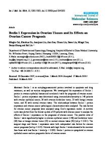
Figure 1 (A) Residual tumour after chemotherapy with low nucleocytoplasmic ratio and irregular nuclear outlines. Tumour cells have abundant eosinophilic cytoplasm. (B) Tumour cell nuclei appear degenerate with chromatin smudging and clumping. (C) Tumour cells with clear vacuolated cytoplasm. (D) Tumour cells with abundant clear foamy cytoplasm.
carboplatin. All available histological sections were reviewed by the histopathologists involved in our study. In nine cases, tissue biopsies were obtained before chemotherapy and these were reviewed. These usually comprised needle biopsies or small biopsies of tumour obtained at laparotomy. In the other
cases, a pre-chemotherapy diagnosis of adenocarcinoma was confirmed by cytological examination of peritoneal fluid. Serum CA125 values had been measured immediately before and after chemotherapy. In all biopsies (both before and after chemotherapy), the mitotic activity index (MAI) was
Figure 2 (A) Numerous cholesterol clefts are surrounded by foamy histiocytes. (B) A large aggregate of foamy histiocytes is present. (C) A collection of multinucleate foreign body-type giant cells. (D) Numerous calcified psammoma bodies are present.
www.jclinpath.com
Morphological effects of chemotherapy on ovarian carcinoma
Table 1
29
Comparison of serum CA125 values before and after chemotherapy and morphological response
Case
Serum CA125 values (U/ml) before chemotherapy
Morphological features Serum CA125 values (U/ml) after Decrease in serum CA125 values suggesting a response to chemotherapy (%) chemotherapy
1 2 3 4 5 6 7 8 9 10 11 12 13 14 15 16 17 18
331 1326 83 274 7300 8 73 NA 1476 4962 570 2351 1400 16 699 3672 2367 566 2000
245 25 47 NA 92 12 23 NA 52 164 17 740 4 350 83 85 10 98
26 98 43 – 99 50* 68 – 96 97 97 69 100 98 98 96 98 95
Yes Yes No Yes Yes Yes Yes Yes Yes Yes No No Yes Yes Yes Yes No Yes
*Serum CA125 value rose by 50%. NA, not available.
measured, as described previously.5 6 This involved counting the total number of mitotic figures in 25 contiguous fields of vision with a conventional light microscope. The volume percentage of epithelium (VPE) and the mean nuclear area (MNA) of tumour cells were also calculated as described previously.5 6 These measurements were performed using the QPRODIT system (Leica, Cambridge, UK).
RESULTS
The preoperative biopsies comprised typical ovarian serous (n = 7) or endometrioid (n = 2) adenocarcinomas. In four cases there were no obvious morphological features suggesting a good response to chemotherapy; these neoplasms comprising typical serous adenocarcinomas. The remaining 14 cases exhibited features suggestive of a good morphological response. Although this varied between individual tumours, in most cases there were pronounced changes. Surviving tumour usually comprised individual cells or small groups of cells, which were often difficult to identify, their identification requiring high power examination. Surviving tumour cells were characterised by both nuclear and cytoplasmic changes, Table 2
which meant that histological typing and grading was not possible. Usually, residual tumour cells had a low nucleocytoplasmic ratio. Nuclear features included extreme enlargement with very irregular outlines (fig 1A). Sometimes, nuclei had such a bizarre outline that they mimicked multinucleate giant cells. Nuclei often appeared degenerate with chromatin smudging or clumping (fig 1B) and there were sometimes prominent nucleoli. In most cases, mitotic figures were not easily identified. The cell cytoplasm was either intensely eosinophilic (fig 1A) or clear with a vacuolated (fig 1C) or foamy appearance (fig 1D). Pronounced stromal changes were also present. These varied between individual cases and from area to area within the same tumour. The stroma was often densely fibrotic and contained inflammatory cells, fibrinous debris, cholesterol clefts (fig 2A), and haemosiderin pigment. Especially within the omentum, there were large areas of fat necrosis with collections of foam cells (fig 2B) and multinucleate foreign body-type giant cells (fig 2C). An additional characteristic finding was the presence of numerous calcified psammoma bodies (fig 2D), which were usually associated with a fibrous stroma with or without residual tumour cells.
MAI, VPE, and MNA of biopsies before and after chemotherapy Before chemotherapy
After chemotherapy
Case
MAI
VPE
MNA
MAI
VPE
MNA
1 2 3 4 5 6 7 8 9 10 11 12 13 14 15 16 17 18
NA NA 114 8 NA 16 NA NA 3 88 3 63 124 NA NA NA NA 26
NA NA 80 12 NA 17 NA NA 70 68 66 56 62 NA NA NA NA 78
NA NA 76 56 NA 43 NA NA 63 100 52 72 65 NA NA NA NA 60
0 3 45 13 35 7 2 0 2 8 3 81 0 9 3 2 6 2
23 13 68 52 54 4 16 6 30 52 53 60 3 51 32 12 12 38
74 133 100 84 102 57 83 79 75 106 124 75 123 89 162 216 200 117
MAI, mitotic activity index; MNA, mean nuclear area; NA, not applicable; VPE, volume percentage of epithelium.
www.jclinpath.com
30
McCluggage, Lyness, Atkinson, et al
Table 1 shows the serum CA125 values immediately before and after chemotherapy, the percent drop in CA125 concentrations and the presence or absence of morphological features suggesting a response to chemotherapy. There was no obvious correlation between the biochemical response to chemotherapy, as determined by serum CA125 values, and the morphological response. Table 2 shows the MAI, VPE, and MNA of all biopsies. As can be seen, in all cases the MNA of tumour cells increased after chemotherapy and in six of nine cases the MAI decreased (in one case the MAI was identical before and after chemotherapy). The mean MAI before chemotherapy was 49 and the mean MAI after chemotherapy was 12. The mean MNA before chemotherapy was 65 and the mean after chemotherapy was 111.
DISCUSSION
The morphological effects of chemotherapy on ovarian cancer have not previously been described in detail. Because preoperative chemotherapy is being used increasingly more often in the management of advanced ovarian malignancy, pathologists should be aware of the resultant morphological effects, which may result in difficulties in tumour typing and grading and in the identification of residual neoplasia. Preoperative chemotherapy has also been used in the management of other malignancies and the morphological effects on epithelial tumours such as breast and lung cancer have been described.7–10 Although in our present study there were only nine cases where pre-chemotherapy and post-chemotherapy tissue biopsies were available for study, the morphological features after chemotherapy in most of the other cases were so characteristic that they were suggestive of a good response. The morphological features of ovarian cancer treated with chemotherapy appear to be similar to those described in other cancers, with both nuclear and cytoplasmic alterations and pronounced stromal changes. Surviving tumour cells tend to have enlarged nuclei (demonstrated by increased MNA) and a low nucleocytoplasmic ratio, with extremely bizarre nuclear outlines and nuclear chromatin smudging and clumping. Nuclear size, as defined by MNA, has been shown to be a useful prognostic indicator in ovarian carcinoma,6 and therefore an increased MNA post-chemotherapy may influence the results if this measurement is used as a predictor of outcome. Post-chemotherapy tumour cells are usually arranged singly or in small groups, often without glandular formation, and mitotic figures are often inconspicuous (this is borne out by a decreased MAI in most cases after chemotherapy). This means that tumour grading, which has important prognostic implications and which depends on the assessment of both cytological and architectural features, including mitotic activity,11 12 is not reliable after chemotherapy. In addition, tumour typing is impossible because in those cases that showed a good morphological response to chemotherapy, the carcinoma could not be classified as either serous, mucinous, endometrioid, or clear cell in type. Tumour type is also of prognostic relevance and may be a predictor of probable chemoresponsiveness.11 In many cases, tumour cells contained clear vacuolated or foamy cytoplasm after chemotherapy, and there was a temptation to diagnose these as clear cell carcinomas. However, we feel that these features are the result of chemotherapy and do not represent clear cell carcinomas. In those cases where pre-chemotherapy tissue was available for histological examination, the tumours could be classified into one of the aforementioned groups. Such problems with tumour typing have been highlighted previously in breast cancer treated with preoperative chemotherapy.8 “Because preoperative chemotherapy is being used increasingly more often in the management of advanced
www.jclinpath.com
ovarian malignancy, pathologists should be aware of the resultant morphological effects” The consistent increase in MNA and decrease in MAI index after chemotherapy was interesting. The decrease in MAI is in keeping with the results of a previous study investigating the effects of chemotherapy on breast carcinoma.10 However, this previous study found no consistent differences between MNA before and after chemotherapy. The number of cases in our study is small and further studies should be performed to ascertain whether chemotherapy treatment results in a consistent increase in MNA in ovarian carcinoma. In addition to problems with the typing and grading of neoplasms, there were in some cases difficulties in detecting residual tumour. Although this indicates a good response to chemotherapy, in some instances it may be impossible to confirm malignancy unequivocally. We feel that in cases where preoperative chemotherapy is being performed, a histological tissue biopsy should be obtained if possible, both to confirm the presence of ovarian cancer and to ascertain the morphological subtype. Confirmation of an ovarian primary tumour may require immunohistochemical studies with antibodies such as CA125 and those directed against cytokeratins 7 and 20.13 14 Although a greatly raised serum CA125 concentration may suggest a primary ovarian malignancy, it is not specific. Raised CA125 concentrations may be seen with other malignancies, especially when widespread pelvic or abdominal peritoneal disease is present. Pronounced stromal changes with little or no residual tumour was especially characteristic of omental involvement. In such cases, multiple tissue sections may need to be examined histologically, possibly with multiple levels, and/or immunohistochemistry to confirm the presence of tumour. If tumour is not definitely identified, then a report of “morphological features consistent with the presence of tumour pre-chemotherapy” can be rendered. Clearly, this has implications for the accurate staging of tumours. In some cases, the resected omentum was unremarkable, with no stromal changes, and in these instances it is probable that there was no previous involvement by tumour. Therefore, the presence of pronounced stromal changes may be a good indicator of previous tumour infiltration. A further potential diagnostic pitfall is the misinterpretation of tumour cells with a low nucleocytoplasmic ratio and abundant foamy cytoplasm as histiocytes. Again, correct interpretation requires careful morphological examination with or without confirmatory immunohistochemistry using anticytokeratin and antihistiocytic antibodies. A characteristic stromal change found in many of the cases that exhibited a good response to chemotherapy was the presence of abundant calcified psammoma bodies, often unassociated with residual tumour. As far as we are aware, this has not been described in other tumours treated with chemotherapy preoperatively. It is possible that the presence of psammoma bodies may indicate a pre-existing serous tumour or alternatively they may be a consequence of the chemotherapy. As with the other stromal changes, the presence of psammoma bodies should alert the pathologist to search carefully for tumour. Clearly, the gynaecological oncologist must always inform the pathologist that preoperative chemotherapy has been administered, and in such cases the pathologist should be aware that definite demonstration of residual neoplasia may be problematic. The comparison of serum CA125 values before and after chemotherapy is often used as an indicator of response to chemotherapy. However, our study showed no obvious correlation between biochemical response, as determined by serum CA125 values, and morphological response. Many of the tumours showed a dramatic biochemical response with a drop in serum CA125 concentrations close to 100%. Clearly, a good biochemical response after chemotherapy is not always
Morphological effects of chemotherapy on ovarian carcinoma
31
REFERENCES
Take home messages + Accurate tumour typing and grading of ovarian cancer is impossible when preoperative chemotherapy is used + It can be difficult to confirm the presence of residual tumour, making it imperative that pre-chemotherapy tissue biopsies are obtained
indicative of a good morphological response. The reasons for this are uncertain. Although most tumours exhibited features suggestive of a good morphological response to chemotherapy, some showed few or none of the histological changes described. The reasons for this are not clear but are presumably linked to resistance to chemotherapy. The number of cases in our study is small and clearly carefully designed prospective studies of large numbers of cases with follow up will be necessary to determine whether a good morphological response to preoperative chemotherapy is indicative of an improved prognosis.
ACKNOWLEDGEMENTS
Thanks to Drs P van Diest and M Broeckaert (Free University Hospital, Amsterdam) for performing the morphometric measurements on these cases. .....................
Authors’ affiliations
W G McCluggage, Department of Pathology, Royal Group of Hospitals Trust, Grosvenor Road, Belfast BT12 6BL, Northern Ireland R W Lyness Department of Pathology, Belfast City Hospitals Trust, Belfast R J Atkinson, Department of Oncology, Belfast City Hospitals Trust S P Dobbs, I Harley, H R McClelland, J H Price, Department of Gynaecological Oncology, Belfast City Hospitals Trust
1 Nguyen HN, Averette HE, Hoskins W, et al. National survey of ovarian cancer part V. Cancer 1993;72:3663–70. 2 Landis SH, Murray T, Bolden S, et al. Cancer statistics, 1999. CA Cancer J Clin 1999;49:8–31. 3 Griffiths CT. Surgical resection of tumour bulk in the primary treatment of ovarian carcinoma. J Natl Cancer Inst Monogr 1975;42:101–4. 4 Junor EJ, Hole DJ, McNulty L, et al. Specialist gynaecologists and survival outcomes in ovarian cancer: a Scottish national study of 1866 patients. Br J Obstet Gynaecol 1999;106:1130–7. 5 Brinkhaus M, Baak JPA, Meijer GA, et al. Value of quantitative pathological variables as prognostic factors in advanced ovarian carcinoma. J Clin Pathol 1996;49:142–8. 6 Brinkhuis M, Baak JPA, van Diest PJ, et al. In Dutch and Danish patients with FIGO III ovarian carcinoma, geographic survival differences are associated with differences in quantitative pathologic features. Int J Gynecol Cancer 1996;6:108–14. 7 Aktepe F, Kapucuoglu N, Pak I. The effects of chemotherapy on breast cancer tissue in locally advanced breast cancer. Histopathology 1996;29:63–7. 8 Carder P. Typing breast cancer following primary chemotherapy. Histopathology 1999;35:579–85. 9 Milano S, Zorzi F, Marini G, et al. Histopathological grading of response to induction chemotherapy in non-small cell lung cancer: a preliminary study. Lung Cancer 1996;15:183–7. 10 Honkoop AH, Pinedo HM, de Jong JS, et al. Effects of chemotherapy on pathologic and biologic characteristics of locally advanced breast cancer. Am J Clin Pathol 1997;107:211–18. 11 Silverberg SG. Histopathologic grading of ovarian carcinoma: a review and proposal. Int J Gynecol Pathol 2000;19:7–15. 12 Shimizu Y, Kamoi S, Amada S, et al. Toward the development of a universal grading system for ovarian epithelial carcinoma. I. Prognostic significance of histopathologic features—problems involved in the architectural grading system. Gynecol Oncol 1998;70:2–12. 13 McCluggage WG. Recent advances in immunohistochemistry in the diagnosis of ovarian neoplasms [review]. J Clin Pathol 2000;53:327–34. 14 Lagendijk JH, Mullink H, van Diest PJ, et al. Immunohistochemical differentiation between primary adenocarcinomas of the ovary and ovarian metastases of colonic and breast origin. Comparison between a statistical and an intuitive approach. J Clin Pathol 1999;52:283–90.
Have your say eLetters If you wish to comment on any article published in the
Journal of Clinical Pathology you can send an
eLetter using the eLetters link at the beginning of each article. Your response will be posted on
Journal of Clinical Pathology online within a few days of receipt (subject to editorial screening). www.jclinpath.com
www.jclinpath.com