MCP Papers in Press. Published on January 25, 2005 as Manuscript M400217-MCP200
hFKBP65 and colorectal cancer, Olesen et al.
1
Human FK506 binding protein 65 is associated with colorectal cancer
Sanne Harder Olesen¤, Lise Lotte Christensen¤, Flemming Brandt Sørensen*, Teresa Cabezon, Søren Laurberg, Torben Falck Ørntoft¤, and Karin Birkenkamp-Demtröder¤¥
¤ Molecular Diagnostic Laboratory, Dept. of Clinical Biochemistry, Aarhus University Hospital, Skejby Sygehus, DK8200 Aarhus N, Denmark; Aarhus C, Denmark;
*
Institute of Pathology, Aarhus University Hospital, Aarhus Sygehus, THG, DK-8000
Institute of Cancer Biology, Danish Cancer Society, Strandboulevarden 49, DK-2100
Copenhagen, Denmark; Surgical Department L, Aarhus University Hospital, Aarhus Sygehus, DK-8000 Aarhus C, Denmark
¥
Corresponding author
Molecular Diagnostic Laboratory, Dept. of Clinical Biochemistry, Aarhus University Hospital, Skejby Sygehus, Brendstrupgaardsvej, DK-8200 Aarhus N, Denmark. Email:
[email protected] Phone: +45 8949 5119, Fax: +45 8949 6018, laboratory’s homepage www.mdl.dk
Keywords: cellular localization, colorectal cancer, gene expression, hFKBP10, hFKBP65, immunohistochemistry, RealTime PCR.
Running title: hFKBP65 and colorectal cancer.
Abbreviations:
EST
expressed
sequence
tag,
hFKBP65:
human
FKBP65,
IHC:
Immunohistochemistry, ER: Endoplasmic Reticulum, mFKBP65: mouse FK506 Binding Protein 65, PPIase: peptidyl prolyl cis/trans isomerase, RealTime PCR: semiquantitative RealTime RTPCR.
Copyright 2005 by The American Society for Biochemistry and Molecular Biology, Inc.
hFKBP65 and colorectal cancer, Olesen et al.
2
SUMMARY We initiated the present study to identify new genes associated with colorectal cancer. In a previously published microarray study an EST (W80763), later identified as the gene hFKBP10 (NM_021939), was found to be strongly expressed in tumors while absent in the normal mucosa. Here we describe this gene hFKBP10 together with its encoded protein hFKBP65 as a novel marker associated with colorectal cancer. Analysis of 31 colorectal adenocarcinomas and 14 normal colorectal mucosa by RealTime PCR for hFKBP10 showed a significant up regulation in tumors, when compared to normal mucosa. Immunohistochemical analysis of 26 adenocarcinomas and matching normal mucosa, as well as benign hyperplastic polyps and adenomas, using a monoclonal anti-hFKBP65 antibody, showed that the protein was not present in normal colorectal epithelial cells, but strongly expressed in the tumor cells of colorectal cancer. The protein was also expressed in fibroblasts of both normal mucosa and tumor tissue. Western blot analysis of matched tumors and normal mucosa supported the finding of increased hFKBP65 expression in tumors compared to normal mucosa, in addition to identifying the molecular mass of hFKBP65 to approximately 70 kDa. Cellular localization and glycosylation studies revealed the hFKBP65 protein to be localized in the ER, and to be N-glycosylated. In conclusion, the protein hFKBP65 is associated with colorectal cancer, and we hypothesize the protein to be involved in fibroblast and transformed epithelial cell specific protein synthesis in the ER.
hFKBP65 and colorectal cancer, Olesen et al.
3
INTRODUCTION Despite progresses made during the last decades, sporadic colorectal cancer remains one of the most frequent neoplasias in the Western world. Approximately 50 % of patients diagnosed with colorectal cancer die within 5 years from diagnosis (1), however, an early diagnosis will improve the patients’ chance of survival dramatically. Multiple genetic alterations are necessary to develop colorectal cancer (2), but very few colorectal cancers have gone through the exact same series of alterations. Furthermore, recent studies have shown a differential gene expression between the proximal and the distal colon (3, 4), which means that the localization of a tumor within the colon also has to be taken into account when investigating this disease. Altogether, new models based on a deeper molecular understanding of the disease are required to improve screening, diagnosis, treatment, and ultimately, survival. In a previous study (5) we identified a subset of genes and ESTs up or down regulated in the four different stages of colorectal cancer (Dukes stages A, B, C, and D) as compared to normal colorectal mucosa (N). The expression of especially one EST (W80763), later identified as the gene hFKBP10 (NM_021939), seemed to be a potentially good marker for colorectal cancer. The gene was not expressed in normal colorectal mucosa and highly expressed in all four Dukes stages. hFKBP10 is a member of the large gene family of immunophilins, comprised of two structurally unrelated subgroups defined by the proteins ability to bind either FK506 (the FK506-binding proteins (FKBPs)) or cyclosporin A (the cyclophilins). The gene hFKBP10 (human FK506-binding protein number 10) encodes a predicted 65 kDa protein named hFKBP65, containing four peptidyl prolyl cis/trans isomerase domains (PPIase domains), an endoplasmic reticulum target sequence, and a putative endoplasmic reticulum (ER) retention signal (6, 7). The mouse homologue of hFKBP65, mFKBP65, has been associated with two different pathways in mice, either being associated with the chaperone Hsp90 and the serine kinase c-Raf-1 in the cytoplasm (8) or,
hFKBP65 and colorectal cancer, Olesen et al.
4
alternatively, being associated with the extracellular matrix protein tropoelastin in the ER (9). Coss et al described the mFKBP65 protein to be both a glycoprotein and a phosphoprotein (10), and later Patterson et al located the mouse protein to ER by immunofluorescent microscopy (11). The mFKBP10 and hFKBP10 are very similar: 84% similar on the nucleotide level, and 89% at the amino acid level (6), with almost identical sizes except for one additional amino acid in hFKBP65 (Val24). To our knowledge, no functional studies have been made on the hFKBP10 or the protein (hFKBP65) encoded by the gene, and neither gene nor protein have been found associated with any form of cancer until now. The aim of the present study was to investigate this gene and its protein further, through analysis of mRNA expression and protein levels in tissue samples. Semiquantitative Real-time PCR confirmed up regulation of hFKBP10 mRNA in tumors compared to normal mucosa in a set of 48 samples, independent of the samples earlier investigated by us (5). Western blot analysis of tissue lysates from colorectal adenocarcinomas and normal colon mucosa as well as cell lysates from COS7 cells overexpressing the protein confirmed an approximate molecular weight of 72 kDa. Immunohistochemistry (IHC) applied to 26 adenocarcinomas and their matching normal mucosa using paraffin specimens confirmed the de novo synthesis of the protein in tumor cells and suggests a localization in the ER, thereby supporting previous findings in mice (9, 11). Cellular localization studies further confirmed the ER localization by immunofluorescent microscopy. In addition, glycosylation studies found the protein to be N-glycosylated. The protein was consistently expressed in fibroblasts in both normal and tumor tissue. The expression of the protein could also be detected in the very early colorectal lesions, comprising benign hyperplastic polyps and benign adenomas. hFKBP65 was found to be absent in normal mucosa of the digestive tract except from an expression in single isolated crypts. This suggests that hFKBP65 is associated with the very early changes in epithelial cells en route to cancer, and colorectal cancer in general.
hFKBP65 and colorectal cancer, Olesen et al.
5
EXPERIMENTAL PROCEDURES Cloning of hFKBP10 – A full-length cDNA sequence was retrieved by BLASTing the target sequence from the 35K chip (Probe RC_W80763_at, sub_B). Primers were designed containing the start and stop codon of the gene (NM_021939) (F: CAACTCCAGGCACCATGTTCCCCGC, R: TCAGAGCTCCTCGTGGA CCCGCTCC). The PCR product was inserted into the pCR®3.1 vector using the Eukaryotic TA Expression Kit, Bidirectional (Invitrogen Corp., Carlsbad, CA).
Transient expression of hFKBP65 in COS7 cells, and tunicamycin treatment – The vector containing the hFKBP10 sequence was transfected into COS7 cells using the Fugene 6 Transfection Reagent (Roche Diagnostics Corp., Indianapolis, IN) or Amaxa Nucleofector System (Amaxa Biosystems, Cologne, DE) following the manufacturers instructions. Tunicamycin (1 µg /mL) was added to the cells 6 hours post-transfection.
Immunofluorescence microscopy – COS7 cells transfected with the hFKBP10 vector were fixed using cold methanol (-20qC), then stained using the following antibodies: mAb mouse anti-FKBP65 (1:50, BD Biosciences, Franklin Lakes, NJ), mAb mouse anti-58k (1:50, Abcam Ltd., Cambridge, UK), or pAb rabbit anti-Calreticulin (1:50, Abcam Ltd., Cambridge, UK). The secondary antibodies were either AlexaFlour 488 goat anti-rabbit IgG highly cross-adsorbed (1:2000, Molecular Probes Inc., Eugene, OR) or AlexaFlour 546 goat anti-mouse IgG1 (1:600, Molecular Probes Inc., Eugene, OR). Mounting was done with Fluorescence Mounting Medium from DakoCytomation (Carpinteria, CA), and the cells were inspected on a Leica DMRS confocal microscope as described previously
(12).
Fluorescent
images
(http://rsb.info.nih.gov/ij/download.html).
were
layered
by
ImageJ
software
hFKBP65 and colorectal cancer, Olesen et al.
6
Tissue samples and patient information – Samples from the cecum (referred to as proximal colon) and the sigmoid, rectosigmoid, or rectum (referred to as distal colon) were obtained fresh from surgery, and immediately transferred to a solution containing SDS and Guanidinium isothiocyanate, snap frozen in liquid nitrogen and stored at -80qC for later RNA extraction. Samples for immunohistochemistry were obtained as paraffin embedded specimens. Samples for protein extraction were embedded in Tissue tek (Sakura Finetek Europe B.V, Zoeterwoude, NL) and frozen in liquid nitrogen directly from surgery. Samples consisted of biopsies from the superficial nonnecrotic part of adenocarcinomas and/or normal mucosa biopsies taken from the macroscopically normal resection margin (TABLEs I and II). Biopsies from hyperplastic polyps, adenomas (pre or post-resection, i.e. before or after resection of primary tumor, TABLE III), and normal mucosa from the gastrointestinal tract (Supplementary TABLE I) were taken through endoscopy. TABLEs I, II, and III show detailed clinico-pathologic information as e.g. the locations of samples within the colon and their TNM status, as well as the analysis performed on each sample.
RNA purification – Total RNA was isolated from approximately 50 mg of single tissue samples using Trizol (Invitrogen Corp., Carlsbad, CA) as previously described (3). RNA quality was determined by measurements of Optical Density (OD) and analysis on an agarose gel.
Semiquantitative Real-time RT-PCR – Semiquantitative Real-Time RT-PCR (Real-Time PCR) was performed on 22 distal tumors, 6 distal normal mucosa (non-matching), 7 proximal tumors, and 7 matching normal mucosa. The analysis was carried out as described in (3) with the primers F: GGAGAATGGAACTGGAGACAAGA, R: GAAGTCAATGACATGGACGTTGAA. All samples were normalized as described previously (3), but normalized to UBC and TPT1, as selected in Andersen et al. (13).
hFKBP65 and colorectal cancer, Olesen et al.
7
Statistical analysis of Real-time PCR – The paired data from matched samples from proximal colon were analyses using a Wilcoxon signed rank test, and the un-paired data from the distal colon samples were analyzed using a Wilcoxon rank sum test. All data were log-transformed before analysis.
Immunohistochemistry – Immunostaining was performed on paraffin-embedded tissue from 26 colorectal adenocarcinomas of Dukes A-D from the proximal and distal colon, as well as on matching normal mucosa from the resection edge. In addition, the antibody was applied to benign hyperplastic polyps, benign tubular or tubulo-villous adenomas, additional malignant primary adenocarcinomas, as well as paraffin embedded endoscopic biopsies from the esophagus, the gastric ventricle, the small intestine, and the appendix. 4 Pm formalin-fixed and paraffin-embedded sections were stained as previously described (3), with monoclonal mouse anti-FKBP65 diluted 1:500 (BD Biosciences, Franklin Lakes, NJ).
SDS-PAGE and western blotting analyses – Western blot analysis was performed on a set of 6 tumors (3 distal and 3 proximal) and matching normal tissue from the resection edge. Cell lysates were prepared by homogenizing between 2 and 48 mg tissue (dependent on the size of the sample) frozen in Tissue tek (Sakura Finetek Europe B.V, Zoeterwoude, NL). Homogenization was carried out using the FastPrep System from Qbiogene, Inc. Carlsbad, USA (Lysing Matrix A, 2 x 20 sec.), and RIPA Lysis buffer (Upstate Group Inc. Charlottesville, VA). Protein concentrations were measured using Bradford Reagent (Dye Reagent Concentrate, Bio-Rad Lab., Inc. Hercules, CA) with BSA as standard. Cell lysates of COS7 cells overexpressing hFKBP65 was prepared by adding 100 µL Lysis buffer, and the protein concentration was measured as described above.
hFKBP65 and colorectal cancer, Olesen et al.
8
For SDS-PAGE the lysates (tissue – 10 µg per lane; COS7 cells – 5 µg per lane, approximately) were denatured by boiling in loading buffer. The samples were electrophoresed on a 12% NuPAGE gel (Invitrogen Corp., Carlsbad, CA) and transferred to a PVDF-plus membrane. The hFKBP65 protein was detected using the monoclonal mouse anti-FKBP65 diluted 1:750 (BD Biosciences, Franklin Lakes, NJ) and followed by the secondary HRP-conjugated goat anti-mouse IgG diluted 1:10,000 (DakoCytomation, Carpinteria, CA). The E-actin protein was detected using mAb anti-Eactin (Sigma-Aldrich Corp., St. Louis, MO) diluted to 0.05µg/mL. The immunoreactive bands were visualized with ECL + Plus Western Blotting Detection System (Amersham Biosciences Europe GmbH, Freiburg Germany).
2D electrophoresis and blotting – 2D electrophoresis was performed as described previously (14), and the proteins were blotted onto a nitrocellulose membrane. Western blot was done with mAb mouse anti-FKBP65 (1:500, BD Biosciences, Franklin Lakes, NJ). The immunoreactive spots were visualized with ECL Western Blotting Detection System (Amersham Biosciences Europe GmbH, Freiburg Germany).
Prediction of signal and target sequences in the hFKBP10 gene – To predict cellular localization of the hFKBP65 protein, the protein sequence (NP_068758) was submitted to both SignalP (v.3.0, http://www.cbs.dtu.dk/services/SignalP/)
and
TargetP
(v.1.01,http://www.cbs.dtu.dk/services/
TargetP/), two protein sorting prediction servers from the Center for Biological Sequence Analysis, Technical University of Denmark, Copenhagen, Denmark.
Prediction of pI, Mw, and glycosylation sites in hFKBP65 – The protein sequence of hFKBP65 (NP_068758) was submitted to ExPASys “Compute pI/Mw tool” (at http://www.expasy.org
hFKBP65 and colorectal cancer, Olesen et al.
9
/tools/pi_tool.html) to get a predicted pI and Mw for hFKBP65. In addition, the protein sequence of hFKBP65 (NP_068758) was submitted to the NetNGlyc Server (v. 1.0, http://www.cbs.dtu.dk /services/NetNGlyc/) and the NetOGlyc Server (v.3.1, http:// www.cbs.dtu.dk/services/NetOGlyc/), two glycosylation site prediction servers from the Center for Biological Sequence Analysis, Technical University of Denmark, Copenhagen, Denmark.
RESULTS hFKBP10 is up regulated in colorectal adenocarcinomas – Semiquantitative Real-Time RT-PCR (RealTime PCR) was performed on 45 samples, both distal tumors and normal mucosa (non-paired samples), and proximal tumors and normal mucosa (paired samples), (TABLEs I and II). Real-Time PCR analysis showed that the expression of hFKBP10 was up regulated in most adenocarcinomas when compared to normal mucosa, both in proximal and distal colon (FIG. 1). Wilcoxon signed rank test yielded a p-value of p=0.036 when comparing hFKBP10 expression in the proximal tumors to the proximal normal mucosa, and p=0.014 when comparing distal tumors to distal normal mucosa (Wilcoxon rank sum test), giving a significant difference in hFKBP10 expression between normal mucosa and colorectal tumors.
The level of hFKBP65 is increased in colorectal adenocarcinomas – Immunohistochemistry was applied to 52 paraffin-embedded tissue sections of colorectal adenocarcinomas and normal mucosa (paired samples), also containing the samples analyzed by RealTime PCR (TABLEs I and II). We identified stained malignant tumor cells (~100%) in all four stages of colorectal cancer (Dukes A, B, C, and D), covering both proximal and distal tumors (FIG. 2, selected specimens; Supplementary FIG. 1 and 2). Inter-tumoral differences in staining were observed, while no major differences were observed within the individual tumor specimens. The staining was localized outside the nucleus,
hFKBP65 and colorectal cancer, Olesen et al.
10
possibly located in the ER (FIG. 2B-C). No staining of epithelial cells was present in normal mucosa (FIG. 2A), documenting an increased level of hFKBP65 protein (de novo synthesis) in the cancer cells compared to the normal epithelial cells. Staining of stromal fibroblasts was present in both normal mucosa and tumor tissue. The stained fibroblasts in normal mucosa were few and scattered, although more fibroblasts were stained in proximal colon than in distal colon tissue. Assuming that the majority of fibroblasts were stained, more fibroblasts were present in the tumor tissue than in the normal mucosa. Staining of fibroblasts and colon epithelial cells /tumor cells are summarized in TABLEs I and II. Staining of each sample is scored as being “no staining”, “low staining”, “medium staining”, “high staining”, or “ND” as not determined.
hFKBP65 is expressed in early lesions of the colon – Immunohistochemistry was applied to a set of paraffin-embedded specimens consisting of malignant primary adenocarcinomas and benign hyperplastic polyps, benign tubular adenomas, or tubulo-villous adenomas from the same patient (TABLE III). Benign hyperplastic polyps as well as adenomas were found to have increased levels of hFKBP65 protein when compared to normal colorectal mucosa (FIG. 3, selected specimens; supplementary FIG. 3). Expression in hyperplastic polyps was detected in fibroblasts and in the epithelial-derived cells (FIG. 3C, arrows). Tubular and tubulo-villous adenomas also showed staining of fibroblasts and epithelial-derived cells (summarized in TABLE III) (FIG. 3A).
hFKBP65 is not expressed in normal mucosa of the gastrointestinal tract – To answer the question whether hFKBP65 expression is expressed at other locations within the digestive tract in addition to the studies on the proximal and the distal colon, we performed immunostainings of paraffin embedded endoscopic biopsies from the esophagus, the gastric ventricle, the small intestine, and the appendix (Supplementary TABLE I). There was no staining for hFKBP65 in the squamous cell
hFKBP65 and colorectal cancer, Olesen et al.
11
epithelium of the esophagus (Supplementary FIG. 4A). Staining was also consistently absent in columnar epithelium of the normal mucosa of the gastric ventricle (e.g. in cardia, antrum, and fundus, Supplementary FIG. 4B-D), in the small intestine epithelium (Supplementary FIG. 4E), the appendix epithelium (Supplementary FIG. 4F), in the proximal colon epithelium, and the distal colon epithelium (comprising sigmoid, rectosigmoid and rectum, FIG. 2). Staining of other cells was detected in the mucosa: stromal fibroblasts, as were found in the normal mucosa of the colon, and lymphocytes were weakly stained. Focal staining for hFKBP65 comprising only a single crypt was found in normal mucosa, e.g. in normal mucosa of the proximal colon (Supplementary FIG. 4G), while no apparent hyperplasia was detected.
Western blot reveals the Mw of hFKBP65 to be approximately 70 kDa – Western blot analysis was performed on a set of six adenocarcinomas and their matching normal mucosas (TABLE I). ȕ-actin staining revealed equal amount of protein in the lanes (FIG. 4). A positive control (hFKBP65 overexpressed in COS7 cells) was run in the SDS-PAGE, and the protein in this sample was detected at approximately 70 kDa. The normal mucosa shows almost no detectable hFKBP65 protein, whereas all of the six tumor samples show expression of the protein. The molecular weight of approximately 70 kDa (predicted Mw: 64.3 kDa) of the protein derived from tumor extracts is in concordance with the control protein derived from overexpression in COS7 cells. Only the hFKBP65 protein is detected on the western blot, showing the monoclonal antibody as specific for FKBP65. Two bands are visible in the lane with protein from COS7 cells, possibly indicating a glycosylated, non-phosphorylated form (~68 kDa) of the hFKBP65 protein, and a glycosylated, phosphorylated form (~72 kDa) of the protein, as reported for the mouse homologue mFKBP65 (10).
hFKBP65 and colorectal cancer, Olesen et al.
12
No apparent splice variants of hFKBP10 – When cloning the hFKBP10 gene through PCR on cDNA, only one band appeared on an agarose gel (FIG. 5), indicating no alternative splice variants with the same start and stop codons as the cloned cDNA sequence.
hFKBP65 is localized in the ER of transfected COS7 cells – COS7 cells transfected with hFKBP10 were immunostained with a combination of anti-FKBP65 / anti-calreticulin or anti-calreticulin / anti-58k, followed by fluorescent secondary antibodies. Calreticulin is a protein located in the ER (15), while 58k is localized to the golgi compartments (16). The staining of COS7 cells with a combination of antibodies against these two proteins revealed that ER and golgi are not overlapping to any great extend in the COS7 cells investigated (FIG. 5A-C). When staining the COS7 cells with a combination of antibodies against hFKBP65 and Calreticulin it is clearly visible that hFKBP65 is co-localized with the Calreticulin antibody in ER (FIG. 5D-F). A z-stack taken through a COS7 cell stained with these two antibodies show that the two proteins co-localize through the entire cell (FIG. 5G-L). These results correlate very well to the predicted location of hFKBP65, using SignalP and TargetP protein sorting prediction servers (Center for Biological Sequence Analysis, Technical University of Denmark, Copenhagen, Denmark). These two servers predicted that the hFKBP65 protein contains a N-terminal 26 amino acid signal peptide, targeting the protein to the ER (17, 18). In addition, the protein was predicted to contain a modified C-terminal ER retention amino acid signal (HEEL) (7, 19).
hFKBP65 is glycosylated when expressed in COS7 cells – COS7 cells transfected with hFKBP10 was subjected to treatment with Tunicamycin. This drug inhibits N-glycosylation of proteins in the cell (20). A clear band-shift was seen when cell lysates from the tunicamycin treated cells were analyzed by western blot along with cell lysates from non-treated cells (FIG. 7). The Mw of
hFKBP65 and colorectal cancer, Olesen et al.
13
hFKBP65 from the non-treated cells was approximately 70 kDa, while hFKBP65 from treated cells had a Mw of approximately 58 kDa. When submitting the protein sequence of hFKBP65 to the NetNGlyc Server and the NetOGlyc Server, hFKBP65 was predicted to contain no O-glycosylation sites but 3 N-glycosylation sites (21). N-glycosylation adds approximately 2-3 kDa per Nglycosylation to the Mw of a protein. Hence the shift of approximately 10 kDa of the tunicamycin treated hFKBP65 corresponds to glycosylation of the 3 predicted N-glycosylation sites.
2D blots – A colorectal adenocarcinomas Dukes C and corresponding normal mucosa was analyzed by 2D electrophoresis and subsequent western blotting for hFKBP65. Two spots were visible on the blots from both tumor and normal tissue (FIG. 8), with different Mw and pI. Mw and pI of the upper left spot corresponds approximately to the predicted values from ExPASys “Compute pI/Mw tool” when submitting the hFKBP65 protein sequence: predicted pI = pH 5.36, predicted Mw = 64.3 kDa. The lower right spot had a lower pI (pH 4.5) and a lower Mw (46 kDa) (possible degradation product of hFKBP65). The upper left spot seemed to contain more protein compared to the lower right spot, both in the adenocarcinomas and the normal mucosa.
DISCUSSION The aim of the present study was to investigate hFKBP10 and its encoded protein hFKBP65, previously identified as a colon tumor associated EST through microarray expression analysis (5). We used a transcriptomic and proteomic approach to identify and characterize this molecule. It is an example of how global gene expression profiling can be used to identify new disease associated proteins.The data indicate that increased transcription and protein expression of hFKBP10 is associated with colorectal cancer. To our knowledge, no previous publications have been made on the human homologue of mFKBP10 and its encoded protein, neither in normal nor in tumor tissues.
hFKBP65 and colorectal cancer, Olesen et al.
14
Real-Time PCR analysis of human distal colorectal samples confirmed the previously described pattern of up regulation in cancers (5). A varying expression level existed within the different stages of disease (Dukes’ A-D compared to Normal), but the tendency towards higher transcript levels in tumors was unambiguous. The p-value of comparing expression of hFKBP10 in tumors to expression in normal mucosa was p=0.014, giving a significant difference between the two groups (at a 5% level). The relatively few samples examined at each stage (Dukes’ A, B, C, D, and Normal) made it impossible to do reliable statistical analyses on comparison of transcript levels in individual Dukes’ stages and normal mucosa. The general expression pattern in proximal tumor samples was even more varied than in the distal colon, but when comparing hFKBP10 expression in tumors to expression in normal mucosa of the same patient (paired samples), the p-value was p=0.036, giving a significant difference between the two groups (at a 5% level). The relatively few samples in the proximal set of samples, and the variation in expression between tumors were the reason for the somewhat higher p-value in the statistical test of proximal colon normal mucosa compared to tumor. The increased levels of hFKBP65 in very early benign lesions compared to normal mucosa leads to the suggestion that hFKBP65 may be involved in initiation of colorectal carcinogenesis, or an early response against malignant transformation. We assume that hFKBP65 may also play a role in carcinogenesis or response towards this in other cancers since preliminary studies using tissue microarrays indicated a differential expression in other cancers. As an example, we found a strong increase of hFKBP65 protein in lung cancers and sarcomas, while the protein was decreased in ovarian cancer (data not shown). Most adenocarcinomas showed an increased expression level of hFKBP65 that was independent of both stage and grade, and no protein expression was detectable in the proliferative zones of normal colon crypts of the gastrointestinal tract. Based on these
hFKBP65 and colorectal cancer, Olesen et al.
15
findings, we conclude that expression of hFKBP65 is probably not associated with either differentiation or proliferation in general. The high number of stromal fibroblasts positive for hFKBP65 in the colon carcinomas might indicate that hFKBP65 is involved in the response against tumor in the tumor microenvironment, and, thus, could be involved in the desmoplastic reaction between stromal fibroblast and cancer cells (22). The phosphorylated and glycosylated 72 kDa protein as well as the non-phosphorylated and glycosylated (68 kDa) protein match the previously described results generated on mouse FKBP65 by Coss et al (10). In addition, human FKBP65 was characterized in this paper as an N-glycosylated protein which is also consistent with the findings on mFKBP65 by Coss et al (10). Fluorescent confocal microscopy confirmed that hFKBP65 is an ER resident in COS7 cells. A zstack of images taken through a cell, showed a co-localization of hFKBP65 and the ER marker Calreticulin at all layers in the z-stack while no co-localization with the Golgi-marker 58K was observed. These findings are in accord with the predicted N-terminal 26 amino acid signal peptide, targeting the protein to the ER. Although the protein does not contain the traditional ER retention signal KDEL, it contains a modified one, HEEL (the last four C-terminal amino acids). According to Sigrist et al. (19), this is within the new consensus for ER retention signals: [KRHQSA][DENQ]-E-L> (> denoting the C-terminus of the protein) (7, 19) and matches the findings perfectly. The fact that the protein is predicted to contain four PPIase domains, and are localized to ER leads us to hypothesize that hFKBP65 could be functioning as a cis/trans isomerase in the ER, assisting in protein folding. The mouse homologue, mFKBP65, has been reported to act as a chaperone to the extracellular matrix protein tropoelastin (9) in the ER.
hFKBP65 and colorectal cancer, Olesen et al.
16
These findings in combination with the fact that the sequences of hFKBP10/hFKBP65 and mFKBP10/mFKBP65 is very much alike (84% and 89% respectively), leads us to conclude that the FKBP65 protein probably is highly conserved between Mus musculus and Homo sapiens.
The functional role of proteins localized to the ER is not always well described, and even less the role of these during carcinogenesis. Several publications have focused on a cancer associated activation of the “ER-stress pathway”, mainly due to the hypothesis that alterations in the homeostasis of the ER could lead to an accumulation of unfolded proteins. As a consequence, the adaptation to ER stress will result in the activation of specific signaling pathways. One novel mechanism of cell adaptation to ER stress proceeds through the inhibition of the apoptotic function of the tumor suppressor p53 as recently reported by Qu et al. (23, 24). Surprisingly, Calreticulin, used as an ER marker in this study, was recently described by Brunagel et al (25) to be a multi-functional protein not only located in the lumen of the ER but also detectable in the nuclear matrix of colon cancer tissue. It may serve as a receptor for nuclear export (26). Recently a novel protein, Leprecan-like 1 (LEPREL1) has been identified to be an ER and Golginetwork resident having four tetratricopeptide repeats (TPRs), a leucine zipper, a P-loop, a prolyl 4hydroxylase alpha domain (P4Halpha), and a C-terminal KDEL ER-retention motif. Leprel1 was found to be overexpressed in round cell liposarcomas (MLS/RCLS) in comparison to normal adipose tissue and its overexpression normal protein disulfide isomerase staining patterns in the ER (27). In conclusion, the de novo expression of hFKBP65 already in the early benign adenoma cells suggests that the molecule may play a role in early carcinogenesis. We demonstrate that hFKBP65 is a glycoprotein located in the ER and hypothesize that hFKBP65 is involved in protein folding through cic/trans isomerization activity.
hFKBP65 and colorectal cancer, Olesen et al.
17
The present data is an example of the use of global microarray based gene expression to identify new genes potentially involved in cancer, followed by a more detailed characterization of the location and potential function of their encoded proteins. Hopefully, this approach will lead to a better understanding of cancer and reveal new targets for therapy.
Acknowledgements -- We are grateful to Pamela Celis and Jette Jensen for their excellent technical assistance as well as Professor Jens Ledet Jensen for assistance with the statistical analysis. We are also very grateful to Jeppe Prætorius og Søren Nielsen, The Water & Salt Research Center, Department of Cell Biology, Institute of Anatomy, Aarhus University, Aarhus, Denmark, for excellent assistance in fluorescent confocal microscopy. The work was supported by funds from the Karen Elise Jensen Foundation, the Danish Research Council, AROS Applied Biotechnology Aps, Aarhus, The University and County of Aarhus, The Nordic Cancer Union, The European Union´s 5th frameprogram (European Community, No. QLG2-CT-2001-01861) and the Eva og Henry Frænkels memorial foundation.
Supplementary data is shown on the MCP homepage.
hFKBP65 and colorectal cancer, Olesen et al.
18
REFERENCES
1.
Bertario, L., Russo, A., Sala, P., Eboli, M., Radice, P., Presciuttini, S., Andreola, S., Rodriguez-Bigas, M.A., Pizzetti, P., and Spinelli, P. (1999) Survival of patients with hereditary colorectal cancer: Comparison of hnpcc and colorectal cancer in fap patients with sporadic colorectal cancer. International Journal of Cancer 80, 183-187
2.
Fearon, E.R. and Vogelstein, B. (1990) A genetic model for colorectal tumorigenesis. Cell 61, 759-67
3.
Birkenkamp-Demtröder, K., Olesen, S.H., Sørensen, F.B., Laurberg, S., Laiho, P., Aaltonen, L.A., and Ørntoft, T.F. (2004) Differential gene expression in colon cancer of the caecum versus the sigmoid and rectosigmoid. GUT (http://gut.bmjjournals.com/future/),
4.
Glebov, O.K., Rodriguez, L.M., Nakahara, K., Jenkins, J., Cliatt, J., Humbyrd, C.J., DeNobile, J., Soballe, P., Simon, R., Wright, G., Lynch, P., Patterson, S., Lynch, H., Gallinger, S., Buchbinder, A., Gordon, G., Hawk, E., and Kirsch, I.R. (2003) Distinguishing right from left colon by the pattern of gene expression. Cancer Epidemiol.Biomarkers Prev. 12, 755-762
5.
Birkenkamp-Demtroder, K., Christensen, L.L., Olesen, S.H., Frederiksen, C.M., Laiho, P., Aaltonen, L.A., Laurberg, S., Sorensen, F.B., Hagemann, R., and Orntoft, T.F. (2002) Gene expression in colorectal cancer. Cancer Res. 62, 4352-4363
6.
Patterson, C.E., Gao, J., Rooney, A.P., and Davis, E.C. (2002) Genomic organization of mouse and human 65 kda fk506-binding protein genes and evolution of the fkbp multigene family. Genomics 79, 881-9
hFKBP65 and colorectal cancer, Olesen et al.
7.
19
Scott, M., Lu, G., Hallett, M., and Thomas, D.Y. (2004) The hera database and its use in the characterization of endoplasmic reticulum proteins. Bioinformatics 20, 937-44
8.
Coss, M.C., Stephens, R.M., Morrison, D.K., Winterstein, D., Smith, L.M., and Simek, S.L. (1998) The immunophilin fkbp65 forms an association with the serine/threonine kinase craf-1. Cell Growth & Differentiation 9, 41-48
9.
Davis, E.C., Broekelmann, T.J., Ozawa, Y., and Mecham, R.P. (1998) Identification of tropoelastin as a ligand for the 65-kd fk506-binding protein, fkbp65, in the secretory pathway. J Cell Biol 140, 295-303
10.
Coss, M.C., Winterstein, D., Sowder, R.C., 2nd, and Simek, S.L. (1995) Molecular cloning, DNA sequence analysis, and biochemical characterization of a novel 65-kda fk506-binding protein (fkbp65). J Biol Chem 270, 29336-41
11.
Patterson, C.E., Schaub, T., Coleman, E.J., and Davis, E.C. (2000) Developmental regulation of fkbp65. An er-localized extracellular matrix binding-protein. Mol Biol Cell 11, 3925-35
12.
Praetorius, J., Kim, Y.H., Bouzinova, E.V., Frische, S., Rojek, A., Aalkjaer, C., and Nielsen, S. (2004) Nbcn1 is a basolateral na+-hco3- cotransporter in rat kidney inner medullary collecting ducts. Am J Physiol Renal Physiol 286, F903-12
13.
Andersen, C.L., Jensen, J.L., and Orntoft, T.F. (2004) Normalization of real-time quantitative reverse transcription-pcr data: A model-based variance estimation approach to identify genes suited for normalization, applied to bladder and colon cancer data sets. Cancer Res 64, 5245-50
14.
Celis, J.E., Ratz, G., Basse, B., Lauridsen, J.B., and Celis, A. (1994) High-resolution twodimensional gel electrophoresis of proteins: Isoelectric focusing and non-equilibrium ph
hFKBP65 and colorectal cancer, Olesen et al.
20
gradient electrophoresis (nephge), in Cell biology: A laboratory handbook, Celis, J.E., Editor.Vol. III, p. 222-230. Academic Press: Orlando, FL 15.
Krause, K.H. and Michalak, M. (1997) Calreticulin. Cell 88, 439-43
16.
Bashour, A.M. and Bloom, G.S. (1998) 58k, a microtubule-binding golgi protein, is a formiminotransferase cyclodeaminase. J Biol Chem 273, 19612-7
17.
Bendtsen, J.D., Nielsen, H., von Heijne, G., and Brunak, S. (2004) Improved prediction of signal peptides: Signalp 3.0. J Mol Biol 340, 783-95
18.
Emanuelsson, O., Nielsen, H., Brunak, S., and von Heijne, G. (2000) Predicting subcellular localization of proteins based on their n-terminal amino acid sequence. Journal Molecular Biology 300, 1005-1016
19.
Sigrist, C.J., Cerutti, L., Hulo, N., Gattiker, A., Falquet, L., Pagni, M., Bairoch, A., and Bucher, P. (2002) Prosite: A documented database using patterns and profiles as motif descriptors. Brief Bioinform 3, 265-74
20.
Elbein, A.D. (1984) Inhibitors of the biosynthesis and processing of n-linked oligosaccharides. CRC Crit Rev Biochem 16, 21-49
21.
Julenius, K., Molgaard, A., Gupta, R., and Brunak, S. (2004) Prediction, conservation analysis and structural characterization of mammalian mucin-type o-glycosylation sites. Glycobiology
22.
Ueno, H., Jones, A.M., Wilkinson, K.H., Jass, J.R., and Talbot, I.C. (2004) Histological categorisation of fibrotic cancer stroma in advanced rectal cancer. Gut 53, 581-6
23.
Qu, L., Huang, S., Baltzis, D., Rivas-Estilla, A.M., Pluquet, O., Hatzoglou, M., Koumenis, C., Taya, Y., Yoshimura, A., and Koromilas, A.E. (2004) Endoplasmic reticulum stress induces p53 cytoplasmic localization and prevents p53-dependent apoptosis by a pathway involving glycogen synthase kinase-3beta. Genes Dev 18, 261-77
hFKBP65 and colorectal cancer, Olesen et al.
24.
21
Qu, L. and Koromilas, A.E. (2004) Control of tumor suppressor p53 function by endoplasmic reticulum stress. Cell Cycle 3, 567-70
25.
Brunagel, G., Shah, U., Schoen, R.E., and Getzenberg, R.H. (2003) Identification of calreticulin as a nuclear matrix protein associated with human colon cancer. J Cell Biochem 89, 238-43
26.
Holaska, J.M., Black, B.E., Love, D.C., Hanover, J.A., Leszyk, J., and Paschal, B.M. (2001) Calreticulin is a receptor for nuclear export. J Cell Biol 152, 127-40
27.
Jarnum, S., Kjellman, C., Darabi, A., Nilsson, I., Edvardsen, K., and Aman, P. (2004) Leprel1, a novel er and golgi resident member of the leprecan family. Biochem Biophys Res Commun 317, 342-51
hFKBP65 and colorectal cancer, Olesen et al.
22
FIGURE LEGENDS
FIG. 1. RealTime PCR. Visualisation of hFKBP10 semi-quantitative RealTime RT-PCR on tissue samples, normalised to UDP and TFT1. Distal colon samples (A). Proximal colon samples (B). n denotes normal mucosa; a, b, c, and d denotes the four Dukes stages respectively.
FIG. 2. Immunohistochemistry of adenocarcinomas. IHC of paraffin sections with anti-FKBP65 mAb, 40x magnification. Normal mucosa from normal resection edge from distal Dukes B (sample 216N) (A); Adenocarcinoma of distal Dukes B (sample 216B) (B); Adenocarcinoma of proximal Dukes B (sample 73B).
FIG. 3. Immunohistochemistry of adenocarcinoma, hyperplastic polyp, and adenoma from a single patient. IHC on paraffin sections of primary adenocarcinoma (B) and endoscopic biopsies of hyperplastic polyp (post-resection) (C) and tubular-adenoma (pre-resection) (A) from patient 2B, with anti-FKBP65 mAb, 40x magnification.
FIG. 4. Western blot analysis of tissue samples. Western blot analysis of the hFKBP65 protein. Tissue samples and a positive control (overexpression in COS7 cells); lane 1: positive control, lane 2-7: matched normal mucosa and tumor samples. Anti-ȕ-actin as control of equal amount of protein in lanes.
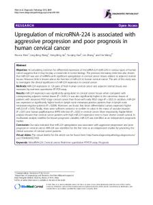
FIG. 5. Agarose gel of hFKBP10 cloning product. The PCR product was run on an 1% agarose gel. Lane 1: hFKBP10 PCR product (1763 bp), lane 2: negative control, and lane 3: GeneRulerTM 100 bp DNA Ladder Plus (Fermentas)
hFKBP65 and colorectal cancer, Olesen et al.
23
FIG. 6. Immunofluorescence microscopy (63x, 2x zoom) of COS7. COS7 cells labelled with anti- 58k (A), anti-Calreticulin (B), and overlay of the 58k and Calreticulin staining (C). Arrow heads indicate staining of the Golgi apparatus. Transfected COS7 cells labelled with anti-FKBP65 (D), anti-Calreticulin (E), and overlay of the FKBP and Calreticulin staining (F). Arrow indicates a non- transfected COS7 cell. Z-stack (0.5µm interval) of the hFKBP65 transfected cell (G-L). The cells were transfected using Amaxa Nucleofector Systems.
FIG. 7. Western blot analysis of hFKBP65 expression in COS7 cells treated with or without tunicamycin. Tunicamycin was added to the cells 6 hours post-transfection and harvested after 24 or 48 hours. The cells were transfected using FUGENE-6.
FIG. 8. 2D blots of colon adenocarcinoma and normal colon mucosa. The blots were reacted with antibody against hFKBP65 and detected using ECL procedure.
TA BLE I Clinical disease stage and analyses of the samples from distal colon IHC h sample no.a
sex
age
loc b
TNM status
d
dif f . grade
e
QPCR f
w estern
g
epithelial
f ibroblast
Norm al (n = 30; m e an age = 67, age range 44-90) 72N
m
57
10
T2N0M0
-
+
112N
f
62
7
T2N0M0
++ luminal
+
216N
f
79
7
T3N0M0
-
+
237N
f
82
7
T3N0M0
-
+
54N
f
81
7
T2N0M0
-
+
127N
m
78
10
T3N0M0
-
+
239N
m
77
7
T2N0M0
-
++
58N
m
81
7
T3N1M0
-
-
74N
m
61
10
T1N2M0
-
+
85N
m
56
9
T4N2M0
-
++
91N
f
53
10
T2N1M0
+
+
96N
m
90
7
T2N1M0
-
-
78N
m
45
9
T3N2M1
ND
ND
92N
m
44
8
T3N2M1
ND
ND
98N
m
76
10
T4N2M1
-
+
114N
m
45
7
T4N0M1
ND
ND
104N
f
66
7
T3N1M1
-
+
201N
m
78
9
T3N1M0
-
+
157N
f
75
7
T1N0M0
x
161N
f
63
10
T3N1M0
x
179N
f
74
10
T4N0M0
x
195N
f
76
7
T3N0M0
x
202N
m
52
7
T3N0M0
x
204N
m
58
8
T3N3M0
x
238N
m
64
10
T3N0M0
x
271N
f
73
10
T3N0M0
x
289N
f
62
10
T3N0M0
x
277N
m
71
9
T2N1M0
x
281N
m
79
9
T3N1M0
x
285N
f
51
7
T3N1M0
x
Duk e s A (n = 5; m e an age = 67.5, age range 57-80) 72A
m
57
10
T2N0M0
2
112A
f
62
7
T2N0M0
1
x
179A
m
76
11
T1N0M0
1
x
180A
m
63
7
T2N0M0¤
2
x
103A
m
80
9
T1N0M0
2
x
+++
++
++
++
+
Duk e s B (n = 10; m e an age = 72.5, age range 52-82) 216B
f
79
7
T3N0M0
3
x
++
237B
f
82
7
T3N0M0
1
x
+++
+
239B
m
77
7
T2N0M0
2
x
++
+
x
+++
++
+++
++
+++
54B
f
81
7
T2N0M0
2
127B
m
78
10
T3N0M0
2
203B
m
77
9
T3N0M0
2
x
202B
m
52
7
T3N0M0
3
x
238B
m
64
10
T3N0M0
2
x
271B
f
73
10
T3N0M0
2
x
289B
f
62
10
T3N0M0
2
x
Duk e s C (n = 10; m e an age = 67.8, age range 53-90) 58C
m
81
7
T3N1M0
2
x
+
74C
m
61
10
T1N2M0
2
x
+++
+
85C
m
56
9
T4N2M0
3
x
++
++
c
91C
f
53
10
T2N1M0
3
x
+
+++
96C
m
90
7
T2N1M0
2
x
++
+++
201C
m
78
9
c
T3N1M0
2
x
+
++
204C
m
58
8
c
T3N3M0
3
x
277C
m
71
9
T2N1M0
2
281C
m
79
9
T3N1M0
2
x
285C
f
51
7
T3N1M0¤
3
x
x
Duk e s D (n = 5; m e an age = 55.2, age range 44-76) 78D
m
45
9
T3N2M1
1
x
++
++
92D
m
44
8
T3N2M1
3
x
+++
++
98D
m
76
10
T4N2M1
2
x
+++
++
114D
m
45
7
T4N0M1
3
x
++
++
104D
f
66
7
T3N1M1¤
2
x
+
++
a
Matching normal and tumour samples have the same number, dif f ering only in the letter code (N= normal tissue; A , B, C, and D = tumours Dukes A , B, C or D).
b
location of samples in the colon (7-9 sigmoid; 10=rectosigmoid, 11=rectum)
c
primary tumour characterized as Dukes C, control CT-scan af ter 3-12 month show ed development of distant metastases
d
"T0" noninvasive. "T1-T4" invasive tumours. (T1 submucosa; T2 tunica muscularis; T3 subserosa; T4 peritoneum or other organs) "N0" no malignant lymphnodes; "N1" 1-3 lymphnode metastases; "N2" >4 lymphnode metastases; "N3" metastatic apical node (marked by surgeon) "M0" no distant metastases; "M1" distant metastasis, in e.g. liver or lung) "¤ " denotes mucinous adenocarcinoma, no "¤ " denotes an adenocarcinoma. TNM given f or normal samples ref ers to the corresponding tumour sample
e
Predominant histologiocal dif f erentiation grade (1=high; 2= moderate; 3= low )
f
an x denotes that RealTime PCR (QPCR) analysis is perf ormed on these samples
g
an x denotes that w estern blot analysis has been perf ormed on these samples
h
Staining in ImmunoHistoChemistry: "-": no staining, "+": low staining; "++": medium staining, "+++": high staining, "ND": not determined
TA BLE II Clinical disease stage and analyses of the samples from proximal colon IHC g sample no.
a
sex
age
loc b
TNM status
d
dif f . grade
e
QPCR f
epithelial
f ibroblast
Nor m al (n = 10; m e an age = 70, age range 47-92) 65N
f
80
1
T3N0M0
x
66N
f
66
1
T3N0M0
x
-
+
73N
f
78
1
T3N0M0
-
++
120N
f
73
1
T4N1M0
x
-
+
137N
m
79
1
T2N0M0
x
-
+
90N
f
58
1
T3N1M0
-
++
126N
f
47
1
T3N2M0
x
-
+
145N
f
72
1
T3N1M0
x
-
+
138N
m
60
1
T3N1M0
x
162N
f
92
1
T3N1M0
x
-
+
x
++
++
Duk e s B (n = 5; m e an age = 75, age range 66-80) 65B
f
80
1
T3N0M0
2
66B
f
66
1
T3N0M0
2
73B
f
78
1
T3N0M0
2
x
+++
++
120B
f
73
1
T4N1M0
3
x
+
++
137B
m
79
1
T2N0M0
1
x
++
++
Duk e s C (n = 5; m e an age = 66 age r ange 47-92 ) 90C
f
58
1
T3N1M0
3
x
ND
ND
126C
f
47
1
T3N2M0
2
x
+++
++
145C
f
72
1
T3N1M0
2
x
++
++
138C
m
60
1
T3N1M0
2
x
162C
f
92
1
T4N1M0¤
3
x
-
+++
c
a Matching normal and tumour samples have the same number, dif f ering only in the letter code (N= normal tissue; B and C = tumours Dukes B and C). b
location of samples in the colon (1=cecum)
c
primary tumour characterized as Dukes C, control CT-scan af ter 3-12 month show ed development of distant metastases d "T0" noninvasive. "T1-T4" invasive tumours. (T1 submucosa; T2 tunica muscularis; T3 subserosa; T4 peritoneum or other organs)
"N0" no malignant lymphnodes; "N1" 1-3 lymphnode metastases; "N2" >4 lymphnode metastases; "N3" metastatic apical node (marked by surgeon) "M0" no distant metastases; "M1" distant metastasis, in e.g. liver or lung) "¤ " denotes mucinous adenocarcinoma, no "¤ " denotes an adenocarcinoma. TNM given f or normal samples ref ers to the corresponding tumour sample e
Predominant histologiocal dif f erentiation grade (1=high; 2= moderate; 3= low )
f
an x denotes that RealTime PCR (QPCR) analysis is perf ormed on these samples
g
Staining in immunohistochemistry: "-": no staining, "+": low staining; "++": medium staining, "+++": high staining, "ND": not determined
TABLE III Clinical disease stage of the adenocarcinom as, with pre and post resection* endoscopic sam ples res ected prim ary adenocarcinom a
pre res ection IHC
pos t res ection IHC
e
IHC
e
IHC
s ex m f f f m
age 67 66 69 68 52
loc b 6 2 1 7 5
TNM s tatus T3N0M0 T4N0M0 T3N0M0 T3N0M0 T3N0M0
c
d
2 2 2 1 3
epi +++ +++ ++ ++ ++
fibro +++ +++ + +++ +
age des cription 65 68
TA HP
f
epi ++ +
fibro ++ +
age des cription f 67 TVA 66 HP 70 HP 68 TVA 52 TA
epi fibro + + + ++ ++ + + + + ++
age des cription f epi fibro
73 58
TA TA
* before or after res ection of the prim ary tum or a
prim ary tum ours characterized as Dukes B
b
location of prim ary adenocarcinom a in the colon (1=cecum , 2=as cending, 5 =s igm oid, 6=rectos idm oid, 7= rectum )
c
"T0" noninvas ive. "T1-T4" invas ive tum ours . (T1 s ubm ucos a; T2 tunica m us cularis ; T3 s ubs eros a; T4 peritoneum or other organs ) "N0" no m alignant lym phnodes ; "N1" 1-3 lym phnode m etas tas es ; "N2" >4 lym phnode m etas tas es ; "N3" m etas tatic apical node (m arked by s urgeon) "M0" no dis tant m etas tas es ; "M1" dis tant m etas tas is , in e.g. liver or lung)
d
Predom inant his tologiocal differentiation grade (1=high; 2= m oderate; 3= low)
e
Staining in Im m unoHis toChem is try: "-": no s taining, "+": low s taining; "++": m edium s taining, "+++": high s taining, "ND": not determ ined epi: epithelial s taining, fibro: fibroblas t s taining
f
e
diff. grade
s am ple no.a 1B 2B 3B 4B 5B
e
HP: hyperplas tic polyp, TA: tubular adenom a, TVA: tubular villous adenom a
+ ++
+ ++
Supplem entary TABLE I Endoscopic norm al b iopsies from the digestive tract s am ple no.
s ex
age
loc
6
m
36
es ophagus
7 8 9
m f f
42 44 64
fundus ventriculi corpus vens triculi antrum ventriculi
10 11
m m
34 59
s m all intes tine appendix