The Journal of Pain, Vol 14, No 4 (April), 2013: pp 371-383 Available online at www.jpain.org and www.sciencedirect.com
Focal Modulation of the Primary Motor Cortex in Fibromyalgia Using 4!1-Ring High-Definition Transcranial Direct Current Stimulation (HD-tDCS): Immediate and Delayed Analgesic Effects of Cathodal and Anodal Stimulation Mauricio F. Villamar,*,y Pakorn Wivatvongvana,*,z Jayanton Patumanond,x Marom Bikson,{ Dennis Q. Truong,{ Abhishek Datta,*,{ and Felipe Fregni* *Laboratory of Neuromodulation, Department of Physical Medicine & Rehabilitation, Spaulding Rehabilitation Hospital and Massachusetts General Hospital, Harvard Medical School, Boston, Massachusetts. y School of Medicine, Pontifical Catholic University of Ecuador, Quito, Ecuador. z Department of Rehabilitation Medicine and xClinical Epidemiology & Clinical Statistics Unit, Faculty of Medicine, Chiang Mai University, Chiang Mai, Thailand. { Department of Biomedical Engineering, The City College of New York of CUNY, New York, New York.
Abstract: Fibromyalgia is a prevalent chronic pain syndrome characterized by altered pain and sensory processing in the central nervous system, which is often refractory to multiple therapeutic approaches. Given previous evidence supporting analgesic properties of noninvasive brain stimulation techniques in this condition, this study examined the effects of a novel, more focal method of transcranial direct current stimulation (tDCS), using the 4!1-ring configuration of highdefinition (HD)-tDCS, on overall perceived pain in fibromyalgia patients. In this patient- and assessor-blind, sham-controlled, crossover trial, 18 patients were randomized to undergo single 20minute sessions of anodal, cathodal, and sham HD-tDCS at 2.0 mA in a counterbalanced fashion. The center electrode was positioned over the left primary motor cortex. Pain scales and sensory testing were assessed before and after each intervention. A finite element method brain model was generated to predict electric field distribution. We found that both active stimulation conditions led to significant reduction in overall perceived pain as compared to sham. This effect occurred immediately after cathodal HD-tDCS and was evident for both anodal and cathodal HD-tDCS 30 minutes after stimulation. Furthermore, active anodal stimulation induced a significant bilateral increase in mechanical detection thresholds. These interventions proved well tolerated in our patient population. Perspective: 4!1-ring HD-tDCS, a novel noninvasive brain stimulation technique capable of more focal and targeted stimulation, provides significant reduction in overall perceived pain in fibromyalgia patients as compared to sham stimulation, irrespective of current polarity. This technique may have other applications in research and clinical settings, which should be further explored. ª 2013 by the American Pain Society Key words: Fibromyalgia, chronic pain, transcranial direct current stimulation (tDCS), high-definition transcranial direct current stimulation (HD-tDCS), electrical brain stimulation, transcranial electrical stimulation (tES).
F
ibromyalgia (FM) is a common cause of diffuse, chronic musculoskeletal pain in adults, with an estimated prevalence between 2 and 5% in the general popula-
tion.6,32,55 Even though its etiology and pathophysiology are not yet fully understood, current evidence strongly suggests that, similarly to other chronic pain syndromes,
Received July 18, 2012; Revised December 1, 2012; Accepted December 10, 2012. Funded by a Translational Research Award from the Wallace H. Coulter Foundation. M.F.V., P.W., J.P., D.Q.T., and F.F. declare no conflicts of interest related to this study. The City University of New York has intellectual property on noninvasive brain stimulation, with M.B. and A.D. as inventors. M.B. and A.D. have equity in Soterix Medical Inc.
Supplementary data accompanying this article are available online at www.jpain.org and www.sciencedirect.com. Address reprint requests to Felipe Fregni, MD, PhD, MPH, Laboratory of Neuromodulation, Spaulding Rehabilitation Hospital, 125 Nashua St #726, Boston, MA 02114. E-mail:
[email protected] 1526-5900/$36.00 ª 2013 by the American Pain Society http://dx.doi.org/10.1016/j.jpain.2012.12.007
371
372
The Journal of Pain
this is a disorder of pain regulation characterized by altered pain and sensory processing in the central nervous system (CNS), likely due to maladaptive plasticity in pain-related neural circuits.22,28,56 FM pain management is often challenging and many cases are refractory to current pharmacological and nonpharmacological approaches, including behavioral interventions and other alternative treatments, frequently leading to a considerable impairment in patients’ quality of life (QOL). There is therefore an unmet clinical need to develop novel interventions for the treatment of FM. Different neuromodulatory and neurostimulatory approaches, both invasive and noninvasive, have been successfully tested as potential therapeutic tools for chronic pain disorders given their ability to modify brain activity in neural networks in the area of stimulation as well as in distant, interconnected regions. Indeed, there has been a growing interest in 2 of these methods—transcranial direct current stimulation (tDCS) and repetitive transcranial magnetic stimulation (rTMS)—particularly due to their noninvasive nature and their ability to modify the excitability of cortical neural circuits. When delivered to the primary motor cortex (M1), these techniques are capable of modifying sensory aspects of pain via modulation of M1-thalamic inhibitory networks,40 as well as of other cortico-cortical and cortico-subcortical projections involved in pain processing pathways.8,40 This is of particular relevance in FM, where pain can be characterized by a lack of inhibitory control over somatosensory processing.13,51 For this reason, modulation of the sensory aspects of pain via M1 stimulation, independent from modulation of affective components, may target one of the pathophysiological mechanisms of FM. Accordingly, an increasing body of evidence supports the analgesic effects of both tDCS4,17,43,44,51 and rTMS30,38 of the M1 in the setting of FM. tDCS may have some additional advantages due to its portability, ease of use, and low cost. However, this technique uses relatively large electrode pads (most commonly 35 cm2) that deliver direct current (DC) in rather diffuse areas of the brain,12,26,45 making focalized stimulation of target regions less feasible. Increased focality would be desirable in tDCS for a number of reasons. First, it may help achieve beneficial clinical effects with larger effect sizes, as occurs with invasive interventions such as epidural M1 stimulation for chronic pain.27 Second, it may also contribute to the understanding of the specific cortical regions implicated in therapeutic action, which is difficult to dissect with diffuse electrical stimulation. This would ultimately contribute to the rational development and optimization of therapy. Hence, a more focal approach may allow for tailoring of stimulation to individual indications and symptoms in a way not possible with diffuse stimulation. Finally, a more focal intervention may also be associated with increased safety. It could potentially reduce the likelihood of side effects due to decreased stimulation of adjacent regions, thus allowing for stimulation with increasing intensity or repetition to enhance efficacy. High-definition (HD)-tDCS is a novel approach that uses arrays of smaller electrodes31 whose configuration
Focal M1 Modulation in Fibromyalgia Using HD-tDCS can be optimized for targeting.14 In particular, the 4!1ring montage of HD-tDCS has been proposed for unidirectional and targeted stimulation, with the polarity (anode or cathode) set by a center electrode and the area of cortical modulation restricted by adjusting the radii of 4 return electrodes.12 The targeting of 4!1-ring HD-tDCS may have important applications in research and eventually in clinical settings. Nevertheless, no studies have been published to date assessing its effects in a patient population. In order to evaluate the analgesic effects of HD-tDCS in patients with FM, we performed a randomized, shamcontrolled, crossover clinical trial where both participants and the assessor were blind to the assigned interventions. Specifically, we were interested in determining whether active anodal and cathodal HD-tDCS could induce a significant reduction in perceived pain as compared to sham stimulation, as evaluated using pain scales and sensory testing.
Methods This trial is reported following 2010 CONSORT guidelines. A participant flow diagram is shown in Fig 1.
Trial Design and Overview This study was a patient- and assessor-blind, randomized, sham-controlled, crossover clinical trial with equal allocation ratio (1:1). The protocol consisted of a total of 5 study visits. After undergoing a telephone screening, patients were scheduled for their first visit, where we further screened them using the 2010 American College of Rheumatology Preliminary Diagnostic Criteria for FM,54 obtained written informed consent, and did a baseline evaluation, as detailed below. On visits 2, 3, and 4, each participant underwent single sessions of anodal, cathodal and sham HD-tDCS delivered to the left M1, which were preceded and followed by pain scales and sensory testing. The order of stimulation was counterbalanced and randomly assigned for each individual. Finally, a follow-up evaluation was conducted on visit 5. All visits were separated by a washout period of at least 7 days to prevent any carryover effects or contamination of the sham stimulation by a preceding active session. Although patients were scheduled for participation at different times of the day based on their availability, each individual patient was scheduled for all of his/her visits at approximately the same time of day. Fig 2 provides an overview of our study design. All study procedures were conducted in the Laboratory of Neuromodulation at Spaulding Rehabilitation Hospital, Harvard Medical School, Boston, MA. The study was approved by the local Institutional Review Board (Protocol # 2010-p-000990) and registered in ClinicalTrials.gov (NCT01402960). This randomized controlled trial was conducted in accordance with the Declaration of Helsinki.
Participants We included patients 1) who were between 18 and 64 years of age; 2) who had a formal diagnosis of FM made by a rheumatologist (FM was chosen as a type of diffuse
Villamar et al
The Journal of Pain
373
Assessed for eligibility (n=46)
Excluded (n=28) ♦ Not meeting inclusion criteria (n=10) ♦ Declined to participate (n=18)
Randomized (n= 18)
Allocated to intervention (n=17) ♦ Received allocated intervention (n=17) ♦
Did not receive allocated intervention (patient could not be contacted) (n=1)
Lost to follow-up (n=0) Discontinued intervention (patient uncomfortable with depression and quality of life questionnaires) (n=1)
Analysed (n=18) ♦ Excluded from analysis (n=0)
Figure 1. CONSORT 2010 patient flow diagram. musculoskeletal pain disorder) for a duration greater than 6 months at the time of enrollment, with an average overall pain intensity of at least 3/10 on a visual numerical scale (VNS); and 3) whose pain was refractory to common analgesics and muscle relaxants. Patients taking analgesics or CNS-active medication were allowed to enroll if dosages had been stable for at least 2 months prior to their screening. Given ethical concerns, they were encouraged to keep taking their usual medication at stable doses for the duration of the trial. A list of CNS-active drug use among study participants is provided as a Supplementary Table. As a safety measure and in order to eliminate variables that could interfere with the effects of the stimulation, our exclusion criteria were as follows: 1) current pregnancy; 2) presence of metallic implants in the head; 3) history of substance abuse within the past 6 months; 4) use of carbamazepine within the past 6 months; 5) severe depression, as defined by a baseline score $29 in the Beck Depression Inventory-II (BDI-II)7; and 6) any history of epilepsy, stroke, moderate-to-severe traumatic brain injury, severe migraines, or brain surgery. A total of 18 patients fulfilled the above-mentioned inclusion criteria and provided written informed consent to participate in our trial. Population characteristics are summarized in Table 1. Participants were recruited through local support groups and online listings over a period of 6 months (see Fig 1).
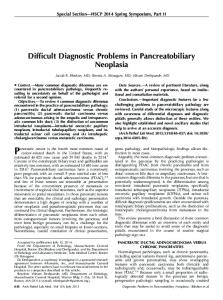
Intervention: 4!1-Ring HD-tDCS We employed a 4!1 Multichannel Stimulation Adaptor (Model 4!1-C2; Soterix Medical Inc, New York, NY)
connected to a conventional tDCS device (Model 1224B; Soterix Medical Inc) to deliver DC to the scalp via Ag/ AgCl sintered ring electrodes (EL-TP-RNG Sintered; Stens Biofeedback Inc, San Rafael, CA), as described by Minhas et al.31 Electrodes were held in place by specially designed plastic casings embedded in a modular electroencephalography (EEG) recording cap. We positioned the center electrode (anode or cathode) over C3 based on the International 10/20 EEG System,42 which corresponds approximately to the location of the left M1. Four return electrodes (cathode or anode, respectively) were placed in a radius of approximately 7.5 cm from the center electrode. Their locations corresponded roughly to Cz, F3, T7, and P3, as depicted in Fig 3. The decision to use this radius aimed at collecting data supporting different possible montages. By changing the position of the return electrodes while keeping the radius constant, the outcomes would not be expected to differ markedly. The hair underlying each electrode was separated in order to expose the scalp, and approximately 1.5 mL of highly conductive gel (Signa Gel; Parker Laboratories, Fairfield, NJ) was placed beneath each electrode to improve conductance. Given that electrode resistance is nonlinear to electrode-interface electrochemical processes,29 electrode resistance (impedance) can be misleading. For example, the resistance apparently measured fully depends on test current.20 Therefore, based on prior experience in set-up and stimulation using HD electrodes, contact quality is normalized to ‘‘quality units’’ by the 4!1-C2 Adapter test circuit. Impedance values were verified to be #1.50 ‘‘quality units’’ for
374
Focal M1 Modulation in Fibromyalgia Using HD-tDCS
The Journal of Pain
Figure 2. Overview of study design. each of the 5 electrodes before the beginning of each stimulation session. During each active 4!1-ring HD-tDCS session, DC was gradually ramped up over a period of 30 seconds until reaching an intensity of 2.0 milliamperes (mA), which were delivered for 20 minutes. These parameters had previously been shown to be well tolerated in healthy subjects.9 The same montage was used for the sham procedure; however, current was applied for 30 seconds only. This duration has been reported to be a reliable method for blinding participants in conventional tDCS trials,19 which induces no effects on cortical excitability.33,35 Borckardt et al9 also reported this blinding strategy to be successful in their HD-tDCS trial.
Demographic Characteristics of Study Participants
Table 1.
CHARACTERISTICS
VALUES
Mean age, years (SD) Mean time since FM diagnosis, years (SD) Gender, n (%) Female Male Self-reported ethnicity, n (%) Caucasian African American Hispanic Education, n (%) Some college Undergraduate degree Graduate degree
50.3 (8.5) 10.7 (6.8) 15 (83.3) 3 (16.7) 15 (83.3) 2 (11.1) 1 (5.6) 12 (66.7) 2 (11.1) 4 (22.2)
Outcomes Participants sat in a chair with back and arm support during all stages of data collection. The following assessments were performed, in order, at the beginning of every visit: 1) baseline VNS for overall pain; 2) VNS for anxiety; 3) Adapted QOL Scale for persons with chronic illness10; 4) BDI-II7; 5) Semmes-Weinstein monofilaments (SWMs) for pain and mechanical detection thresholds; 6) pressure pain thresholds (PPTs); and 7) diffuse noxious inhibitory controls (DNICs). Additionally, patients were asked to keep a pain and medication diary for the duration of the study. On visits 2, 3, and 4, these assessments were followed by the assigned HD-tDCS sessions, as described above. Patients were again asked to rate their overall level of pain using the VNS immediately and 30 minutes after active or sham HD-tDCS. The VNS for anxiety, SWMs, PPTs, and DNICs were also evaluated immediately after the intervention, and a questionnaire was used to inquire about potential adverse effects related to the stimulation. Changes in VNS for pain constituted our primary outcome measure. All other outcome measures were considered secondary.
VNS for Pain Patients were asked to rate their current overall level of pain on a visual scale from 0 to 10 divided at .5point intervals, with 0 being ‘‘complete absence of pain’’ and 10 ‘‘the worst pain imaginable.’’
VNS for Anxiety Since anxiety levels might act as a confounder for changes in pain perception, we evaluated them using
Villamar et al
The Journal of Pain
375
Figure 3. Equipment for 4"1-ring HD-tDCS. (A) Multichannel Stimulation Adaptor (left) connected to conventional tDCS device (right). (B) Electrode positioning used for left primary motor cortex stimulation.
a similar visual scale, where 0 corresponded to ‘‘completely calm’’ and 10 to ‘‘extremely anxious.’’
Adapted QOL Scale for Persons With Chronic Illness10 This tool uses a 7-point scale (1 = terrible; 7 = delighted) to assess 16 QOL-related domains among chronic illness populations.
BDI-II7 This questionnaire consists of 21 multiple-choice questions for evaluating the presence and severity of depression in adults.
SWMs Calibrated esthesiometers (Touch-Test Sensory Evaluators; North Coast Medical Inc, Morgan Hill, CA) were used for assessing mechanical detection and pain thresholds. While patients kept their eyes closed, filaments of increasing thicknesses were sequentially applied to the thenar region of both hands. For each hand, we registered the values at which patients first perceived the stimulus touching their skin (mechanical detection threshold) and reported it as painful (pain threshold).
PPTs PPTs were evaluated by delivering blunt pressure in 4 paired points of the body using the standard 1-cm2 hard-rubber nozzle of a Commander Algometer (JTECH Medical, Salt Lake City, UT). Pressure was gradually increased at a rate of approximately 2 lb/second, and patients were instructed to inform the assessor as soon as they experienced a sensory transition from pressure to pain. When this occurred, the device was immediately removed and the value recorded. If no pain was reported, we recorded the threshold maximum of 25 lb. This procedure was performed 3 times in each point of interest, and their average was used for statistical analyses. Petzke et al39 reported that examination of tenderness by dolorimetry at only 3 paired sites is a reliable and clinically useful assessment that highly predicts overall pain threshold in FM patients and healthy subjects. Therefore, we measured PPTs in 1) the area of the
forearm 2 cm distal to the lateral epicondyle; 2) the supraspinatus muscle, above the medial border of the scapular spine; and 3) the occiput, at the suboccipital muscle insertions. These are the sites that showed the highest correlation with overall pain threshold in the above-mentioned study and have also been documented by Tastekin et al48 as having high discriminative ability for FM syndrome. In addition, we measured PPTs in the thenar area as a control site. In all cases, assessments in the right side of the body preceded the left.
DNICs Participants were asked to immerse their left hand in cold water (10–12! C) for the duration of the test (1 minute total). Once the initial 20 seconds had elapsed, patients rated their local level of pain on a VNS, which had to be $4.0 in order to proceed. If pain levels were lower, ice was added until this threshold was reached. During the last 30 seconds, PPT assessments were performed in the right thenar area as previously described. After allowing for normalization of cutaneous temperature, the same procedure was performed in the opposite hand.
Pain and Medication Diary Patients were given preformatted forms where they were asked to rate their average levels of overall pain on a daily basis using a VNS. They were encouraged to do so at approximately the same time each day. An average of each week’s values was used for statistical analyses. Additionally, any changes in their medication (analgesics in particular) were recorded.
Randomization Participants were consecutively assigned to a randomization scheme generated on the website Randomization. com (Dallal GE, http://www.randomization.com, 2008). We used the second generator, with random permutations for a 3-group trial. The randomization sequence was concealed until interventions were assigned. Generation of the random allocation sequence and assignment of participants were performed by a research coordinator not involved with any other aspect of the trial.
376
The Journal of Pain
Blinding In order to prevent introduction of bias,52 all participants and the trained physiatrist (P.W.) who performed the assessments were blind to the type of stimulation. Once baseline assessments had been performed on each visit, a different researcher (M.F.V.) was allowed to check the randomization code for that patient. He then set up the montage, operated the device, and delivered the stimulation accordingly. The assessor (P.W.) was not present in the room during delivery of the stimulation. Once the HD-tDCS session had ended and the operator (M.F.V.) removed the equipment, the assessor came back to the room and finished the assessments. Blinding of participants was discussed as part of the Intervention section.
Statistical Analyses Patients’ characteristics were summarized using descriptive statistics. Differences in baseline values between the 3 stimulation conditions were analyzed by 1-way analysis of variance. A multilevel regression model was used to analyze global differences in changes over time in VNS for pain across stimulation conditions, allowing for random individual patient responses (the intercepts) and autocorrelation of repeated responses within patients, while simultaneously adjusting for baseline values. Significant global differences (2-tailed P < .05) were further analyzed with Wald tests under the multilevel regression model to detect any significant differences between the 3 possible pairs of stimulation conditions (sham versus anodal, sham versus cathodal, and anodal versus cathodal. P values were corrected for multiple comparisons (Bonferroni) and then converted in order to maintain a level of significance of .05. Effect sizes of the active interventions were calculated. A similar model was conducted for each of our secondary outcomes. Sample size was estimated based on results from a previous study on patients with refractory chronic pain.15 In this trial, active anodal tDCS induced a significant reduction (P < .05) in perceived pain of .797 points (SD .5) in a visual analog scale, as compared to sham stimulation. Given a power of 80% and an alpha of 5%, 16 patients would be needed. We increased the sample to 18 patients to account for potential dropouts. All statistical analyses were performed using STATA (StataCorp 2009. Stata Statistical Software: Release 11. StataCorp LP, College Station, TX). Two-tailed P values