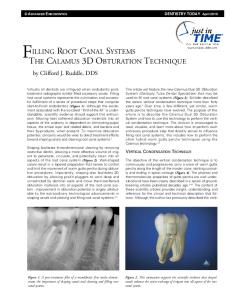
Endodontics Colleagues for Excellence Fall 2009
Obturation of Root Canal Systems
Published for the Dental Professional Community by the
American Association of Endodontists
Cover artwork: Rusty Jones, MediVisuals, Inc.
EndodoNtics: Colleagues for Excellence
T
he purpose of the obturation phase of endodontic treatment is to prevent the reinfection of root canals that have been biomechanically cleaned, shaped and disinfected by instrumentation, irrigation and medication procedures. Successful obturation requires the use of materials and techniques capable of densely filling the entire root canal system and providing a fluid tight seal from the apical segment of the canal to the cavo-surface margin in order to prevent reinfection. This also implies that an adequate coronal filling or restoration be placed to prevent oral bacterial microleakage. It has been shown that endodontic treatment success is dependent both on the quality of the obturation and the final restoration.1 The quality of the endodontic obturation is usually evaluated using radiographic images upon completion. Additionally, during the root canal preparation and obturation phases of treatment, clinical criteria can be identified that are essential for achieving an adequate root canal obturation.2,3,4,5 It’s All About Cleaning, Shaping and Disinfection of Root Canal Systems
Prior to the obturation phase, the clinician must establish the proper shape and size of the root canal. Figure 1 is an example of a retreatment case; it demonstrates the importance of locating all canals then cleaning, shaping and sealing them to the proper working length. Proper canal preparation provides an apical resistance form for the adequate adaptation of filling materials and the prevention of excessive apical extrusion of these materials. Remember, what is removed from the root canal durFig. 1. Retreatment case demonstrating the importance of locating all canals then cleaning, shaping and sealing them to the proper working ing cleaning, shaping and disinfection is more important than what is length. placed during obturation. Biomechanical preparation and disinfection are achieved through mechanical instrumentation and copious irrigation to remove bacterial and tissue debris, and by shaping the root canal space to allow for a three-dimensional obturation and seal of the root canal system. The importance of maintaining the original shape of a root canal during and after cleaning and shaping in order to promote periapical healing in endodontic cases has been demonstrated in several studies.6,7,8 The clinician’s inability to maintain the original shape and to develop the proper taper of canals can result in procedural errors such as ledges and perforations. When preparing root canals for obturation the clinician must maintain the working length during the instrumentation phase of treatment. Figure 2 shows original, working length and final obturation image with post and core in place. Maintaining working length is essential for preparing and sealing the root canal to its proper apical extent. It is Fig. 2a. 5 November 2007: 2b. 10 March 2009: Working 2c. 3 April 2009: Post and Core Original Image Length Image Image also necessary to create an apical resistance form to obtain adequate compaction of the filling materials and also to prevent excessive overextension of materials into the periapical tissues. Today’s clinicians have a number of methods, materials and technologically advanced instruments at their disposal to achieve these goals. Examples of these are electronic apex locators, nickel-titanium rotary instruments, various irrigation systems, newly formulated sealers, and microscopic magnification and illumination. Poor obturation quality as judged by radiographs has been associated with nonhealing in 65% of retreatment cases.9 The radiographic appearance of a completed case should show the obturation material: (1) at the apical terminus without excessive material overextending into periapical tissues; (2) completely filling the root canal system in three dimensions; and (3) appearing as a dense radiopaque filling of the root canal system.4 Obturation errors often are a result of inadequate cleaning and shaping (ledges, perforations, inaccurate working lengths, and underprepared or overprepared canals). If inadequate obturation is not a result of an instrumentation error, the clinician should recognize this reversible procedural error on the obturation check film. The obturation material should then be removed and the canal re-obturated prior to restoration. If the procedural error is gross overextension of material into the periapical tissues, removal by conventional means may not be possible and periapical surgery may be necessary. Many good techniques are available to the clinician for the instrumentation phase of root canal treatment. Smooth, clean, and properly shaped and sized canals will allow for efficient removal of debris with instruments and irrigants, and insertion
2
EndodoNtics: Colleagues for Excellence of sealers and placement of root filling material. If a clinician is having difficulty with the obturation phase of endodontic therapy, their cleaning and shaping technique should be reevaluated prior to consideration of changing obturation techniques. Why Obturate Canals? Microorganisms and their byproducts are the major cause of pulpal and periapical disease.11 However, it is difficult to consistently and totally disinfect root canal systems.10 Therefore, the goal of three-dimensional obturation is to provide an impermeable fluid tight seal within the entire root canal system, to prevent oral and apical microleakage.12 Preparation of Dentin Surface (Irrigation) The purpose of endodontic irrigation is to remove debris created during instrumentation, and to dissolve and/or flush out inorganic and organic remnants of the pulp system, bacteria and bacterial byproducts that are not removed by mechanical instrumentation. With the introduction of obturation materials designed to bond with dentin, irrigation solutions must be used with consideration of the condition of the dentin surface that is most suitable for bonding. Figure 3 shows the dentin surface before and after preparation for bonding. Attempts to eliminate pulp space infection with instrumentation only, without the use of antimicrobial agents, have proven to be unsuccessful.13 Modern root canal treatment requires the use of both mechanical and chemical preparation and disinfection of the canal system. During cleaning and shaping procedures, a superficial amorphous layer of tissue remnants, organic and inorganic materials, and bacteria and their byproducts accumulate on the canal walls. This “smear layer” may interfere or prevent adhesion of sealers to the canal wall and serve as a substrate for bacterial growth. Evidence tends to support removing the smear layer prior to obturation.32 This removal supports reduction of potential irritants and permits better adaptation of sealer to the canal walls. Removal of the smear layer is easily accomplished by irrigating the canal with 17% disodium EDTA for one minute, followed by a final rinse of sodium hypochlorite. Chelators remove the inorganic components and sodium hypochlorite is necessary for removal of the remaining organic components. Adequate irrigation of root canals requires an effective irrigant as well as an efficient delivery system. The characteristics of an ideal irrigant and irrigation system are listed in Table I.14 The irrigating system should be one that does not allow the irrigant to extrude beyond the apical foramen into the periapical tissues. Fig. 3a. Dentin surface before and Fig. 3b. after preparation for bonding.
Table I: Characteristics of an Ideal Irrigation System Physical flushing of debris Biocompatible Bactericidal agent Sustained effect Disinfect and detoxify dentin and tubules of all microbial substances
Table II list factors that can influence the efficacy of irrigating root canal systems.15
Table II: Factors Influencing Efficacy of Irrigation Diameter of the irrigating needle Depth of the irrigating needle engaged in root canal Size of enlarged root canal (radius of tube)
Tissue solvent
Viscosity of the irrigating solution (surface tension)
Lubricant
Velocity of the irrigating solution at the tip of the needle (ultrasonics, sonics)
Smear layer removal Not affect physical properties of dentin
Orientation of the bevel of the needle Temperature
3
Continued on p. 4
EndodoNtics: Colleagues for Excellence Traditionally, irrigation has been done using a syringe and side-slotted needle placed 3mm short of working length. Other techniques such as sonic, ultrasonic and apical negative pressure devices have been introduced and are currently available to the clinician. Research has indicated some of these systems may be safer and more efficient than others.16,17 Research evidence and experience should be used when deciding which system to incorporate into your technique based on its effectiveness and safety. Criteria for Judging Technical Success of the Obturation Phase of Endodontic Treatment Techniques vary by the way they accomplish a three-dimensional obturation of the canal system. The basic principles and criteria for technical success must be achieved regardless of the technique. 1. Clinical Evaluation. For a case to be considered successful, normal findings to routine tests such as percussion, palpation,
periodontal probing and visual inspection of the final restoration should be obtained and recorded in the patient’s record at an appropriate follow-up visit. If the clinician is concerned about some aspect of therapy or the prognosis, the reevaluation visit should be scheduled in a few weeks. Routine reevaluation periods may be 6 months and 1 year. Patients should be informed that if symptoms occur they should call the office for an appointment. 2. Radiographic Evaluation (Length, Shape and Density). (overfilled, overextended and underextended). Three qualities that should
be observed, are length, shape and density. The length of an ideal fill should be from the canal’s apical minor constriction to the canal orifice unless a post is planned. The core restoration should complete this seal to the cavo-surface margin. The shape of the completed case is somewhat dependent on the obturation technique being used. Some require a more tapered canal than others. Voids should not be visible on the radiographic image. In terms of percentage rates of success, a meta-analysis of the literature showed that obturations 0 to 1mm short of the apex were better than obturations 1 to 3mm short of the apex; both were superior to obturations beyond the apex.18 Coronal Seal Regardless of the quality of the sealing of the root canal space, unless this space is protected against ingress of oral microorganisms, success may not be achieved. Figure 4 shows the preoperative status and postoperative image of #29 adequately restored with a post, core and crown. Notice the presence of an obturated lateral canal. A study of failed cases found that 59.4% of endodontically treated teeth failed due to restorative reasons, 32% for periodontal reasons and 8.6% for endodontic reasons.19 It Fig. 4a. Preoperative and Fig. 4b. postoperative images with post, core and crown. is apparent from the literature that prevention of coronal microleakage is critical to success. The use of intra-orifice barriers, which are restorative materials placed over the canal orifices and covering the pulp chamber floor, has been strongly recommended. Criteria have been proposed for the ideal intra-orifice barrier. They should: (1) be easily placed, bond to tooth structure (retentiveness); (2) seal effectively against coronal microleakage; (3) be easily distinguished from the natural tooth color; and (4) not interfere with the final restoration of the access preparation. This material can be any material that will bond or seal the dentin and have a distinguishable color from dentin.20 Ideal Mechanical, Physical and Biological Properties of Obturation Materials Many materials and techniques for obturation are available on the market. Dr. Louis I. Grossman, one of the founders of the specialty of endodontics, determined the ideal properties of obturation materials listed in Table III.21 Taking these into consideration, the clinician should realize that material: (1) content; (2) toxicity; and (3) physical properties are controlled by the manufacturer. The clinician’s choice of obturation materials should be based on: (1) purchasing materials meeting the American Dental Association and the American National Standards Institute specifications; (2) assuring their compatibility with patient’s medical history; and (3) those that best match the instrumentation and obturation techniques being used. These materials are divided into two basic groups—sealers and core materials—each of which can be found in a large variety of materials and brands. Materials approved by the International Standards Organization and the American Dental Association should be used. 4
EndodoNtics: Colleagues for Excellence Table III: Grossman’s Ideal Properties of Root Canal Obturation Materials It should be easily introduced into the root canal system. It should seal the canal laterally as well as apically. It should not shrink after being inserted.
It should be radiopaque. It should not stain tooth structure. It should not irritate periapical tissue.
It should be impervious to moisture.
It should be sterile or easily and quickly sterilized immediately before insertion.
It should be bacteriostatic or at least not encourage bacterial growth.
It should be easily removed from the root canal if necessary.
A. Sealers
Sealers are used between dentin surfaces and core materials to fill spaces that are created due to the physical inability of the core materials to fill all areas of the canal. Traditionally desirable characteristics were to adhere to dentin and the core material as well as to have adequate cohesive strength. Newer generation sealers are being engineered to improve their ability to penetrate into dentinal tubules and bond to, instead of just adhering to, both the dentin and core material surfaces. Various types of delivery systems such as auto-mix syringes have improved not only the efficiency of mixing, but also the quality of the mix and ultimately the properties of the set material. Various types of sealers include zinc oxide-eugenol, as well as polymer resins, glass ionomer, bio-glass and silicon-based materials. B. Core Materials 1. Gutta-Percha: This material was first used in dentistry in the late 1800s as a temporary restorative material and
then to obturate root canal systems. During the Civil War, a material called Hill’s stopping (which contained gutta-percha, quick lime, quartz and feldspar) and gutta-percha were advocated by Taft and Harris as temporary filling materials. Its use as a temporary filling material continued until 1950.22 Used without sealer, gutta-percha does not provide a seal. It is derived from the Taban tree (Isonandra perchas). The natural chemical form of gutta-percha is 1, 4-polyisoprene.23 It is an isomer of natural rubber and has been used for various purposes such as coating the first trans-Atlantic cable and for the cores of golf balls. Gutta-percha undergoes phase transitions when heated from beta to alpha phase at around 115° F (46° C). At a range between 130° to 140° F (54° to 60° C) an amorphous phase is reached. When cooled at an extremely slow rate the material will recrystalize to the alpha phase. However, this is difficult to achieve and under normal conditions the material returns to the beta phase. The softening point of gutta-percha was found to be 147° F (64° C).24 The phase transformation is important in thermoplastic obturation techniques. Gutta-percha is soluble in chloroform, eucalyptol, halothane and less well in turpentine. This property of gutta-percha allows it to be removed for post preparation and in the retreatment of nonhealing cases. Any method manipulating gutta-percha using heat or solvent will result in some shrinkage (1-2%) of the material. Shrinkage of the core material is not desirable when attempting to seal a canal. Dental gutta-percha is not pure or even mostly gutta-percha. Its major component is zinc oxide (50-79%), heavy metal salts (1-17%), wax or resin (1-4%) and only 19-22% actual gutta-percha. The variations in content are because of different manufacturers and distributors desiring different handling properties. Some formulations are softer than others. Some clinicians choose the brand of gutta-percha depending on the technique being used. Compaction with spreaders, condensers or carriers is usually the means used to attempt to compensate for this shrinkage of the core material.25 In any case, some means of compensation for this shrinkage must be incorporated into the technique being used. An important characteristic of gutta-percha and of clinical importance is the fact that when it is exposed to air and light over time it becomes more brittle.26 Storage of gutta-percha in a refrigerator extends the shelf life of the material. 2. Resilon: Resilon™, a new, synthetic resin-based polycaprolactone polymer has been developed as a gutta-percha
substitute to be used with Ephiphany®, (Pentron® Clinical Technologies, Wallingford, Conn.) a new resin sealer in an attempt to form an adhesive bond at the interface of the synthetic polymer-based core material, the canal wall and the sealer. Advocates of this technique propose that the bond to the canal wall and to the core material creates
5
Continued on p. 6
EndodoNtics: Colleagues for Excellence a “monoblock.” It is capable of being supplied in standardized ISO sizes and shapes, conforms to the configuration of the various nickel-titanium rotary instruments, and is available in pellet form for injection devices. The manufacturer states that its handling properties are similar to those of gutta-percha and therefore it can be used with any obturation technique. Resilon contains polymers of polyester, bioactive glass and radiopaque fillers (bismuth oxychloride and barium sulfate) with a filler content of approximately 65%. It can be softened with heat or dissolved with solvents like chloroform. This characteristic allows the use of various current treatment techniques. Being a resin-based system makes it compatible with current restorative techniques in which cores and posts are being placed with resin-bonding agents.27,28 3. Coated Cones: This process has been developed in an attempt to achieve similar results as those claimed by Resilon—a
bond between the canal wall, the core and the sealer. Two versions of coating gutta-percha are available. Ultradent Corporation has surface coated their gutta-percha cones with a resin (Ultradent, South Jordan, Utah). A bond is formed when the resin sealer contacts the resin-coated gutta-percha cone. The manufacturer claims this will inhibit leakage between the solid core and sealer. The technique calls for the use of EndoRez™ sealer (Ultradent, South Jordan, Utah) with this new coated solid core material. Another company has coated their gutta-percha cones with glass ionomer (Brasseler USA, Savannah, Ga.) and is designed for use with their glass ionomer sealer. Their system is called Active GP Plus™. Techniques for Obturating Root Canal Systems A. Lateral Compaction: A master cone corresponding to the final instrumentation size and length of the canal is coated with
sealer, inserted into the canal, laterally compacted with spreaders and filled with additional accessory cones. B. Vertical Compaction: A master cone corresponding to the final instrumentation size and length of the canal is fitted, coated
with sealer, heated and compacted vertically with pluggers until the apical 3-4mm segment of the canal is filled. Then the remaining root canal is back filled using warm pieces of core material. C. Continuous Wave: Continuous wave is essentially a vertical compaction (down-packing) of core material and sealer in the
apical portion of the root canal using commercially available heating devices such as System B (SybronEndo, Orange, Calif.) and Elements Obturation Unit™ (SybronEndo, Orange, Calif.), and then back filling the remaining portion of the root canal with thermoplasticized core material using injection devices such as the Obtura (Obtura Spartan, Earth City, Mo.), Elements Obturation Unit™ (SybronEndo, Orange, Calif.) and HotShot (Discus Dental, Culver City, Calif.). D. Warm Lateral: A master cone corresponding to the final instrumentation size of the canal is coated with sealer, inserted into the canal, heated with a warm spreader, laterally compacted with spreaders and filled with additional accessory cones. Some devices use vibration in addition to the warm spreader. E. Injection Techniques:
1. A preheated, thermoplasticized, injectable core material is injected directly into the root canal. A master cone is not used but sealer is placed in the canal before injection, with either the Obtura (Obtura Spartan, Earth City, Mo.), or Ultrafil (Coltene Whaledent, Cuyahoga Falls, Ohio) or Calamus® (DENTSPLY Tulsa Dental Specialties, Tulsa, Okla.) filling systems. 2. A cold, flowable matrix that is triturated, GuttaFlow® (Coltene Whaledent, Cuyahoga Falls, Ohio), consists of guttapercha added to a resin sealer, RoekoSeal. The material is provided in capsules for trituration. The technique involves injection of the material into the canal and placing a single master cone. F. Thermomechanical: A cone coated with sealer is placed in the root canal, engaged with a rotary instrument that frictionally warms, plasticizes and compacts it into the root canal. G. Carrier-Based:
1. Carrier-Based Thermoplasticized: Warm gutta-percha on a plastic carrier, is delivered directly into the canal as a root canal filling. Examples are: ThermaFil® (Dentsply Tulsa Dental Specialties, Tulsa, Okla.), Realseal 1™ (Sybron, Orange, Calif.), Densfil™ (DENTSPLY Maillefer, Tulsa, Okla.) and Soft-Core® (Axis Dental, Coppell, Texas). 2. Carrier-Based Sectional: A sized and fitted section of gutta-percha with sealer is inserted into the apical 4mm of the root canal. The remaining portion of the root canal is filled with injectable, thermoplastized gutta-percha using an injection gun. An example is SimpliFill (Discus Dental, Culver City, Calif.). 6
EndodoNtics: Colleagues for Excellence H. Chemoplasticized: Chemically softened gutta-percha, using solvents such as chloroform or eucalyptol, is placed on
already fitted gutta-percha cones, inserted into the canal, laterally compacted with spreaders and the canal filled with additional accessory cones. I. Custom Cone/Solvents: Solvents such as chloroform, eucalyptol or halothane are used to soften the outer surface of the cone as if making an impression of the apical portion of the canal. However, since shrinkage occurs, it is then removed and reinserted into the canal with sealer, laterally condensed with spreaders and accessory cones. J. Pastes: Paste fills have been used in a variety of applications. When used as the definitive filling material without a
core, they are generally considered to be less successful and not ideal. K. Apical Barrier: Apical barriers are important for the obturation of canals with immature roots with open apices. Min-
eral trioxide aggregate is generally considered the material of choice at this time. Diagnosis and Assessment of Degree of Difficulty Avoiding procedural errors by a clinician performing endodontic treatment is ultimately based on adherence to the scientific evidence, and biological and technical principles considered to be the standard of care. The clinician who is able to correctly diagnose and assess case difficulty before initiating irreversible procedures will experience a higher rate of success. The AAE has developed a form, the AAE Case Difficulty Assessment Form and Guidelines (AAE Web site: www.aae.org), to assist clinicians in accomplishing at least part of this goal of assessing the case prior to treatment or referral. 29,30 Summary If healing of pulpal and periapical disease is to be predictable, a proper diagnosis and treatment plan is essential. The clinician should also utilize an evidence-based approach to treatment applying knowledge of anatomy and morphology, and endodontic techniques to the unique situations each case presents. It is crucial that all canals are located, cleaned, shaped, disinfected and sealed from the apical minor constriction of the root canal system to the orifice and the cavosurface margin. Clinicians should know their level of competency and experience levels when performing endodontic treatment, and work within these parameters or refer the case to an endodontist.
References
8. Gorni F, Gagliani M: The Outcome Of Endodontic Retreatment: A 2-Yr Follow-Up, J Endodon 2004; 30:1-4.
1. Ray HA, Trope M. Periapical status of endodontically treated teeth in relation to the technical quality of the root filling and the coronal restoration. Int Endod J 1995; 28:12-18.
9. Hoen MM, Pink, Frank E. Contemporary Endodontic Retreatments: An Analysis based on Clinical Treatment Findings. J Endodon 2002; 28:834836.
2. Burch JG, Hulen S. The relationship of the apical foramen to the anatomic apex of the tooth root. Oral Surg Oral Med Oral Pathol 1972; 34:262-268.
10. Siqueira JF, Arujo MCP, Garcia PF, Fraga RC, Saboia Dantas CJ. Histologic evaluation of the effectiveness of five instrumentation techniques for cleaning at the apical third of root canals. J Endodon 1997; 23:499-502.
3. Pineda F, Kuttler Y. Deviation of the apical foramen from the radiographic apex. Oral Health 1972; 62:10-13. 4. Chugal NM, Clive JM, Spångberg LSW. Endodontic infection: some biologic and treatment factors associated with outcome. Oral Surg Oral Med Oral Pathol Oral Radio and Endod 2003; 96:81-90.
11. Kakehashi S, Stanley HR, Fitzgerald RJ. The effects of surgical exposures of pulps in germ-free and conventional rats. Oral Surg Oral Med Oral Pathol 1965; 20:340-349.
5. Sjogren U, Hagglund B, Sundqvist G, Wing K. Factors affecting the longterm results of endodontic treatment. J Endodon 1990;16:498-504.
12. Delivanis PD, Mattison GD, Mendel RW. The survivability of F43 strain of Streptococcus sanguis in root canals filled with gutta-percha and Procosol cement. J Endodon 1983; 9:407-410.
6. Pettiette M, Metzger Z, Phillips C, Trope M. Endodontic Complications of Root Canal Therapy Performed by Dental Students with Stainless-Steel K-Files and Nickel-Titanium Hand Files. J Endodon 1999;25:230-234.
13. Byström A, Sundqvist G. Bacteriologic evaluation of the efficacy of mechanical root canal instrumentation in endodontic therapy. Scand J Dent Res 1981; 89:321-328.
7. Pettiette M, Delano E, Trope M. Evaluation of Success Rate of Endodontic Treatment Performed by Students with Stainless-Steel K-Files and Nickel-Titanium Hand Files. J Endodon 2001; 27:124-127.
14. Harrison JW. Irrigation of the root canal system. Dent Clin North Am 1984; 28:797-808. 15. Hsieh YD, Gau CH, Kung SF, Shen EC and Hsu PW. Dynamic recording of irrigating fluid distribution in root canals using thermal image analysis. Int Endod J 2007; 40:11-17. 7
Continued on p. 8
EndodoNtics: Colleagues for Excellence 16. Fukumoto Y, Kikuchi I, Yoshioka T, Kobayashi C, Suda H. An ex vivo evaluation of a new root canal irrigation technique with intracanal aspiration. Int Endod J 2006; 39:93-99. 17. Nielsen DMD, Baumgartener J, Benjamin A. Comparison of the Endovac system to needle irrigation of root canals. J Endodon 2007; 33:611-615. 18. Schaeffer MA, White RR, Walton. Determining the Optimal Obturation Length: A Meta-Analysis of Literature. J Endodon, 2005; 4:271-274. 19. Vire DE. Failures of endodontically treated teeth: classification and evaluation. J Endodon 1991; 7:338-342. 20. Wolcott JF, Hicks M, Himel VT. Evaluation of pigmented intraorifice barriers in endodontically treated teeth. J Endodon 1999; 25:589-592.
25. McElroy DL. Physical properties of root canal filling materials. J Am Dent Assoc 1955; 50:433. 26. Wong M, Peters DD, Lorton L, Bernier WE. Comparison of gutta-percha filling techniques: three chloroform-gutta-percha filling techniques, part 2. J Endodon 1982; 8:4. 27. Oliet S, Sorin SM. Effect of aging on the mechanical properties of handrolled gutta-percha endodontic cones. Oral Surg Oral Med Oral Pathol 1977; 43:954. 28. Shipper G, Ørstavik D, Teixeira F, Trope M. An evaluation of microbial leakage in roots filled with a thermoplastic synthetic polymer-based root canal filling material (Resilon™). J Endodon 2004; 30:342-347.
21. Grossman L. Endodontic Practice. Philadelphia, Lea & Febiger 1981; 10:279.
29. Shipper G, Trope M. In vitro microbial leakage of endodontically treated teeth using new and standard obturation techniques. J Endodon 2004; 30:154-158.
22. Glenner RA, Willey P. Dental Filling Materials in the Confederacy. Journal of the History of Dentistry. 1998; 46:71-75.
30. AAE Web site: www.aae.org
23. Schilder H, Goodman A, Aldrich W. The thermomechanical properties of gutta-percha. I. The compressibility of gutta-percha. Oral Surg Oral Med Oral Pathol. 1974; 37;946. 24. Goodman A, Schilder H, Aldrich W. The thermomechanical properties of gutta-percha. Part IV. A thermal profile of the warm gutta-percha packing procedure, Oral Surg Oral Med Oral Pathol 1981; 51:544.
31. Law AS, Withrow JC. Endodontic Case Difficulty Assessment and Referral. ENDODONTICS: Colleagues for Excellence Spring/Summer 2005. 32. Clark-Holke D, Drake D, Walton R, Rivera E, Guthmiller JM. Bacterial penetration through canals of endodontically treated teeth in the presence or absence of the smear layer. J Dent 2003; 31:275-81.
AAE COLLEAGUES ONLINE Exclusive Bonus Materials This issue of the ENDODONTICS: Colleagues for Excellence newsletter is available online at www.aae.org/colleagues with the following exclusive bonus materials: • Additional Clinical Cases • Online Study Guide from the Journal of Endodontics • Bonus Recall Summary • “Ask the Author” Discussion Board for all of your questions and comments This issue, as well as all back issues of this newsletter, are available for your ongoing reference. The AAE wishes to thank Drs. Van T. Himel and Peter M. DiFiore for authoring this issue of the newsletter, as well as the following article reviewers: Drs. James A. Abbott, Michael J. Feldman, Gerald N. Glickman, William T. Johnson and James F. Wolcott. Do you have questions for the author? Visit the Dental Professionals section of the AAE Web site at www.aae.org/colleagues and click on the link for this issue of ENDODONTICS: Colleagues for Excellence. Questions and comments for the author can be posted to a special discussion board dedicated to this topic.
The information in this newsletter is designed to aid dentists. Practitioners must use their best professional judgment, taking into account the needs of each individual patient when making diagnosis/treatment plans. The AAE neither expressly nor implicitly warrants against any negative results associated with the application of this information. If you would like more information, consult your endodontic colleague or contact the AAE. Did you enjoy this issue of ENDODONTICS? Are there topics you would like ENDODONTICS to cover in the future? We want to hear from you! Send your comments and questions to the American Association of Endodontists at the address below.
American Association of Endodontists 211 E. Chicago Ave., Suite 1100 Chicago, IL 60611-2691
[email protected] • www.aae.org