Pain Mechanisms Section Editor: Tony L. Yaksh
Needlestick Distal Nerve Injury in Rats Models Symptoms of Complex Regional Pain Syndrome Sandra M. Siegel, PhD Jeung W. Lee, PhD Anne Louise Oaklander, MD, PhD
BACKGROUND: Complex Regional Pain Syndrome (CRPS)-I consists of chronic limb pain and dysautonomia triggered by traumas that sometime seem too trivial to be causative. Several pathological studies have identified minor distal nerve injuries (DNIs) in CRPS-I patients, but retrospective studies cannot establish causality. Therefore, we, prospectively investigated whether DNIs are sufficient to cause CRPS-like abnormalities in animals. We used needlestick, a cause of human CRPS, to evaluate lesion-size effects. METHODS: Left tibial nerves of male Sprague–Dawley rats were transfixed once by 30G, 22G, or 18G needles. Unoperated and sham-operated rats provided controls. Hindpaw sensory function, edema, and posture were measured. RESULTS: At Day-7 postoperatively, thresholds for ipsilateral-hindpaw withdrawal from Semmes–Weinstein monofilaments were reduced by ⱖ51% in 0% of shamoperated controls; 67% of rats that received 18G-DNI, 88% that received 22G-DNI, and 89% that received 30G-DNI. Fifty-seven percent of all DNI rats had contralateral hindpaw “mirror” changes. The prevalence and severity of allodynia appeared independent of lesion size. Hyperalgesic responses to cold and pinprick applied to the plantar hindpaw were less common and were ipsilesional only, as was neurogenic hindpaw edema. Ipsilesional-only, tonic, dystonic-like hindpaw postures were evident in 42% of 18G-DNI, 6% of 22G-DNI, and no 30G-DNI or sham-operated control rats. The prevalence of postural abnormalities correlated with needle diameter (P ⫽ 0.001). Counting protein gene product 9.5immunolabeled axons in skin biopsies from rats’ ipsilesional hindpaws demonstrated mean reductions of 0% after 30G-needlestick, 15% after 22G-needlestick, and 26% after 18G-needlestick, which closely reproduces the 29% mean epidermal neurite losses of CRPS-I patients. CONCLUSIONS: Needlestick DNI models several clinical and pathological features of human CRPS and provides direct prospective evidence that even minor DNI can cause CRPS-like abnormalities in rats. (Anesth Analg 2007;105:1820 –9)
C
omplex regional pain syndrome (CRPS) consists of chronic limb pain and vascular dysregulation (edema, color, and/or temperature abnormalities). CRPS symptoms are defined as disproportionately severe relative to the causative or remaining tissue injury. For the majority of patients who lack identified nerve injuries (currently defined as CRPS-I and also
From the Departments of Neurology, Anesthesiology, and Pathology, Massachusetts General Hospital, Harvard Medical School, Boston, Massachusetts. Accepted for publication September 12, 2007. Supported by Public Health Service grant NINDS R01NS42866, The Reflex Sympathetic Dystrophy Association of America, and the National Organization for Rare Disorders. Presented to the Society for Neuroscience and the American Pain Society in abstract form. This study does not concern commercial products, and the authors have no conflicts of interest in the outcome of the study. Address correspondence and reprint requests to Anne Louise Oaklander, MD, PhD, Massachusetts General Hospital Department of Neurology, 275 Charles St./Warren 310, Boston, MA 02114. Address e-mail to
[email protected]. Copyright © 2007 International Anesthesia Research Society DOI: 10.1213/01.ane.0000295234.21892.bc
1820
known as reflex sympathetic dystrophy) these unexplained symptoms contribute to concerns about malingering or psychiatric causality, and can complicate treatment of an already difficult condition. We suggest that both subtypes of CRPS involve chronic, partial, injuries to the small-diameter axons that mediate painful sensations and autonomic function (small fibers). Axonopathy has been identified in most neuropathological studies of CPRS-I tissues (1–3). The other pathological abnormalities present: blood vessel dilation and hypertrophy, muscle atrophy, osteopenia, and synovial abnormalities (4) are also consistent with, and explicable by, axonopathy. Quantitative analyses suggest that small fiber loss is often less severe in CRPS-I than in other neuralgias studied (5,6). Pathological examination of nerves from amputated legs of eight CRPS-I patients has identified subtle axonal degeneration, predominantly affecting small fibers (1). Skin biopsies from 18 CRPS-I subjects (including one needlestick patient) have revealed 29% ⫾ 15% fewer protein gene product (PGP) 9.5-immunoreactive neurites in biopsies from painful CRPS-I-affected skin than in biopsies from unaffected, same-subject control Vol. 105, No. 6, December 2007
sites (2). A detailed study of skin from two amputated limbs of CRPS-I patients has identified A-␦ and C-fiber losses, as well as reduced and altered autonomic innervation of sweat glands and cutaneous blood vessels (3). However, these retrospective studies cannot prove causality, so we felt it important to evaluate prospectively in animals whether small distal nerve injuries (DNIs) that cause minor reductions in smallfiber density similar to those of human CRPS-I are sufficient to cause CRPS symptoms. The second goal was to translate clinical experience into an animal model of CRPS-I. The leading rodent nerve injury models reproduce many CRPS symptoms (7–9), but use lesions too proximal, too extensive and permanent, or too temporary to model CRPS. Several more relevant models have been proposed (10,11), but it is not yet known if they are pathologically similar to CRPS-I. We sought to model CRPS epidemiology by having only a proportion of lesioned rats develop abnormal phenotypes that resolve in some, but not all, rats. Most preclinical evaluations of potential pain medications use reductions in severity of pain behaviors as the primary outcome, but we used symptom prevalence instead. Collecting prevalence data in preclinical trials facilitates identification of treatments that have long-term disease-modifying effects such as prevention and cure, rather than those that simply provided temporary reduction of symptom severity. Also, comparing biological data from rats who are asymptomatic versus symptomatic after lesioning (12) may help differentiate pain-specific mechanisms from nonspecific effects of injury.
METHODS Sprague–Dawley rats (200 –250 g; Charles River Labs, Wilmington, MA) were studied using procedures approved by our Institutional Animal Care and Use Committee and conforming to ethical guidelines (13). Three needle sizes, common in clinical practice (Becton Dickinson, Franklin Lakes, NJ), were studied: 30G (0.32 mm outer diameter), 22G (0.72 mm), and 18G (1.28 mm). Needle diameters were selected to approximately double between groups. Experimental groups comprised 12–16 rats, unless otherwise specified. Same-age, sham-operated (n ⫽ 4) or unoperated (n ⫽ 8) rats were purchased, housed, tested, and euthanized among experimental rats. Food and water were available ad libitum; a 12 h light– dark cycle was used, and cages had smooth bottoms.
Minor DNI by Needlestick Using aseptic technique, the left common sciatic trifurcation was surgically exposed in anesthetized rats (50 mg/kg intraperitoneal sodium pentobarbital, Abbott Labs, North Chicago, IL). The tibial nerve was identified based on size (largest) and location. In sham-operated rats the epineurium was touched with a cotton swab before closing the muscle and skin. In DNI rats, the wooden handle of a surgical swab Vol. 105, No. 6, December 2007
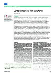
Figure 1. (A) Cartoon depiction of needlestick-distal nerve injury (DNI). (B) The innervation of the rat hindpaw. The tibial innervation territory is autonomous in the center of the hindpaw, and overlaps somewhat with the sural territory laterally (14). (Baxter, Deerfield, IL) was whittled into a flat platform and placed under the nerve while a needle bevel was inserted once entirely through the nerve with the bevel perpendicular to the nerve axis (Fig. 1A). Muscles were approximated with 5.0 silk (Ethicon, Somerville, NJ), and the skin was closed with 9 mm wound clips (Becton Dickinson, Sparks, MD).
Testing Plantar Hindpaw Sensory Function Rats were rested for at least one day after arrival and then underwent three consecutive days of sensory testing, the median of which defined the baseline. Sensory testing was performed in random order by a single investigator who was unaware of rats’ experimental groups. Before testing, rats were weighed. Gait, foot position, and color were observed, and they were habituated to the testing environment for 15–30 min. Sensation was measured by stimulating the tibial and sural innervated areas of each hindpaw through a wire mesh floor on Days 1, 3, 7, 10, and 14 postoperatively. The tibial innervated study-site was in the middle of the plantar surface proximal to the two distal tori (Fig. 1B). The sural innervated study-site was at the lateral edge of glabrous skin. Because not all rats develop abnormalities, group means of symptom severity could not fully represent the data, so the primary outcome was the prevalence of abnormality (measured as percent of tested rats). Static, low-threshold mechanosensation was measured by stimulating rats’ plantar hindpaws with a set of balance-calibrated Semmes–Weinstein monofilaments (Stoelting, Wood Dale, IL) held vertically and applied once until the filament bent. The threshold for paw withdrawal was defined as the thinnest monofilament that produced withdrawal on at least two of five consecutive trials for two consecutive filaments © 2007 International Anesthesia Research Society
1821
(15). A rat was classified as hyperalgesic at a particular site if the postoperative threshold force required to elicit paw withdrawal was decreased from the baseline threshold force by 51% or more, which in this range corresponds roughly to an increase of at least one Semmes–Weinstein filament. Punctate mechanical nociception was tested using a stopwatch to time the duration of hindpaw withdrawal after one safety-pin prick. A normal response was no withdrawal or a withdrawal too brief to time. Abnormal withdrawals comprised those lasting two or more seconds. To assess the response to cold sensation, a drop of acetone was applied to rats’ plantar hindpaw using a syringe that did not contact the rat. A normal response was no withdrawal or a withdrawal too brief to time. Abnormal withdrawals were those lasting two or more seconds. In preliminary experiments, heat sensation was evaluated by radiant heating through a transparent plastic floor (16), but abnormal hindpaw postures precluded full paw contact with the floor, and these measurements were discontinued.
Testing Hindpaw Position and Edema Rats were observed from below when they were relaxed with their weight evenly distributed over their hindpaws. Posture was rated as abnormal if one hindpaw was consistently positioned differently from the other, or differently from the hindpaws of unoperated rats. Plantar hindpaw color was assessed by visually comparing left and right plantar hindpaws from below. Hindpaw edema was assessed by gently holding the rats around the torso to elevate their front paws and applying an electronic caliper (Fisher Scientific, Hanover Park, IL) from above across each hindpaw at the base of the first digit (17).
Neuropathological Data about Axonal Injury After administering terminal pentobarbital anesthesia, one 2-mm skin punch was removed from each of the four hindpaw study sites, fixed in 2% paraformaldehyde lysine periodate (PLP), cut into 50 m vertical sections, immunolabeled against PGP9.5 (Chemicon, Temecula, CA) using standard clinical methods (18) identical to those used to study skin biopsies from CRPS-I patients (2). Axonal localization of epidermal PGP9.5 immunolabeling has been verified ultrastructurally (19). Quantitative data are obtained from the epidermis where axons individuate. Almost all PGP9.5-immunoreactive epidermal neurites are transient receptor potential vanilloid 1 (TRPV1)⫹ nociceptive small-fiber axonal endings (20). Slides were masked and randomized, then all epidermal PGP9.5⫹ neurites were quantified by a single skilled morphometrist. Our laboratory reports neurite densities per mm2 skin surface area rather than per linear mm to factor in section thickness. Rats were then injected transcardially with 0.1 mL heparin and perfused by pump with 200 mL 0.9% 1822
Needlestick Model of CRPS
saline (4°C) followed by 200 mL of freshly prepared 4% paraformaldehyde, 0.13% para-picric acid in 0.1 M phosphate buffer solution (pH 7.4, 4°C). The common sciatic and major branches were dissected and postfixed overnight in freshly prepared 5% glutaraldehyde in Sorenson’s buffer (4°C). Tibial nerve samples whose proximal ends were 5 mm distal to the site of DNI were processed through graded alcohols, propylene oxide, and epoxy resin using a Leica Lynx tissue processor and standard methods. Nerves were oriented longitudinally and polymerized overnight in epoxy resin (60°C). One-m transverse sections were cut by glass knife using a Leica UltracutR ultramicrotome. Sections were placed on coated microscope slides, stained with 1% borate-buffered toluidine blue, coverslipped, and photographed at 40⫻ using a Zeiss Axiophot microscope equipped with a Spot digital camera system.
Data Analysis Statistical significance represented P ⬍ 0.05. SAS 9.1 and Statistica 7.1 software were used. Symptom prevalence provided the primary outcomes and where feasible, symptom severity provided secondary outcomes. Means ⫾ se summarize data from groups. Four postoperative Semmes–Weinstein threshold change values that were more than 3.5 sd from group means were considered errors and excluded from analysis. Paw width, weight gain, and neurite density were compared using signed rank tests for withingroup comparisons and Wilcoxon’s ranked sum test for between-group comparisons. Categorical outcomes, such as symptom prevalence, were compared using McNemar’s tests for within-group comparisons and 2 tests for between-group comparisons. Fisher’s exact test was used when expected frequencies were less than five. Analysis of variance and Tukey’s Honestly Significant Difference Procedure were used to evaluate severity of allodynia.
RESULTS Evidence of Sensory Abnormalities Preoperative baseline threshold forces for hindpaw withdrawal from monofilament stimulation at the left (ipsilesional) tibial innervated testing site were as follows: Unoperated (94 ⫾ 19 g), sham-operated (88 ⫾ 21 g), 18G-DNI (105 ⫾ 15 g), 22G-DNI (88 ⫾ 18 g), 30G-DNI (112 ⫾ 17 g). Mean values from the other three test sites (ipsilesional-sural, and contralesional tibial-innervated and sural-innervated sites) were similar. These high values reflect the fact that many rats only met the withdrawal criteria (at least two withdrawals in five trials for two consecutive filaments) for the stiffest filaments, or did not meet them for any filament and so were assigned the 168 g value of the highest filament used (#20). At the Day-7 time point after surgery, the prevalence of hypersensitivity to low threshold mechanical ANESTHESIA & ANALGESIA
Figure 2. Group mean prevalence of static punctate mechanical hyperalgesia (⬎50% reduction in threshold for hindpaw withdrawal from Semmes– Weinstein monofilaments). No shamoperated rats developed this. In distal-nerve injury (DNI) rats, intraterritorial (tibial-innervated) and extraterritorial (sural-innervated) mechanical hyperalgesia was prevalent bilaterally.
stimuli at any one or more hindpaw site (e.g., at least a 51% reduction in threshold as compared to presurgery baseline) was 0% in unoperated (n ⫽ 8) and sham-operated (n ⫽ 4) rats, 67% among 18G-DNI rats (n ⫽ 12), 88% among 22G-DNI rats (n ⫽ 16), and 89% among 30G-DNI rats (n ⫽ 16). Figure 2 stratifies the Day-7 and Day-14 prevalences at each of the four testing sites. There were no correlations between prevalence and needle diameter at any site. Prevalence of hypersensitivity to low threshold mechanical stimuli was similar in the extraterritorial sural innervated and intraterritorial tibial innervated sites. Prevalence was almost as high contralesionally as ipsilesionally. At postoperative Day 14, the prevalence at any ipsilesional site was reduced to 58% in 18GDNI, 63% in 22G-DNI, and 51% in 30G-DNI rats. The prevalence at any contralesional site was 42% in 18G-DNI, 25% in 22G-DNI, and 19% in 30G-DNI rats. In the entire study, 57% of DNI rats met criteria for mechanical hypersensitivity at a contralesional (right) tibial or sural test site at one or more timepoints. Contralesional hypersensitivity developed virtually exclusively in rats with concomitant same-site ipsilesional hypersensitivity. In 78 instances of contralesional hypersensitivity, there were only two exceptions (2.6%). The prevalence of contralesional hypersensitivity was similar at the tibial and sural test sites, and appeared independent of needle diameter. Because CRPS patients treated by pain specialists probably represent the subset of most severely affected individuals, we performed a secondary analysis to assess prevalence of the most profound reductions in low threshold mechanosensation (⬎75% decrease from baseline at any site). A rat Vol. 105, No. 6, December 2007
with a baseline threshold of 100 g would have to have a post-DNI threshold less than 25 g to meet these criteria. On Day-7 postoperatively these criteria were met by 42% of 18G-DNI rats, 19% of 22G-DNI rats, and 70% of 30G-DNI rats. On Day-14 postoperatively, these criteria were met by 58% of 18G-DNI rats, 19% of 22G-DNI rats, and 13% of 30G-DNI rats. In another secondary analysis, group means of hindpaw-withdrawal thresholds (Fig. 3) were analyzed to permit comparison of our data to similar data collected from other rat models. This analysis was limited to rats defined as having abnormal lowthreshold mechanosensation specifically at their ipsilesional tibial study site on Day-7 postoperatively. Among 18G-DNI rats mean thresholds at the ipsilesional tibial- and sural-innervated sites were each 22 ⫾ 9 g. Thresholds at the contralesional tibial- and sural-innervated sites were 59 ⫾ 17 g and 57 ⫾ 17 g. Among 22G-DNI rats mean thresholds at the ipsilateral tibial- and sural-innervated sites were 30 ⫾ 7 g and 36 ⫾ 10 g. Thresholds at the contralateral tibial- and sural-innervated sites were 38 ⫾ 9 g and 52 ⫾ 13 g. Among 30G-DNI rats mean thresholds at the ipsilateral tibial- and sural-innervated sites were 33 ⫾ 7 g and 22 ⫾ 8 g. Thresholds at the contralesional tibial- and sural-innervated sites were 56 ⫾ 8 g and 68 ⫾ 12 g. These results also showed no correlation between severity of abnormal mechanosensation and lesion size. Prolonged withdrawal from pinprick and cold were uncommon (Fig. 4), never developed contralesionally, and among 30G-injured rats, each developed in only one rat (not the same one). Prevalence was too low to justify secondary analysis of the duration of paw withdrawal. © 2007 International Anesthesia Research Society
1823
Figure 3. Group mean changes in hindpaw withdrawal thresholds from Semmes–Weinstein monofilaments among rats defined as hyperalgesic. Data from six 18G- distal nerve injuries (DNIs), 13 22G-DNI, and eight 30G-DNI rats qualified for inclusion.
Figure 4. Prevalence of abnormal hindpaw withdrawal responses to (A) pinprick and (B) acetone stimulation; group means ⫾ asymptotic standard errors. These abnormal responses were not prevalent, and developed only in ipsilesional hindpaws.
Evidence of Neurogenic Edema Ipsilesional hindpaw widths did not change significantly from baseline in four unoperated or five shamoperated rats. A trend toward reduced paw width in 18G-injured rats (n ⫽ 4) was interpreted to reflect neurogenic muscle atrophy (Fig. 5). In DNI rats, hindpaw width measurements were statistically significantly higher than baseline in 22G-DNI (n ⫽ 8) and 1824
Needlestick Model of CRPS
30G-DNI rats (n ⫽ 8), but only 30G-DNI rats had values significantly higher than sham-operated rats (P ⫽ 0.032; Day 10), consistent with an additional neurogenic contribution. Contralesionally, hindpaw width increases were never different after DNI and sham-operation and so were considered nonspecific perioperative changes. No hindpaw skin color asymmetry was detected. ANESTHESIA & ANALGESIA
Figure 5. Analysis of hindpaw edema; group means ⫾ sem. Statistically significant differences between 30G- distal nerve injuries (DNIs) and sham-operated rats (*) were interpreted as neurogenic edema.
shown), were present in 14% of all DNI rats (42% among 18G-, 6% among 22G-, and 0% among 30Ginjured rats) at Day-7 postoperatively (Fig. 6B). These positions were tonic, independent of activity, and appeared to persist during sleep and anesthesia. The prevalence of abnormal posture at Day-7 postoperatively correlated directly with needle diameter (P ⫽ 0.001). Usually these postures gradually improved before resolving, and on Day-14 postoperatively only one 18G-injured rat with persistent profound pain behaviors still had abnormal hindpaw posture.
Pathological Severity of Small Fiber Injuries After DNI
The mean density of PGP9.5⫹ epidermal neurites in tibial innervated plantar hindpaw skin of unoperated rats (n ⫽ 5) was 802 ⫾ 72. Ten 18G-injured rats (n ⫽ 10) had 26% ⫾ 8% fewer neurites (591 ⫾ 64; P ⫽ 0.078), 22G-injured rats (n ⫽ 8) had 15% ⫾ 11% fewer neurites (678 ⫾ 85; P ⫽ 0.52), and 30G-injured rats (n ⫽ 6) had 0% ⫾ 9% fewer neurites (801 ⫾ 184; P ⫽ 0.92). Thus, the mean severity of small fiber axonal injury after 18G-needlestick closely reproduced the severity of losses described in CRPS-I patients (2). Light microscopic evaluation of cross-sections of distal tibial nerve (Fig. 7) demonstrated that, as expected, the extent of Wallerian degeneration of large myelinated fibers appeared proportional to needle diameter.
DISCUSSION
Figure 6. Analysis of ipsilesional abnormal tonic hindpaw posture. (A) A representative rat with ipsilesional lateral hindpaw elevation (circled) 10 days after 18G needlestick of its left tibial nerve. (B) Prevalence of ipsilesional abnormal tonic hindpaw posture. This appears early and usually remitted spontaneously by Day 14. Risk of abnormal hindpaw posture was directly proportional to needle diameter.
Evidence of Postural Abnormalities Behavioral explorality of lesioned rats appeared normal, and feeding was judged unimpaired because weight gain at postoperative Day 14 in all groups of DNI rats was similar to weight gain of unoperated rats (P ⱖ 0.15). Neither autotomy nor tremors were detected. Abnormal postures never developed in unoperated or sham-operated rats, or in DNI rats’ contralesional hindpaws. Ipsilesionally, abnormal postures, specifically lateral hindpaw margin elevation with paw eversion (Fig. 6A) or plantar-flexion of all digits with ambulation on the volar surface of the digits (not Vol. 105, No. 6, December 2007
This study demonstrates prospectively that a single needle puncture through one distal nerve is sufficient to cause some rats to develop abnormalities that resemble the behavioral and neuropathological abnormalities of human CRPS. Needlestick DNI reproduces aspects of CRPS interpreted by some as “nonbiological” or psychogenic, including pain-behaviors disproportionate to lesion severity, spread of pain behaviors extraterritorially and contralesionally, and dystoniclike hindpaw postures. These findings provide indirect prospective support for the hypothesis that the residual DNI found in chronic human CRPS-I patients are sufficient to be causal. If reproducible, they may provide a new model of CRPS based on needlestick, an established cause of human CRPS (21). In this study, the risk of developing several chronic pain phenotypes after nerve injury appeared stochastic and independent of the number of axons cut, over a considerable range of needle diameters. Thus, hypersensitivity to pinprick or cold (Fig. 4) was very rare after injury with all needle sizes. The prevalence (Fig. 2) and severity (Fig. 3) of mechanical allodynia, as well as the likelihood of ipsilesional and contralesional spread, appeared random with respect to needle diameter. To further investigate the effects of lesion size, we sought to compare our sensory testing results to data from other models with larger nerve injuries. This proved difficult because of methodological differences © 2007 International Anesthesia Research Society
1825
Figure 7. Light photomicrograph of cross-sections of toluidine-blue stained tibial nerve stump (5–7 mm distal from injury site) from rats euthanized 14 days after distal nerve injuries (DNIs). (A) A representative nerve from a 30G-injured rat has two areas undergoing Wallerian degeneration (arrows) and occasional degenerating fibers scattered throughout. (B) A representative nerve from a 22G-injured rat demonstrating more widespread partial Wallerian degeneration (arrows). (C) A representative nerve from an 18G-injured rat showing Wallerian degeneration of about 1/3 of axons (arrows). in lesions, sensory testing, timepoints, and data analysis. For instance, in many studies (including ours) rats that do not withdraw from any monofilament are assigned the value of the highest filament used. Because we used all of the stiffest filaments including #20, this default value was 168 g in our study. Because of the logarithmic increases in force applied by subsequent monofilaments, laboratories that do not use the thickest filaments will generate much lower default value for rats that do not withdraw from any filament. This may partly explain the much lower baseline and postinjury thresholds reported by groups that use subsets of monofilaments with much lower maximum values, e.g., 19 g, (8,22) 21 g, (23) and 51 g (24). Other groups’ use of less stringent criteria such as 1/5 withdrawals (22,23) may be another factor. To help compensate for these different baseline thresholds, we used changes from baseline as our primary outcome variable for mechanical testing. We identified several studies methodologically similar enough to compare to this current one. Hofmann et al. (25) found that their rats’ tibial-territory baseline thresholds near 70 g were reduced to about 58 g on day 13 after complete tibial nerve transection. The spared nerve injury (SNI) lesion (tibial and peroneal ligation and transection) produced monofilament hypersensitivity at the sural-innervated territory in 100% of 200 –250 g male Sprague–Dawley rats (source unspecified) in Cambridge, Massachusetts (9) but the same lesion produced hypersensitivity in only 79% of 200 g male Sprague–Dawley rats from Mo¨llegaard, Denmark (24). No matter how defined, fewer rats develop mechanical hypersensitivity after tibial nerve needlestick, particularly at the Day-14 timepoint, where there was a suggestion of decreased prevalence and resolution in some rats. Perhaps smaller axotomies cause more rapid resolution of abnormalities than do larger lesions, presumably because of the greater possibility of axonal regeneration into the distal nerve stump and target tissues. Combined behavioral/pathological study of the partial sciatic nerve transection model has linked resolution of hyperalgesia to axonal regeneration (26). 1826
Needlestick Model of CRPS
Our needlestick model permits direct comparison of the effects of lesion size with all other known variables held constant. No suture or other foreign material remains near the nerve to potentially cause inflammatory pain. Although an 18G needle has a diameter similar to a rat’s tibial nerve, examination of cross-sections of nerve distal to 18G-needlestick (Fig. 7) showed that less than half of myelinated fibers had been cut and were degenerating. This implies that many axons had been laterally displaced but not transected, despite the perpendicular needle bevel orientation. Although 18G needlestick produces a severity of PGP9.5⫹ neurite losses most similar to those of human CRPS-I, all three needle diameters studied were sufficient to produce CRPS-like abnormalities in some rats, even though they caused statistically insignificant distal losses of small fiber axons. This models the very definition of CRPS-I, “continuing pain, allodynia, or hyperalgesia with which the pain is disproportionate to any inciting event” (27). These results support the clinical observation that factors other than numbers of axons injured influence which individuals do or do not have persistent pain after a particular injury. The characteristics of the postinjury inflammation are certainly relevant, because some animal models of neuralgia are purely inflammatory, without any known distal loss of PGP9.5⫹ nociceptive axons (28). In contrast to mechanical hypersensitivity after DNI, prevalence of abnormal hindpaw posture correlated highly with needle diameter, which suggests independent mechanisms. Although skin biopsy results are similar in DNI rats and CRPS-I patients, a very different story emerged from study of postherpetic neuralgia (PHN) patients with these same methods. PHN pain after shingles was restricted to patients with profound distal axonal losses within their skin biopsies (28,29). Posttraumatic neuralgia may therefore have different mechanisms than PHN pain. This study suffers from the common limitation of inability to detect stimulation-independent pain, the single most important CRPS symptom. No rats ANESTHESIA & ANALGESIA
developed autotomy, but some licked or groomed their left paws intermittently, consistent with stimulusindependent sensations. Another limitation was a short 2-wk postoperative follow-up during which time some, but not all, symptomatic rats recovered. Abnormal hindpaw posture was most likely to remit, whereas sensory abnormalities and edema appeared slower to resolve. Pinprick hyperalgesia developed particularly slowly and was more prevalent at 14 days postoperatively than at 7 days, whereas most other abnormalities appeared to be most prevalent or severe at 7–10 days postoperatively. A preliminary long survival study of DNI rats in our laboratory suggested that even persistent pain behaviors usually resolve at about 6 mo postoperatively, in accord with epidemiological evidence that most CRPS cases resolve spontaneously (30). Models in which not all animals develop pain phenotypes after standardized injuries complicate data analysis, requiring unfamiliar analyses and exposing limitations of familiar statistics. In most rodent pain studies, central tendency (group means or medians) plus variability is the primary outcome variable for ordinal or ratio scale data pertaining to withdrawal from sensory stimuli. This accurately represents data sets in which most or all individuals change their behavior in the same direction (e.g., reduced withdrawal thresholds), as after larger or more proximal nerve injury models such as chronic constriction injury (7) or SNI (9). Even here, these analytic methods can obscure data from rare animals with different outcomes. One group (24) found a 79% prevalence of hypersensitivity to von Frey monofilaments in SNI model rats, with remaining rats’ thresholds unchanged or increased from baseline. In humans, nerve injuries produce this same trichotomy. Unchanged or increased sensory thresholds (numbness) are more common than reduced thresholds (allodynia/hyperalgesia) after nerve injuries. Prevalence data offer advantages for analysis of these complex outcomes. Prevalences are standard in human medical research but are unfamiliar and often rejected by animal researchers. Prevalences work best with dichotomous outcomes that are either present or absent (e.g., abnormal hindpaw posture). With ordinal or ratio scale data, prevalence values are highly influenced by where one arbitrarily divides normal from abnormal. Using more stringent criteria for “abnormal” will decrease its prevalence but increase its apparent severity. Clinicians and clinical researchers struggle with these same definitional issues. Although statistically significant, the evidence of neurogenic hindpaw edema after DNI (Fig. 5) was not as strong as for the other symptoms studied. We attribute this to the use of means from the entire group, because not all rats become abnormal after DNI, and to using sham-operated rather than unoperated rats as the control group. Our measurement of hindpaw width may have also been compromised by Vol. 105, No. 6, December 2007
inadequate power, neurogenic muscle atrophy, and relative lack of hydrostatic pressure in short rodent limbs. Thus we recommend other methods for future studies. CRPS is characterized by spread of pain outside the territory of individual peripheral nerves. This has led some to discount focal nerve injuries as a possible cause. But nerve injury studies in animals prove that extraterritorial pain behaviors are the rule rather than the exception (9,31). In the current study, extraterritorial (sural-site) mechanical allodynia was statistically no less prevalent than intraterritorial (tibial-site) allodynia. Spread of CRPS symptoms to the homonymous mirror location on patients’ uninjured contralesional limb is also common and poorly understood. Many animal studies document “mirror” sensory, motor, and autonomic changes after unilateral limb injuries (32). Our group has identified profound, long-lasting bilateral homonymous reductions in PGP9.5⫹ cutaneous neurites in neuralgic humans (5) and rats (33). In both cases, there were no behavioral correlates of these contralesional neuropathological changes; therefore, the relationship remains obscure. Comparing after studies suggests that mirror mechanicalhypersensitivity is more prevalent after needlestickDNI than SNI (9) or chronic constriction injury (7). Does small lesion size or partial penetrating injury enhance development of mirror symptoms? In the current study, three abnormalities never spread contralesionally; abnormal hindpaw posture, cold hyperalgesia, and pinprick hyperalgesia. Further study of CRPS-I patients is needed to determine if there are similar patterns of contralesional symptoms. Motor abnormalities are common but not required for CRPS diagnosis (34). Some are secondary (disuse weakness, atrophy, contractures) and not specific to CRPS. We investigated a primary motor symptom of CRPS, tonic limb dystonia (35), which reportedly affects 1/4 to 1/3 of patients (34,36,37). CRPS/dystonia is different from the childhood-onset dystonias or acquired, adult dystonias that usually affect proximal muscles and are caused by basal ganglia dysfunction. It also differs from limb dystonias that are phasic or activity-triggered (38). Some, but not all, CRPS/dystonias remain fixed even when the patient is asleep or anesthetized, and these can be difficult to distinguish from secondary contractures. Many movement-disorder specialists consider toniclimb dystonias psychogenic, particularly when they affect the lower limbs (39), however, study of 103 tonic-limb dystonia patients (20% of whom also fulfilled CRPS criteria) found that 45% of patients had no evidence of psychogenic causality (40). The foot eversion and toe flexion that develop in most animal models of neuralgia cannot be psychogenic, although it is unclear if dystonia rather than weakness or allodynia is the cause. If DNI rats’ hindpaw positions do model CRPS/dystonia, our current data predict that CRPS/dystonia should be more prevalent after larger © 2007 International Anesthesia Research Society
1827
than smaller nerve injuries, should be ipsilesional-only, and should often resolve spontaneously. On a final note, this model highlights the interindividual variability in pain-related responses to standardized injuries. Such responses appear to be complex genetic traits, influenced not only by the lesion, but also by intrinsic factors, both inherited (41) and acquired (42). Perhaps the variable response to needlestick injury might serve as a point of departure for identifying pain-related genetic differences within outbred rodent strains and, ultimately, in human patients. ACKNOWLEDGMENTS We gratefully acknowledge the statistical support of Yuchiao Chang, PhD, the technical assistance of Heather Downs, Ralph Gott, Li Zheng, and Yangfeng Li, and the contributions of the anonymous reviewers and editors of Anesthesia and Analgesia. REFERENCES 1. van der Laan L, ter Laak HJ, Gabreels-Festen A, Gabreels F, Goris RJ. Complex regional pain syndrome type I (RSD): pathology of skeletal muscle and peripheral nerve. Neurology 1998;51:20 –5 2. Oaklander AL, Rissmiller JG, Gelman LB, Zheng L, Chang Y, Gott R. Evidence of focal small-fiber axonal degeneration in complex regional pain syndrome-I (reflex sympathetic dystrophy). Pain 2006;120:235– 43 3. Albrecht PJ, Hines S, Eisenberg E, Pud D, Finlay DR, Connolly MK, Pare M, Davar G, Rice FL. Pathologic alterations of cutaneous innervation and vasculature in affected limbs from patients with complex regional pain syndrome. Pain 2006;120: 244 – 66 4. Kozin F, McCarty DJ, Sims J, Genant H. The reflex sympathetic dystrophy syndrome. I. Clinical and histologic studies: evidence for bilaterality, response to corticosteroids and articular involvement. Am J Med 1976;60:321–31 5. Oaklander AL, Romans K, Horasek S, Stocks A, Hauer P, Meyer RA. Unilateral postherpetic neuralgia is associated with bilateral sensory neuron damage. Ann Neurol 1998;44:789 –95 6. Lauria G, McArthur JC, Hauer PE, Griffin JW, Cornblath DR. Neuropathological alterations in diabetic truncal neuropathy: evaluation by skin biopsy. J Neurol Neurosurg Psychiatr 1998;65:762– 6 7. Bennett GJ, Xie YK. A peripheral mononeuropathy in rat that produces disorders of pain sensation like those seen in man. Pain 1988;33:87–107 8. Kim SH, Chung JM. An experimental model for peripheral neuropathy produced by segmental spinal nerve ligation in the rat. Pain 1992;50:355– 63 9. Decosterd I, Woolf CJ. Spared nerve injury: an animal model of persistent peripheral neuropathic pain. Pain 2000;87:149 –58 10. Coderre TJ, Xanthos DN, Francis L, Bennett GJ. Chronic postischemia pain (CPIP): a novel animal model of complex regional pain syndrome-type I (CRPS-I; reflex sympathetic dystrophy) produced by prolonged hindpaw ischemia and reperfusion in the rat. Pain 2004;112:94 –105 11. Guo TZ, Offley SC, Boyd EA, Jacobs CR, Kingery WS. Substance P signaling contributes to the vascular and nociceptive abnormalities observed in a tibial fracture rat model of complex regional pain syndrome type I. Pain 2004;108:95–107 12. Oaklander AL, Lee JW, Siegel SM, Downs HM. Searching for pain markers: ipsilesional and contralesional nerve morphometry in rats with or without pain behaviors after unilateral minor distal nerve injuries (MDNI). Society for Neuroscience Abstracts 2006 13. Zimmermann M. Ethical guidelines for investigations of experimental pain in conscious animals. Pain 1983;16:109 –10 14. Bajrovic F, Sketelj J. Extent of nociceptive dermatomes in adult rats is not primarily maintained by axonal competition. Exp Neurol 1998;150:115–21
1828
Needlestick Model of CRPS
15. Anseloni VC, Weng HR, Terayama R, Letizia D, Davis BJ, Ren K, Dubner R, Ennis M. Age-dependency of analgesia elicited by intraoral sucrose in acute and persistent pain models. Pain 2002;97:93–103 16. Hargreaves K, Dubner R, Brown F, Flores C, Joris J. A new and sensitive method for measuring thermal nociception in cutaneous hyperalgesia. Pain 1988;32:77– 88 17. Sharma JN, Samud AM, Asmawi MZ. Comparison between plethysmometer and micrometer methods to measure acute paw oedema for screening anti-inflammatory activity in mice. Inflammopharmacology 2004;12:89 –94 18. Lauria G, Cornblath DR, Johansson O, McArthur JC, Mellgren SI, Nolano M, Rosenberg N, Sommer C. EFNS guidelines on the use of skin biopsy in the diagnosis of peripheral neuropathy. Eur J Neurol 2005;12:747–58 19. Hilliges M, Wang L, Johansson O. Ultrastructural evidence for nerve fibers within all vital layers of the human epidermis. J Invest Dermatol 1995;104:134 –7 20. Simone DA, Nolano M, Johnson T, Wendelschafer-Crabb G, Kennedy WR. Intradermal injection of capsaicin in humans produces degeneration and subsequent reinnervation of epidermal nerve fibers: correlation with sensory function. J Neurosci 1998;18:8947–59 21. Horowitz SH. Peripheral nerve injury and causalgia secondary to routine venipuncture. Neurology 1994;44:962– 4 22. Shir Y, Zeltser R, Vatine JJ, Carmi G, Belfer I, Zangen A, Overstreet D, Raber P, Seltzer Z. Correlation of intact sensibility and neuropathic pain-related behaviors in eight inbred and outbred rat strains and selection lines. Pain 2001;90:75– 82 23. Roytta M, Weig H, Pertovaara A. Spinal nerve ligation-induced neuropathy in the rat: sensory disorders and correlation between histology of the peripheral nerves. Pain 1999;80:161–70 24. Erichsen HK, Blackburn-Munro G. Pharmacological characterisation of the spared nerve injury model of neuropathic pain. Pain 2002;98:151– 61 25. Hofmann HA, De Vry J, Siegling A, Spreyer P, Denzer D. Pharmacological sensitivity and gene expression analysis of the tibial nerve injury model of neuropathic pain. Eur J Pharmacol 2003;470:17–25 26. Lindenlaub T, Sommer C. Partial sciatic nerve transection as a model of neuropathic pain: a qualitative and quantitative neuropathological study. Pain 2000;89:97–106 27. Merskey H, Bogduk N. Classification of chronic pain: descriptions of chronic pain syndromes and definitions of pain terms. 5th ed. Seattle: IASP Press, 1994 28. Spataro LE, Downs H, Wieseler J, Watkins LR, Oaklander AL. The persistent sciatic inflammatory neurotherapy (SIN) rat model of neuropathic pain does not involve small-fiber axon damage. J Neuropathic Pain 2006;2:41–7 29. Oaklander AL. The density of remaining nerve endings in human skin with and without postherpetic neuralgia after shingles. Pain 2001;92:139 – 45 30. Sandroni P, Benrud-Larson LM, McClelland RL, Low PA. Complex regional pain syndrome type I: incidence and prevalence in Olmsted county, a population-based study. Pain 2003;103:199 –207 31. Tal M, Bennett GJ. Extra-territorial pain in rats with a peripheral mononeuropathy: mechano-hyperalgesia and mechanoallodynia in the territory of an uninjured nerve. Pain 1994;57: 375– 82 32. Koltzenburg M, Wall PD, McMahon SB. Does the right side know what the left is doing? Trends Neurosci 1999;22:122–7 33. Oaklander AL, Brown JM. Unilateral nerve injury produces bilateral loss of distal innervation. Ann Neurol 2004;55:639 – 44 34. Schwartzman RJ, Kerrigan J. The movement disorder of reflex sympathetic dystrophy. Neurology 1990;40:57– 61 35. Verdugo RJ, Ochoa JL. Abnormal movements in complex regional pain syndrome: assessment of their nature. Muscle Nerve 2000;23:198 –205 36. Veldman PH, Reynen HM, Arntz IE, Goris RJ. Signs and symptoms of reflex sympathetic dystrophy: prospective study of 829 patients. Lancet 1993;342:1012– 6 37. Birklein F, Riedl B, Sieweke N, Weber M, Neundorfer B. Neurological findings in complex regional pain syndromes—analysis of 145 cases. Acta Neurol Scand 2000;101:262–9
ANESTHESIA & ANALGESIA
38. Hochberg FH, Harris SU, Blattert TR. Occupational hand cramps: professional disorders of motor control. Hand Clin 1990;6:417–28 39. Fahn S, Williams DT. Psychogenic dystonia. Adv Neurol 1998;50:431–55 40. Schrag A, Trimble M, Quinn N, Bhatia K. The syndrome of fixed dystonia: an evaluation of 103 patients. Brain 2004;127:2360 –72
Vol. 105, No. 6, December 2007
41. Lacroix-Fralish ML, Rutkowski MD, Weinstein JN, Mogil JS, DeLeo JA. The magnitude of mechanical allodynia in a rodent model of lumbar radiculopathy is dependent on strain and sex. Spine 2005;30:1821–7 42. Shir Y, Ratner A, Raja SN, Campbell JN, Seltzer Z. Neuropathic pain following partial nerve injury in rats is suppressed by dietary soy. Neurosci Lett 1998;240:73–76
© 2007 International Anesthesia Research Society
1829