2008
19
67-71
Colon Cytomegalovirus Infection A Case Report with Rare Endoscopic Presentations Chih-Hung Chen, Chung-Mou Kuo, Tsung-Hui Hu, Seng-Kee Chuah, and Chi-Sin Changchien
Abstract Cytomegalovirus ( CMV ) infection can occur in severely immunocompromised populations, such as people suffering from acquired immunodeficiency syndrome ( AIDS ), patients receiving immunosuppressive therapy after transplantation or undergoing chemotherapy for malignanies, and long-term corticosteroid users. CMV frequently occurs in the gastrointestinal tract of such immunocompromised individuals, but only a few of them develop clinically apparent CMV disease. The gold standard of diagnosis for CMV infection is the presence of viral inclusion bodies in infected cells, after the exclusion of other viral, fungal, parasitic, and bacterial infections. CMV colitis results in lesions varying from segmental to extensive mucosal ulcerations. We report a rare endoscopic feature of severe and extensive colitis, resembling pseudo-polyp lesions, observed in a 72-year-old woman with myelomatosis who had suffered from progressive bloody diarrhea and abdominal pain for one month. Histological examination of biopsies from the ulcer bases, stained with hematoxylin and eosin ( H&E ) and an immunohistochemical stain for anti-CMV monoclonal antibody confirmed the presence of CMV inclusion bodies. The patient expired despite treatment with ganciclovir. ( J Intern Med Taiwan 2008; 19: 67- 71 ) Key Words
Cytomegalovirus ( CMV ), Severe gastrointestinal tract infection, Pseudopolyp colonic lesions
therapy, and cancer patients receiving chemotherapy
Introduction
1, 2
. Although CMV can be detected in the gastroin-
Infection with cytomegalovirus ( CMV ) has
testinal tracts of 30-43 % of the immonocompromised
been reported in immunocompromised patients with
patients, only about 7 % of them develop clinically
acquired immunodeficiency syndrome ( AIDS ), sol-
apparent CMV disease 1 . Early detection of CMV
id organ transplant recipients on immunosuppressive
antigen in infected cells by means of monoclonal an-
Correspondence and requests for reprints : Dr. Seng-Kee Chuah Address : Division of Hepatogastroenterology Department of Internal Medicine Chang Gung Memorial Hospital, Kaoshiung 123 Ta-Pei Road, Niaosung Hsiang Kaohsiung County, 833 Taiwan, R.O.C.
68
C. H. Chen, C. M. Kuo, T. H. Hu, S. K. Chuah, and C. S. Changchien
tibody E13 provides a sensitive, specific, and rapid test for CMV infections, but this means may not be 3
available in many hospitals . Therefore, a diagnosis of gastrointestinal CMV disease can be confirmed by the presence of the cytomegalic inclusion bodies with immunohistochemical staining 4. We report a case of pathologically proven CMV colitis in a multiple myeloma patient with symptoms of progressive bloody stool and abdominal pain, and a rare endoscopic feature of severe and extensive colitis resembling pseudo-polyp lesions.
Case Report This 72-year-old female had a 20 year history of
Fig.1.LGI series showing severe ir regularity and ri gidity of mucosa over the right side and transverse colon, especiall y in the hepatic flexure (arrowed) .
hypertension well-controlled with regular medication. She presented with general bone pain in June 2000, but did not pay it any further attention after symptomatic relief was provided by a non-steroidal anti-inflammatory agent. In February 2001, she began to suffer from chest tightness, shortness of breath, fever and abdominal pain, and was referred from a local hospital in southern Taiwan. Upon entering our hospital, the patient was initially diagnosed with right lower lobe pneumonia and advanced multiple myeloma , sta ge I II A ( I gG , la mb da ) ligh t ch ain . Hematochezia and diffuse abdominal pain occurred
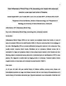
Fig.2.Severe infection of the colon, with extensive nodular sur faces and ul cer ations throughout the involved segments resembling an exacerbation of ulcerative col itis, and wi th rar e presentations of pseudo-polyps formation at the hepatic flexure site (arrowed).
10 days after admission, but the patient was reluctant
biopsies were taken from nodular mucosa to provide
to undergo colonscopic examination. However, due to
sufficient tissue for diagnosis. H&E staining of biop-
intermittent episodes of bloody stool, she finally
sies revealed the presence of cytomegalic inclusion
agreed to a lower gastrointestinal tract ( LGI ) series
bodies ( Figure 3 ), and CMV was confirmed by im-
examination, which revealed severe irregularity and
munohistochemical staining ( Figure 4 ) after exclu-
rigidity of mucosa over the right side and the trans-
sion of herpes simplex or other viral, fungal, para-
verse colon, especially the hepatic flexure ( Figure 1 ).
sitic, and bacterial infections. A complete blood count
The patient was persuaded to undergo colonscopy,
showed hemoglobin at 8.4 g/dl, hematocrit at 25.9 %,
which revelaed nodular, erythematous, and edema-
platelets at 126000/cmm and white blood cells at
tous mucosa with mild oozing extending from the pre-
5000/cmm ( neutrophils, 76.2 %; lymphocytes, 14.5 %;
cecal area to the splenic flexure, which was compat-
monocytes, 7.5 %; basophils, 1.2% and eosinophils,
ible with the LGI findings. A rare endoscopic feature
0.6%). Blood urea nitrogen, creatinine, aspartate
of severe and extensive colitis, resembling pseudo-
aminotransferase, alanine aminotransferase, alkaline
polyp lesions was, observed at the site of the hepatic
phosphatase, total bilirubin, sodium and potassium
flexure of the colonic mucosa ( Figure 2 ). Multiple
levels were within normal limits.
Rare Endoscopic Presentations of Severe Cytomegaloviral Colitis
69
risk. CMV is a member of the herpes virus family 8 . In immunosuppressed adults, CMV infection can affect the lungs, adrenal glands, liver, and to a lesser extent, the gastrointestinal tract 1 . CMV infection may arise from reactivation of latent infection, or from a 1
new infection in an immunocompromised host . The colon is the most commonly affected region of the gastrointestinal tract, but CMV can involve other regions, such as the esophagus or stomach 8 . The endoscopic morphology of our patient showed extensive nodular surfaces, ulcerations throughout the Fig.3.Bi opsies fr om the col onic l esi ons r eveal cytomegalovirus inclusion body, H&E stained ( arrowed, or iginal magnification, 400).
involved segments resembling an exacerbation of ulcerative colitis, and a presentation of pseudo-polyps formation at the hepatic flexure site ( Figure 1 ). The most commonly encountered colonoscopic features of CMV colitis are multiple ulcers with persistent inflammation and fibrotic changes followed by large, deep ulcers in the colon resulting in lumen stricture. However, presentations of pseudo-polyp formations are rarely mentioned in the literature. Such morphologic presentations suggest imply the severity of the infection producing such peculiar colonic mucosal damage 9,10. In the American population, the majority of adults have evidence of previous CMV infection,
Fig.4.Intra-nucl ear i ncl usion body of CMV in gastrointestinal tract, immunohistochemical staini ng by anti-CMV monoclonal antibody (arr owed, origi nal magnification, 400).
with 53-79 % positive for IgG antibodies 11 . However, IgM antibodies, indicating active CMV infection, are uncommon. The only reliable marker of CMV infection in AIDS patients is the presence of typical viral
The patient received 250 mg ganciclovir intra-
inclusion bodies, although they may be few in num-
venously twice daily immediately after diagnosis of
ber and atypical in appearance . Among immuno-
CMV infection was confirmed, but the treatment was
compromised patients who present with gastroin-
unfortunately too late. Her condition deteriorated and
testinal symptoms, CMV disease must be suspected
she expired 2 weeks after beginning treatment.
for a successful early diagnosis. Multiple biopsies are
Discussion
8
necessary to assist in making the pathologic diagnosis. Cytomegalic inclusion bodies may be detected
Gastrointestinal CMV infections in Taiwan oc-
with H&E stain, and confirmed by an immunohisto-
cur most often among HIV patients 5-7. However, pa-
chemical stain, after the exclusion of other viral, fun-
tients receiving immunosuppressive therapy after
gal, bacterial and parasitic infections.
transplantation or chemotherapy for malignancies,
For immunocompromised individuals, ganci-
and long-term corticosteroid users are also at high
clovir is the choice of treatment for CMV disease,
70
C. H. Chen, C. M. Kuo, T. H. Hu, S. K. Chuah, and C. S. Changchien
with a response rate of up to 83 % in gastrointestinal 12
CMV infections . Our patient's poor response to intravenous ganciclovir therapy was probably due to the delayed diagnosis and the severity of the disease. Foscarnet, a virostatic agent reported to advance a rapid resolution of symptoms and healing of ulcers in esophageal disease, is also an effective therapy for CMV dise a se o f the ga str ointe stina l tra c t1 3 . Reactivation of CMV can occur after a course of treatment, so maintenance therapy is recommended in cases of rapid relapse of CMV esophageal ulceration after the initial treatment 13. A high level of suspicion for the presence of
tomegalovirus infection using monoclonal antibody. J Formos Med Assoc 1990; 89: 199-204. 4.Reichert CM, O'Leary TJ, Levens DL, et al. Autopsy pathology in the acquired immunodeficiency syndrome. Am J Pathol 1983; 12: 357-82. 5.Yeni PG, Hammer SM, Carpenter CCJ, et al. Antiretroviral treatment for adult HIV infection in 2002: updated recommendations of the International AIDS Society-USA Panel. JAMA 2002; 288: 222-35. 6.Wei SC, Hung CC, Chen MY, et al. Endoscopy in acquired immunodeficiency syndrome patients with diarrhea and negative stool studies. Gastrointest Endosc 2000; 51: 427-32. 7. Chiu H M, Wu MS, Hung CC , e t al. Low prevalence of Helicobacter pylori but high prevalence of cytomegalovirus-associated peptic ulcer disease in AIDS patients: comparative study of symptomatic subjects evaluated by endoscopy and CD4
CMV disease is required when diagnosing immuno-
counts. J Gastroenterol Hepatol 2004; 19: 423-8. 8.Lai IR, Chen KM, Shun CT, et al. Cytomegalovirus enteritis
compromised patients with gastrointestinal symp-
causing massive bleeding in a patient with AIDS. Hepatogastro-
toms, and endoscopic biopsies must be taken from all
enterology 1996; 43: 987-91. 9.Lin WR, Su MY, Hsu CM, et al. Clinical and endoscopic fea-
gastrointestinal tract lesions to detect CMV inclusion bodies.
Acknowledgments The authors would like to acknowledge Mr. JC
tures for alimentary tract cytomegalovirus disease: report of 20 cases with gastrointestinal cytomegalovirus disease. Chang Gung Med J 2005; 28: 476-84. 10.Kelly JK, Langevin JM, Price LM, et.al. Giant and symptomatic inflammatory polyps of the colon in idiopathic inflammatory bowel disease. Am J Surg Pathol 1986; 10: 420-8.
BARTIMUS for the English revisions.
11 . Wilcox CM, D ie hl D L, Cello JP, e t a l. Cytomega lovirus esophagitis in patients with AIDS: a clinical, endoscopic and
References
pathologic correlation. Ann Intern Med 1990; 113: 589-93. 12.Buhles WC, Mastre BJ, Tinker AJ, et al. Ganciclovir treatment
1.Klatt EC, Shibata D. Cytomegalovirus infection in the acquired immunodeficiency syndrome. Arch Pathol Lab Med 1988; 112:
of life- or site-threatening cytomegalovirus infection: experience in 314 immunocompromised patients. Rev Infect Dis 1988;
540-4. 2.Gorensek MJ, Stewart RW, Keys TF. A multivariate analysis of
10: 495-504. 13.Nelson MR, Connolly GM, Hawkins DA, et al. Foscarnet in
the risk of cytomegalovirus infection in heart transplant recipients. J Infect Dis 1988; 157: 515-22.
treatment of cytomegalovirus infection of the esophagus and colon in patients with acquired immune deficiency syndrome.
3.K ao CL, Lee CN. Rapid laboratory diagnosis of human cy-
Am J Gastroenterol 1991; 86: 876-81.
Rare Endoscopic Presentations of Severe Cytomegaloviral Colitis
71
( CMV )
( inclusion body ) 72 hematoxylin & eosion stain antibody ) clovir
( anti-CMVmonoclonal ganci-