3 Clinical Symptoms of Gastric Cancer Introduction – 21 Symptomatology – 31 Conclusion
– 43
Introduction
The clinical symptoms of gastric cancer were established on the basis of the concept dominant in the 1960s, according to which the intestinal forms of gastric cancer are representative of the entire diagnostic aspects of this problem, the clinical symptoms included. We have already stated that this concept should be corrected. We plan to discuss two main questions: 1. What new can be added to the known clinical symptoms of gastric cancer? (This chapter deals with this problem.) 2. What signs of gastric cancer can currently be detected using X-ray methods? (This problem will be covered in more detail in ▼ Chap. 5.) If we describe the currently accepted clinical symptoms of gastric cancer, based on the analysis of a great number of monographs, manuals, papers, etc., we must conclude that they need serious revision and correction. This is connected with the rapidly changing morphological signs of gastric cancer. No
one today doubts that the intestinal forms of gastric cancer have switched with its diffuse and mixed forms; the latter two have become prevalent, whereas the intestinal forms have decreased in incidence accordingly. Beginning in the 1960s in connection with the development of endoscopy, which is recognized as an indispensable tool for diagnosing gastric diseases, the use of X-ray examination of the stomach has decreased substantially [31]. This has not improved the diagnosis of gastric cancer. The reason is that endoscopy is useful mainly in cases of intestinal-type gastric cancer, which is known to show actively on the surface of the gastric mucosa (⊡ Fig. 18). It is also useful in advanced cases of mixed gastric cancer, when changes in the mucous membrane are accessible for endoscopic visualization. Tissue specimens taken from such patients are suitable for histological verification of gastric cancer. Meanwhile, the diffuse forms of gastric cancer that originate in the deep parts of the mucosa develop inside the stomach wall and remain inaccessible for endoscopic visualization for a long period of time [24, 38, 98, 185].
22
Chapter 3 · Clinical Symptoms of Gastric Cancer
3
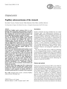
▲ Fig. 18 a.
▼ Fig. 18 b.
▲ Fig. 18 c.
▼ Fig. 18 d.
⊡ Fig. 18a–o. Female patient A., age 58. Diagnosis: gastric cancer. a Stomach X-ray (tight filling, vertical position, anterior projection): contours of the lesser and greater curvatures of the stomach body are uneven (arrows). b, c Series of X-rays (tight filling, vertical position, anterior projection): the contour of the lesser curvature is uneven and eroded (arrows). d Stomach X-ray (tight filling, vertical position, anterior projection) taken after additional portion of barium meal. The contour of the greater curvature is not changed; uneven contour of the lesser curvature (considerable depression) remains stable (arrow). e Stomach X-ray (tight filling, vertical position, left oblique projection): so-called filling defect with a small niche which does not extend beyond the stomach contour (arrow). f Stomach X-ray (double contrast, horizontal position, anterior projection): significant thickening of
23 Introduction
▲ Fig. 18 e.
▼ Fig. 18 f.
3
▲ Fig. 18 g
the lesser curvature wall depressed into the cavity, with a small depot of contrast medium (arrow). g Stomach X-ray (double contrast, horizontal position, left oblique projection): large tumor of the upper third of the stomach body, bypassed by the contrast medium (black arrows), with insignificant ulceration (white arrow). Conclusion: Cancer of the stomach body with mixed growth. The patient was examined by computed tomography to verify tumor spread.
24
Chapter 3 · Clinical Symptoms of Gastric Cancer
3
▲ Fig. 18 h.
▼ Fig. 18 i.
▲ Fig. 18 j.
▼ Fig. 18 k.
h Computed tomogram of the stomach (tight filling with E-Z-CAT DRY, supine position): distinctly visualized is thickened wall of the upper third of the stomach body; uneven inner contour (arrow). i Computed tomogram of the stomach (tight filling with E-Z-CAT DRY, supine position): The infiltrative component spreads to the lesser omentum to form a tumor conglomerate together with the regional and para-aortic lymph nodes (white arrows); an enlarged lymph node is differentiated (black arrow). j, k, l Series of CT images: distinct visualization of enlarged lymph nodes (arrow). Conclusion: gastric cancer extending onto the lesser omentum with metastases into the lymph nodes of the abdominal cavity and the retroperitoneal space. m Macrospecimen of a resected stomach: exophytic component of the tumor with insignificant ulceration. n Fragment of a macrospecimen (strip): thick gastric wall, tumor infiltration extends onto all layers; area of necrosis in center of tumor (arrows). o Macrospecimen of a resected stomach wall: moderately differentiated adenocarcinoma
25 Introduction
▲ Fig. 18 m.
▲ Fig. 18 l.
3
▼ Fig. 18 o.
▼ Fig. 18 m.
During about the same period, diffuse and mixed forms of gastric cancer started their ascending march, to slowly but steadily displace the intestinal forms of cancer. For this reason, in this and subsequent chapters we place special emphasis on the clinical symptoms of the diffuse (⊡ Fig. 19) and mixed morphological forms of gastric cancer (⊡ Fig. 20). If we evaluate the symptoms based on modern ideas, we note that the discussion concerns mainly the clinical symptoms, characterizing a whole group
of diseases of the so-called gastroenterological profile. These symptoms are almost unsuitable for the differential diagnosis of gastric cancer and non-cancerous diseases of the stomach. We have already noted that during the last decades of the past century, diffuse and mixed forms of gastric cancer became the dominant morphological forms. Accordingly, the »old« set of clinical symptoms of gastric cancer became even more useless for diagnostic purposes (⊡ Fig. 21).
26
Chapter 3 · Clinical Symptoms of Gastric Cancer
3
▲ Fig. 19 b.
▲ Fig. 19 a.
▲ Fig. 19 c.
▼ Fig. 19 d.
⊡ Fig. 19a–e. Female patient T., aged 60. Diagnosis: gastric cancer. a Stomach X-ray (tight filling, vertical position, anterior projection): lower third of stomach body irregularly narrowed by circles; the walls are rigid, the angular notch is straightened, the lesser curvature is depressed over a significant length (arrow). b Target X-ray of the stomach (tight filling, vertical position, anterior projection): stable narrowing, uneven and eroded contours. c Stomach X-ray (double contrast, horizontal position, anterior projection): circular infiltration of lower third of the stomach body; the wall of the lesser curvature at the notch and the proximal antral part is thickened due to expansion of intramural infiltration in the distal direction; the wall of the lesser curvature is thickened over a long distance because of predominant spread of the tumor in the proximal direction (arrows). Conclusion: infiltrative cancer of the antral part and the body of the stomach. d Macrospecimen of a resected stomach: part of the intramural infiltration of 3–4 cm on the greater curvature (arrows); relief of the lesser curvature with no visible changes (longitudinal folds persist). e Fragments of a macrospecimen (strips): gastric wall is thick over a long distance due to intramural infiltration (arrows). Histologically, a signet-ring cell carcinoma
27 Introduction
▲ Fig. 19 e.
▼ Fig. 20 a.
3
▼ Fig. 20 b.
⊡ Fig. 1.
⊡ Fig. 20a–g. Female patient K., age 72. Diagnosis: gastric cancer. a Stomach X-ray (tight filling, horizontal position, left oblique projection): markedly uneven contour of the anterior wall of upper third of the stomach body with an ulcer niche not extending beyond the stomach contour (arrow). b Target X-ray of the stomach (tight filling, vertical position, anterior projection): in projection of the lesser curvature of the anterior wall in the upper third of the stomach body, there is a depot of contrast medium surrounded by areas with changed relief (arrow).
28
Chapter 3 · Clinical Symptoms of Gastric Cancer
3
▲ Fig. 20 c.
▼ Fig. 20 d.
▲ Fig. 20 e.
c Stomach X-ray (double contrast, horizontal position, left oblique projection): the anterior wall of the upper third of the stomach body is thickened due to intramural infiltration (arrow). d Stomach X-ray (double contrast, vertical position, left oblique projection): infiltrative component of the tumor with ulceration (arrows). e Stomach X-ray (double contrast, vertical position, left oblique projection): pronounced thickening of the anterior wall of the body and the upper part due to intramural infiltration (arrow). Conclusion: infiltrative-ulcerative cancer of the anterior wall in the upper third of the stomach body and the upper part of the stomach. f Macrospecimen of a resected stomach: a portion showing infiltration of the anterior wall and the upper part with ulcerated surface coated occasionally with fibrin. Peripheral convergence of the folds toward smooth parts of the relief near the infiltration ridge slightly elevated over the surface of the mucous membrane (arrows).
▼ Fig. 20 f.
29 Introduction
3
▲ Fig. 20 g Fragment of a macrospecimen (strip): stomach wall is thickened due to intramural infiltration over a length of 9 cm (arrows). Histologically, an adenocarcinoma with the signet-ring cell component
▲ Fig. 21 a.
▲ Fig. 21 b.
⊡ Fig. 21a–d. Female patient B, age 46. Diagnosis: gastric cancer. Complained of weakness and epigastric pain unassociated with meals. Endoscopy: ulcerous defect in middle third of the stomach body. Histolological studies failed to reveal tumor cells. Pain persisted for about 6 months and intensified despite active outpatient treatment. In the absence of improvement, X-ray examination was done. a Stomach X-ray (tight filling, vertical position, anterior projection): ulcer niche in the lower third of the stomach body (arrow) and uneven contour of the lesser curvature in the form of a small depression in the middle third of the stomach body. b Target stomach X-ray (tight filling, vertical position, anterior projection), dosed compression: a flat niche with converging folds in the lower third of the stomach body (arrows).
30
Chapter 3 · Clinical Symptoms of Gastric Cancer
3
▲ Fig. 21 c. c Stomach X-ray (double contrast, horizontal position, anterior projection): wall of the lesser curvature is thick and rigid, with converging folds, due to intramural infiltration (arrows). Conclusion: infiltrative-ulcerous cancer of the stomach body. d Endophotograph: disfigured stomach lumen; the lesser curvature is straightened and rigid, with a rounded ulcer sized at 1 cm, with even, sloping edges. Histologically, a nondifferentiated cancer
� Fig. 21 d.
31 Symptomatology
Symptomatology What are the main clinical manifestations of gastric cancer nowadays? In other words, what should be changed and what should be added to the known clinical symptoms? In the clinical diagnosis of early gastric cancer it is necessary to consider the patient’s age and the presence of the symptom of rapid satiability while taking food. It should be noted that this symptom, which is more characteristic of advanced gastric cancers, can also occur in the early stages. True, this symptom may be discovered only by very thorough observation of the patient by a gastroenterologist or general clinician, or else by the family doctor. A further symptom is transient epigastric pain which rapidly subsides in response to the treatment of endoscopically diagnosed gastritis or peptic ulcer. It should be noted that the clinical picture of gastric cancer, which is far from being conspicuous, is now blurred by medications which suppress gastric secretion. Drugs such as Omez, Losec, Ortanol, Promezol (omeprazole) Nexium (esomeprazole, S-isomer of omeprazole), Pariet (rabeprazole), omeprazole and its analogues inhibit the H+, K+-ATPase in the parietal cells of the stomach to block the final stage of gastric juice secretion. In addition, the following important aspect should be mentioned. During recent years, the primary focus of cancer has changed its location in the stomach; the incidence of affections of the upper part have increased significantly. The situation is the same with the primary site of cancer on the greater curvature and the anterior wall. It therefore follows that in characterizing the clinical symptoms of gastric cancer these factors will be taken into consideration [38, 125]. Generally speaking, the problem of gastric cancer inevitably requires a revision of the screening strategy. Radical improvement in the early diagnosis of gastric cancer is infeasible without mass screening [144]. It is also important to note that, despite the existing opinion that the incidence of gastric cancer is decreasing, epidemiological data convincingly show that gastric cancer remains one of the leading oncological pathologies, and that in most regions of the world it constitutes 12–13% of all oncological diseases.
3
In our concise review of the clinical symptoms of gastric cancer, it is necessary to note that the centuries-old concept of a malignant tumor as a rapidly progressing pathology is unacceptable today. The concept was based on the fact that in earlier times the tumor was diagnosed at the advanced stage when its size was quite significant and the progress appeared to be »rapid«. As a matter of fact, up to a certain moment a tumor grows slowly, sometimes for 3 or 4 years, and does not extend beyond the limits of the mucous and submucous membranes, nor does it manifest itself clinically [206]. For this reason, any clinical symptom may be regarded as the late manifestation of the tumor. In order to avoid misunderstanding, it is necessary to be more accurate: The presence of clinical symptoms of gastric cancer should not be interpreted as a sign of advanced cancer. It was established that anorexia (the absence of appetite) and decreasing body weight (and some other symptoms), which were formerly regarded as symptoms specific for advanced and neglected cancer and, according to some authors, occur in 19–25% and 20–88% of patients, respectively, may appear in patients with relatively early forms of the disease [99, 131, 189, 201, 217]. The rate of tumor growth can be estimated beginning only from the moment of its discovery on; and it is impossible to tell the age of a particular tumor before this moment. There have been times when, during the clinical examination of a patient who had no complaints or any clinical symptoms that might suggest pathologies of the gastrointestinal tract, the X-ray examination discovered minor gastric cancer which had been resolutely precluded by preceding endoscopy and histological studies. A year or more later, repeated endoscopic and histological examinations confirmed the radiological diagnosis. At its earliest stages, gastric cancer does not show itself clinically for a long time. This time varies depending on the primary location of the tumor, and no one can say (even approximately) how long it may last. But it is precisely during this period that surgical treatment may result in complete cure [66]. The first clinical signs of the disease appear only when the tumor growth is so significant that it interferes with normal function of the stomach by decreasing its capacity, narrowing the gastroesoph-
32
3
Chapter 3 · Clinical Symptoms of Gastric Cancer
ageal junction, and evacuating its contents. As the tumor starts to disintegrate, the destroyed vessels begin to bleed. This, in turn, evokes gradually increasing hypochromic anemia. Absorbed products of tumor decay cause toxicity. Infection develops in the tumor and the surrounding tissues, and the bacterial toxins are also absorbed, causing specific symptoms. Nerve trunks become involved in the process and the tumor invades the adjacent organs to result in the development of an unbearable and persistent pain syndrome. Remote metastatic tumor nodes discovered accidentally in persons who are regarded as being »in full health« are sometimes the first symptoms of the disease. These nodes may be revealed on the X-ray images of thoracic organs (PA chest, for example). Sometimes, an ultrasonographic follow-up examination for some pathology which by no means is connected with the stomach reveals free fluid in the abdominal cavity, which is also an indirect sign of carcinomatosis. The search for the primary focus then reveals gastric cancer. Even when the presence of distant metastases suggests infeasibility of radical intervention, the patient’s complaints might not give grounds to suspect gastric disease. In some cases, even in the presence of specific complaints the physician does not think it reasonable to suspect a blastomatous process in the stomach, and only steadily intensifying clinical symptoms force the physician to continue the diagnostic search. The first clinical symptoms of gastric cancer are quite varied and depend on many factors, the main ones being location of the tumor in a particular part of the stomach, the nature of tumor growth, its morphological structure, involvement of the adjacent organs, and bodily dysfunctions. The common clinical symptoms are connected with the intoxication syndrome (non-motivated weakness, flaccidity, decreased working capacity, fatigue, impaired appetite, gastric discomfort, loss of body weight in the absence of visible reason, depression). These symptoms may be so insignificant that it is only their persistent character that makes the patient seek medical aid. It is easy to understand that many diseases are connected with intoxication, but the diagnostic investigation should always be conducted with the awareness of possible oncological involvement [6, 189] . While physicians should be alert, they should not exaggerate the danger of tu-
mor and prescribe X-ray or endoscopic examination to all their patients during influenza epidemics, because influenza is manifested by marked intoxication.
Epigastric Pain The most typical symptom of gastric cancer is disturbed patency of the gastrointestinal canal, which is narrowed by the growing tumor. It is evident that such disorders will be the relatively early and more pronounced signs of tumor in the cardiac and prepyloric parts, the pylorus included, because these are the most narrow portions of the stomach cavity. And these symptoms may be absent if the tumor is located in the stomach body or the fundus, especially on the greater curvature or the anterior wall of the stomach. The feeling of an overfilled epigastrium is believed to be the sign of the initial process of obstruction of the pylorus, the antral, and the prepyloric parts. In other words, this feeling is characteristic of cancer. At the same time, development of the obstructive symptoms in the presence of invasive carcinoma in the pyloric part, which causes its steno-
⊡ Fig. 22 a.
33 Symptomatology
3
⊡ Fig. 22 b.
sis, may be associated with a relatively insignificant invasion of the submucous membrane of the stomach. In addition, even in the presence of cancer infiltration over a short length, marked clinical symptoms may appear. But this relates only to tumors, the primary location of which is the pylorus proper [31, 222]. In patients with exophytic carcinoma, evacuation disorders develop mostly in cases where the tumor passes from the expansive growth stage to mixed-type cancer, which is associated with a strongly advanced process. First, the evacuation disorders may occur only occasionally, especially in severe abuse of foods, especially bitter dishes or alcohol. The disorders are especially marked in patients who suddenly stop drinking strong alcoholic drinks to replace them with soft ones. Such patients may develop obstruction all of a sudden, while they are »totally healthy«. Epigastric pain and vomiting may develop. All these symptoms may persist for several days or weeks, and then subside as a result of dietary restrictions. The patient may feel healthy for several
weeks or months, until another attack of obstruction occurs. In some patients, obstruction fails to be removed, but rather progresses steadily. Gradually increasing symptoms are more common. Even small meals cause an unsurpassable feeling of gravity in the stomach, and sometimes eructation. Voiding of gas through the mouth is explained by congestion of food in the stomach. The wind is first odorless, but later, as the congestion is characterized by putrefactive decomposition of the stomach contents, eructation becomes fetid, and the patient complains of »rotten-egg eructation«. Pyrosis (heartburn) is also not infrequent. This is due to irritation of the esophageal mucosa with regurgitated acid stomach contents. This may occur as well in the case of normal gastric secretion and also when organic acids are formed during decomposition of gastric contents. Therefore, hypoacidity patients with this complaint should undergo both an X-ray and endoscopic examination immediately, regardless of how recent their previous examination was (⊡ Fig. 22).
⊡ Fig. 22a–d. Patient D, age 54. Diagnosis: gastric cancer. Complained of overfilled stomach after eating very small meals, and of heartburn which persisted for 8 months. Slow but steady intensification of the symptoms led the patient to seek medical aid. a Stomach X-ray (tight filling, vertical position, anterior projection): the stomach contains much liquid, the antral part is disfigured, narrow, uneven walls are rigid (arrows), markedly disordered evacuation. b Stomach X-ray (double contract, horizontal position, right oblique projection): the pyloric and antral parts are disfigured and narrow, the walls are thick and rigid due to intramural circular infiltration (arrows). Conclusion: infiltrative cancer of the pyloric and antral parts of the stomach. c Macrospecimen of a resected stomach: the distal part is narrow, the walls are thick and firm (arrows). d Fragment of the macrospecimen (strip): the stomach wall is thick due to intramural infiltration (arrows). Histologically, an adenocarcinoma with the signet-ring cell component
34
Chapter 3 · Clinical Symptoms of Gastric Cancer
3
▲ Fig. 22 c.
▼ Fig. 22 d.
35 Symptomatology
If the evacuation dysfunction is severe and the stomach is not emptied, the patient starts vomiting. A typical picture of the so-called stenosed pyloric part of the stomach develops [28]. It is characterized by the patient’s relatively good condition with a fasting stomach (in the morning). But after the first meal, the patient feels epigastric discomfort and dull pain. After the next meal the discomfort and pain increases. By the evening, these symptoms intensify, becoming unbearable after supper. The patient vomits all food taken during the day, and sometimes the erupted gastric contents include residues of »old« food ingested the day before. The odor is fetid. The patient feels better. Improvement is so significant that if vomiting does not occur spontaneously, the patient tries to provoke it artificially. Despite an increasing feeling of hunger, the patient refuses much food in fear of the painful consequences, and an aversion to food may develop. Daily lavage of the stomach is indicated for such patients. After this procedure the patient feels much better, but the amounts of water and nutrients in the body remain deficient, which leads to loss of weight and dehydration. Some patients with an involved pyloric part do not develop obstruction; rather, the pylorus loses the ability to contract, and all food leaves the stomach through an open passage. The same situation can develop when a part of the pylorus wall remains uninvolved, but the pylorus does not contract and fails to close the outlet from the stomach, owing to affection of the relevant nerve endings or because of the lost reflex in the presence of disordered secretion of gastric juice and the absence of hydrochloric acid in it. The rapid passage of non-digested food into the intestine results in a permanent feeling of hunger. The patient begins to eat a large amount of food but continues to lose weight rapidly because the food is inadequately absorbed in the intestine. Stools become frequent and liquid, containing much undigested food. Involvement of the pancreas is of great importance: Digestion becomes upset not only in the stomach but also in the intestine. If the tumor affects the middle third of the stomach, especially on its anterior and posterior walls, and also on the greater curvature, the patient does not have any complaints associated with gastric dysfunction for a long time. Epigastric pain may
3
develop against the background of general symptoms. Pain develops after meals and abates after defecation. In contrast, the patient may experience pain which develops only in the fasting stomach and abates after food is ingested (just like fasting pain in duodenal ulcer). If the tumor does not grow beyond the limits of the stomach, fasting fain is either mild or completely absent. Strong permanent pain not connected with meals and radiating to the back, at times dull or acute, almost incurable by medication, is often the result of tumor propagation to the retroperitoneal organs and anatomical structures, e.g., the pancreas, solar plexus, nerve trunks. Appetite is often absent; the patient frequently develops an aversion to food in general, and sometimes to a particular food, most often to meat. Stomach capacity may decrease significantly in patients with endophytic tumors which are difficult to diagnose endoscopically, especially the linitis plastica type tumor. The rapid satiability symptom develops in such patients (35–45%). The patient feels epigastric discomfort and an overfilled stomach, with the symptoms developing following a small meal. According to patients’ complaints, they can eat only half, one fourth, or even less of the normal amount of food. If the tumor grows circularly, the stomach can be narrowed to the extent where it resembles an hourglass in scirrhous carcinoma, or it may occupy considerable space in fibrous carcinoma, to form a rigid tube, which is usually characterized by expansive infiltration along the lesser curvature reaching the esophagus. The stomach capacity decreases, evacuation of its contents from the upper part, and sometimes from the esophagus, becomes difficult. This condition develops not only if the tumor encircles the stomach to diminish its lumen, but much earlier, because the infiltrated muscular coat becomes unable to contract and interferes with the peristaltic movement of the gastric wall (⊡ Fig. 23). Symptoms of gastric involvement with tumors of this localization usually develop at late stages of the disease when the tumor becomes very large and begins to disintegrate and bleed. The symptoms of tumor bleeding (melena, hypochromic anemia) may thus become the first clinical symptoms of gastric disease.
36
Chapter 3 · Clinical Symptoms of Gastric Cancer
3
▲ Fig. 23 a.
▼ Fig. 23 b.
▲ Fig. 23 c.
▼ Fig. 23 d.
⊡ Fig. 23a–h. Patient A., age 58. Diagnosis: gastric cancer. Complained of rapid fatigue, rapid satiability after eating small meals, moderate dysphagia. Diseased for 2 years (from the time when he first felt epigastric discomfort after meals). Received outpatient treatment with no positive results. Anamnesis: the first X-ray examination revealed infiltrative cancer of the stomach. Multiple endoscopy, including the taking of 6–8 tissue specimens, failed to reveal organic involvement. Histological studies did not reveal tumor cells either. The first symptom of dysphagia had occurred about a month earlier. The patient was given another Xray examination. a Stomach X-ray (vertical position, anterior projection): symptom of gas redistribution in the stomach, the gastric air bubble is extended (arrows); b Stomach X-ray (tight filling, vertical position, anterior projection) at the moment of contrast medium passage through the gastroesophageal junction; the abdominal segment of the esophagus is irregularly narrow,
37 Symptomatology
▲ Fig. 23 e.
▼ Fig. 23 g.
3
▲ Fig. 23 f.
⊡ Fig. 1.
with uneven contours (white arrow), stomach capacity is decreased considerably, uneven contours (black arrows). c Stomach Xray (double contrast, horizontal position, anterior projection): the walls are rigid, the capacity decreased significantly (arrows), markedly accelerated evacuation, contrast medium drops straight to the duodenum; d Stomach X-ray (double contrast, horizontal position, left oblique projection) after additional intake of gas-producing mixture and barium meal: thick walls of the stomach body and the upper part; rigid due to circular infiltration (arrows). Conclusion: infiltrative cancer of the stomach spreading to the abdominal segment of the esophagus. Control endoscopy and histological examination of tumor samples failed to reveal tumor cells. e Computed tomogram of the stomach (native study in supine position) at the level of the abdominal segment of the esophagus: walls of the abdominal esophagus are thick due to intramural infiltration (arrow). f Computed tomogram of the
38
Chapter 3 · Clinical Symptoms of Gastric Cancer
stomach (native study, supine position) at the level of the upper third of the stomach body: the walls of the upper part are thick due to circular intramural infiltration (white arrow); infiltration extends to the esophagus (black arrow). Conclusion: infiltrative cancer of the stomach with spread to the abdominal segment of the esophagus. g Macrospecimen of a resected stomach: inner cavity is diminished, walls are thick and firm due to circular intramural infiltration which extends onto the esophagus (arrows). h Fragment of a macrospecimen (strip): stomach wall is thick due to intramural infiltration (arrows). Histologically, a signet-ring cell carcinoma ▼ Fig. 23 h.
3
Ulceration Clinical symptoms become more pronounced in the presence of ulceration. This helps in the differentiation of non-differentiated and signet-ring cell carcinoma from the great variety of other forms, among which infiltrative cancers dominate. The clinical picture of the disease depends mainly on infiltrative-ulcerous carcinomas, which now prevail. This, in turn, interferes with the differential diagnosis of malignant and benign ulcers. Ulcer in the anamnesis makes a clinical diagnosis even more difficult. Correct diagnostic and therapeutic tactics are therefore decisive for the prognosis. According to some authors, symptoms specific for peptic ulcer are absent [117, 177]. This means that if epigastric discomfort and pain intensify after meals, and the feeling of full stomach, nausea, and vomiting develop, this suggests a very high probability of malignancy (⊡ Fig. 24).
At the same time, the clinical picture of malignant ulcers, which often attack the lesser curvature, is not specific either, as distinct from scirrhous affections of the greater curvature and the anterior and posterior walls of the stomach. Meals intensify pain in such patients, but the feeling of a full stomach is the main symptom. The symptoms intensify at a comparatively rapid pace and, if therapy is not given in due time, the pronounced clinical picture of gastric cancer develops with prevalence of the obstructive signs [37].
Dysphagia Dysphagia is another symptom connected with difficult patency. Since dysphagia is a symptom of blastomatous affection of the cardia, which according to the topographic classification refers to the proximal part of the stomach (the most complicated part from the anatomical standpoint), it should be regard-
39 Symptomatology
▲ Fig. 24 a.
▼ Fig. 24 c.
3
▲ Fig. 24 b. ⊡ Fig. 24a–d. Patient B., age 64. Diagnosis: gastric cancer. Complained of epigastric discomfort which intensified after meals. The symptom had increased steadily during the preceding month. Nausea occurred periodically. a Stomach X-ray (double contrast, horizontal position, right oblique projection): depot of irregular shapes with converging folds on the lesser curvature. b Stomach X-ray (double contrast, horizontal position, anterior projection): after the ulcer crater is emptied of the contrast medium, thick wall of the lesser curvature with converging folds is visualized (arrow). Conclusion: infiltrative ulcerous cancer of the stomach body. c Macrospecimen of a resected stomach: firm walls, flat ulcer on the lesser curvature (arrow). d Fragments of the macrospecimen (strips): white tumor tissue underlying the ulcer crater and infiltrating the stomach wall (arrows). Histologically, an adenocarcinoma with the signet-ring cell component
40
Chapter 3 · Clinical Symptoms of Gastric Cancer
3
� Fig. 24 d.
ed together with all clinical symptoms of cancer of the upper part of the stomach. Clinical symptoms of proximal gastric cancer and the time of their development depend even more on the primary (initial) site of tumor location within the limits of this anatomical part, as compared with tumors developing in other parts of the stomach. Note that, in addition to the general clinical symptoms, an important symptom such as dysphagia develops in some patients with cancer of the upper part of the stomach only at late stages of the disease, and in some patients not at all. The idea that dysphagia is an obligatory companion of cancer of the upper part of the stomach is therefore incorrect. A tumor originating in the immediate vicinity of the cardiac sphincter very soon spreads to the abdominal part of the esophagus to cause dysphagia. At the same time, a tumor of the upper part of the stomach located at a distance from the cardiac rosette (cardioesophageal junction), e.g., on the posterior wall, the greater curvature, or the fundus, produces these symptoms either much later or not at all. Dysphagia occurs in about 89.4% of cases of proximal gastric cancer. First the patient may feel only scratching, burning, or pain when swallowing food. Passage of food through a narrowed canal becomes more difficult with time. A sudden complete obstruction of the esophagus may also be the first sign, but dysphagia usually develops when the tumor size is considerable enough to affect half of the
canal circumference or even greater. Marked dysphagia can also develop in the presence of very small tumors, owing to spasm of the stomach wall at the moment of food passage. However, sometimes dysphagia does not develop in patients with extensive disease. This may happen when the involved stomach wall becomes rigid and incapable of contraction. Food drops freely through the open tube of the esophagus in such patients (⊡ Fig. 25). Cardiac impatency develops more often in patients with tumors infiltrating the wall of the stomach. Blastomatous infiltration spreads onto the muscular coat to involve nerve endings. First this increases the sensitivity of the muscular membrane and induces mild spasms, but later, the contractility of the muscles becomes impaired. Exophytic tumors, which rarely occur now, interfere with food passage to the stomach less frequently and at later stages of the disease. Ingested food in most cases passes freely over a mushroom-like tumor, provided the latter does not affect the rosette proper, where the mucous membrane is immobile relative to the muscular coat and the tumor can obstruct the entrance to the stomach. Most often, dysphagia first manifests as a difficulty in swallowing solid food, and the patient has to drink water after each portion is ingested. Later it becomes difficult to swallow semi-liquid food. As cardiac impatency gradually increases, the portion of the esophagus located above the affected
41 Symptomatology
▲ Fig. 25 a.
▼ Fig. 25 b.
▲ Fig. 25 c.
3
▼ Fig. 25 d.
⊡ Fig. 25a–d. Patient Z., age 68. Diagnosis: gastric cancer. a Stomach x-ray (tight filling, vertical position, anterior projection): the stomach body is disfigured to a rigid tube with uneven contours (arrows). b Stomach X-ray (double contrast, horizontal position, anterior projection): walls of the body and the upper part are thick and rigid; the inner surface is tuberous. c Stomach X-ray (double contrast, vertical position, left oblique projection): posterior wall of the body and the upper part is thick due to infiltration extending to the cardia; the walls of the esophagus are rigid, the lumen is wide open (arrows). d X-ray of the stomach and abdominal segment of the esophagus (double contrast, vertical position, left oblique projection): relief of the cardiac rosette (cardioesophageal junction) is changed, cardia is wide open, the abdominal esophagus is a rigid tube owing to spreading of the intramural infiltration (arrows). Conclusion: infiltrative cancer of the stomach body and its upper part, with extension of the tumor onto the esophagus
42
3
Chapter 3 · Clinical Symptoms of Gastric Cancer
site distends to accumulate food masses, which are partly propelled to the stomach by esophageal contractions and partly evacuated by vomiting. The patient rapidly loses weight and water. The skin becomes dry and flaccid, tissue turgor decreases markedly. When tested, skin folds do not straighten for a long time. This condition may develop very soon if the tumor is found in the cardia proper. As the tumor grows, it soon obstructs the entrance to the stomach to account for the typical symptoms of cancer before development of general disorders in the patient. If the tumor is at a distance from the rosette (cardioesophageal junction), then, before the tumor closes the lumen, it affects considerable areas of the gastric wall. Ulcers often develop on the tumor, which begins to disintegrate and bleed. In view of this, long before the development of cardiac impatency, the patient develops general disorders due to chronic blood loss and intoxication with products of tumor decay and bacterial toxins. The ESR is high. The patient may experience pain owing to involvement of the nerve elements of the stomach wall or the adjacent organs. If the tumor resides in the upper part of the stomach, the patient feels retrosternal pain or pain between the shoulder blades. Very often this is interpreted as heart pain. This pain may be the result of intensified contractions of the esophagus in patients with disordered cardiac patency. As a rule, pain develops at the moment of swallowing when the esophagus must contract. These contractions may be very painful and the patient has to drink water, to take a deep breath, assume a special position, etc. If the esophagus is strongly distended above the point of obstruction, pain may be absent immediately after small portions of food are ingested. Pain develops only after a lapse of time in connection with distension of the esophagus by the accumulated food, mucus, swallowed saliva, all of which irritate the mucosa of the esophagus, causing it to contract vigorously. In such cases the patient does not associate pain with meals but relates its development to disorders of his/her cardiovascular system. Tumors with their primary location in the region of the fundus, on the posterior or anterior wall, or on the greater curvature may manifest for a long time only as general disorders. Gastric disorders may be either absent or so mild that the patient disregards
them. The disease is often identified correctly only when the tumor attacks the stomach wall close to the entrance, which manifests as dysphagia, or when pain of specific localization develops. Hiccup is the most common clinical symptom of cancer in the zone of the fundus and the supracardiac part; it is due to involvement of the diaphragm. Among other symptoms may be pain irradiating by the diaphragmatic nerve; diaphragmatic pleurisy is not infrequent either. Left-sided pleurisy occurs in tumors not only of the stomach fundus but of its body as well, which spread onto the cardiac part. This is sometimes the first sign of the disease. When located on the greater curvature, a tumor more often grows into the splenic portal. In patients with affections of the posterior wall, the tumor
▲ Fig. 26 a. ⊡ Fig. 26a–e. Patient S., age 59. Diagnosis: gastric cancer. a Stomach X-ray (tight filling, vertical position, anterior projection): stomach inner volume is diminished, the stomach is disfigured (rigid tube type); the contours are uneven. b Target stomach X-ray (tight filling, vertical position, anterior projection): the angular notch is straightened, the lesser curvature is depressed, the walls are rigid. c Stomach X-ray (double contrast, horizontal position, anterior projection): walls of the distal part and the body are thick and rigid due to circular infiltration. Conclusion: infiltrative cancer of the stomach. d Macrospecimen of a resected stomach: walls are thick due to intramural infiltration (arrow). e Fragments of the macrospecimen (strips): the wall is thickened due to the white tumor tissue (arrows)
43 Conclusion
3
spreads to the retroperitoneal space and first of all into the pancreas, sometimes also into the left kidney [158]. Of course, it should be understood that we are speaking here about neglected or advanced cancers, and that involvement of the diaphragm and the adjacent organs is more frequent in diffuse cancers than in the intestinal form.
Conclusion To conclude our survey of the symptoms which are currently acknowledged as characteristic of gastric cancer, it should be noted that the markedly high frequency of latent cancer (which is diagnosed only at its advanced stages) is due to the ▲ Fig. 26 b. prevalence of the diffuse forms. In other words, the absence of clinical symptoms is more typical of endophytic cancer of the stomach (disregarding the terminal stage of the disease). The special features of clinical symptoms of infiltrative tumors (which often develop in the absence of specific symptoms of malignancy due to tumor development in the submucous membrane and often with only microscopic signs on the mucosal surface) have been known for several centuries. The changes in the stomach wall in such cases were interpreted as the result of inflammation. Lilientaud was the first to describe this pathology (1779). Andral (1829) interpreted the discovered thickening of the stomach wall as a result of hypertrophy. This standpoint was supported by W. Brinton (1864), who gave the name of linitis plastica to the discovered pathological changes. The name was deemed quite appropriate, and it has persisted up to the present to designate new growth inside the submucous membrane, which markedly thickens the
▼ Fig. 26 c.
stomach wall (⊡ Fig. 26) [88]. If this disease runs an asymptomatic course, the tumor may spread over a significant area and become manifest only when the entire stomach has already been involved. Early diagnosis of gastric cancer in the young is especially difficult. The time from appearance of the first complaints till the moment of establishing the diagnosis in aged patients is 6 months on average, whereas for persons aged 40–45 this period extends
44
Chapter 3 · Clinical Symptoms of Gastric Cancer
3
▲ Fig. 26 d.
▼ Fig. 26 e.
45 Conclusion
3
▲ Fig. 27 c.
▲ Fig. 27 a.
▼ Fig. 27 b.
to 10 months, even if the patient visits the doctor within the first 5–10 weeks. These patients are usually subjected to an endoscopic examination which establishes the diagnosis of gastritis, and the patient is given relevant treatment, whereas the true character of their complaints is not established [45].
⊡ Fig. 27a–c. Patient T., age 19. Diagnosis: gastric cancer. Anamnesis: the patient complained of epigastric pain for about 6 months. Primary endoscopy revealed ulcerous defect on the anterior wall of the stomach. Examination of bioptates did not reveal signs of inflammation or tumor cells. The absence of improvement suggested X-ray examination. a Stomach X-ray (tight filling, vertical position, anterior projection): markedly disfigured stomach body, uneven contours, eroded at some points (arrows). b Stomach X-ray (double contrast, horizontal position, anterior projection): stomach walls are thick and rigid due to circular intramural infiltration (arrows), with some irregular rounded spots with contrast medium at the periphery (ulcers). Conclusion: infiltrative-ulcerous cancer of the stomach. c Endophotograph (following X-ray examination): 2.5-cm ulcer with rough floor covered with necrotic tissues, with thick converging folds. Infiltration into adjacent mucous membranes. Histologically, non-differentiated cancer
This situation can be explained as follows: First, the physician is not on the alert while examining the young; cancer occurs far less frequently in young patients than in the aged. But physicians should never underestimate the danger of cancer even in persons under 20 (⊡ Fig. 27). Second, most carcinomas of the young are characterized by intramural growth (in 70–75% of cases). Therefore, any patient presenting with gastrointestinal complaints must be examined by both X-ray and endoscopy [45, 53]. To complete our discussion of the clinical symptoms of gastric cancer we should return to the problem of its early diagnosis. When the diagnostic search begins only after the appearance of clinical symptoms, radical change is necessary. In most cas-
46
Chapter 3 · Clinical Symptoms of Gastric Cancer
es gastric cancer need not be diagnosed only at the clinical stage of its development; indeed, this must be done earlier, because gastric cancer belongs to the group of diseases that are diagnosed mainly by instrumental methods. The situation with early diagnosis of gastric cancer may be improved only if a complex of diagnostic tools, including a primary X-ray survey of the entire stomach and subsequent
3
endoscopy, is used. In other words, risk group patients should be examined. It must also be noted that in view of recent advances in technology, the diagnostic algorithm of patient examination for gastric cancer must include ultrasonography, computed tomography, and magnetic resonance tomography. These should be regarded as methods additional to the traditional X-ray investigation and endoscopy.