Wo m e n ’s I m a g i n g • O r i g i n a l R e s e a r c h Hassen et al. Ultrasound of Ovarian Masses
Downloaded from www.ajronline.org by 37.44.207.116 on 01/23/17 from IP address 37.44.207.116. Copyright ARRS. For personal use only; all rights reserved
Women’s Imaging Original Research
W O M E N ’S IMAGING
Kamel Hassen1 Michel A. Ghossain2 Pascal Rousset 1 Catherine Sciot 1 Danielle Hugol 3 Rafic Baddoura4 Dominique Vadrot 1 Jean-Noël Buy 1 Hassen K, Ghossain MA, Rousset P, et al.
Keywords: color Doppler imaging, ovarian neoplasm, ovary, papillary projection DOI:10.2214/AJR.10.5014 Received May 18, 2010; accepted after revision November 29, 2010. 1 Department of Radiology, Hôtel Dieu de Paris, Assistance Publique des Hôpitaux de Paris, Université Paris Descartes, 1 Pl du Parvis Notre Dame, 75004, Paris, France. Address correspondence to P. Rousset (
[email protected]). 2 Department of Radiology, Hôtel Dieu de France CHU, Université Saint-Joseph, Beirut, Lebanon. 3 Department of Pathology, Hôtel Dieu de Paris, Assistance Publique des Hôpitaux de Paris, Université Paris Descartes, Paris, France. 4 Faculty of Medicine, Hôtel Dieu de France CHU, Université Saint-Joseph, Beirut, Lebanon.
AJR 2011; 196:1444–1449 0361–803X/11/1966–1444 © American Roentgen Ray Society
1444
Characterization of Papillary Projections in Benign Versus Borderline and Malignant Ovarian Masses on Conventional and Color Doppler Ultrasound OBJECTIVE. The purpose of our study was to evaluate on endovaginal ultrasound the morphologic and color Doppler characteristics of papillary projections in benign compared with borderline and malignant epithelial stromal ovarian tumors. MATERIALS AND METHODS. A total of 283 women (mean age, 52 years; age range, 20–85 years) with 343 operated adnexal masses comprising 167 epithelial stromal tumors of the ovary with 76 tumors containing papillary projections at pathology were retrospectively studied on ultrasound. We systematically evaluated the topography of the papillary projections, the morphologic features of the largest papillary projection, and the presence or absence of color Doppler findings. All these findings were correlated with macroscopic and microscopic features. RESULTS. Ultrasound detected papillary projections in 78% of tumors. Papillary projections were disseminated in 33% of malignant, 20% of borderline, and 0% of benign tumors (p = 0.0049). The mean size of the papillary projections was 9.6, 15.7, and 35.3 mm in benign, borderline, and malignant tumors, respectively (p = 0.0007). An acute angle was present in 68% of benign tumors and an obtuse angle in 40% of borderline and 89% of malignant tumors (p = 0.0001). The surface was regular in 77% of benign tumors and irregular in 50% of borderline and 88% of malignant tumors (p = 0.0000). Calcifications were present only in benign tumors (18%). For papillary projections ≥ 10 mm, color flow was present in all malignant, in 86% of borderline, and absent in all benign tumors. CONCLUSION. Association of morphologic and vascular ultrasound findings can highly suggest the diagnosis of benign or malignant papillary projection.
A
t pathology, papillary projections in an ovarian mass are considered diagnostic of epithelial stromal tumor, commonly referred to as epithelial tumors, which can be benign, borderline, or malignant [1–4]. This statement applies to pathology because on ultrasound a clot or any other amorphous material can simulate a true papillary projection. Papillary projection is defined at pathology as folding of the proliferating neoplasmic epithelium growing over a central fibrovascular stromal core as opposed to more nondescript solid tissue [1]. Papillary projections are mainly located against the inner wall of the cyst (endocystic papillary projections) or more rarely on the septa or the outer wall of the mass (exocystic papillary projections). Although a definite diagnosis of benignity versus malignancy or borderline malignancy can only be made at microscopy, macroscopic findings of these papillary projections can highly suggest the
diagnosis [3, 4]. Papillary projections in benign cystic tumors have already been reported by different imaging techniques but, to our knowledge, no attempt was made to differentiate them from their borderline or malignant counterparts on the basis of morphologic findings [5, 6]. Thus, the primary purpose of this article was to retrospectively evaluate on ultrasound a detailed analysis of the morphologic findings of papillary projections in these tumors correlated with macroscopic and microscopic findings. We also reevaluated the color Doppler findings previously described [5, 7, 8] in the differentiation of benign versus borderline and malignant tumors. Materials and Methods From January 2003 to January 2009, with the approval of our institutional review board that waived the need to obtain patient consent, 345 consecutive women (mean age, 50 years; age range, 18–85 years) with a suspicion of an adnexal mass on a previous
AJR:196, June 2011
Downloaded from www.ajronline.org by 37.44.207.116 on 01/23/17 from IP address 37.44.207.116. Copyright ARRS. For personal use only; all rights reserved
Ultrasound of Ovarian Masses ultrasound or at pelvic examination were retrospectively studied with endovaginal ultrasound and color Doppler imaging. All ultrasound studies were made by two radiologists with more than 15 and 20 years’ experience in gynecologic ultrasound on an ATL HDI 5000 (Philps Healthcare) sonography scanner with a 5–2MHz transabdominal probe and an 8–4-MHz transvaginal probe. On color Doppler imaging, color flow was evaluated with a pulse repetition frequency between 1000 and 1500. The presence or absence of Doppler flow, arterial or venous, was identified by spectral Doppler imaging to rule out artifact. All gross and histologic examinations were done by one pathologist who specialized in gynecology or his assistant using the same standardized protocol as the search for papillary projections and their description. Two hundred eighty-three women (mean age, 52 years; age range, 20–85 years) with 343 adnexal masses underwent surgery (Table 1). Among these patients, 132 had 167 ovarian epithelial stromal tumors at pathology; 76 of 167 of these ovarian tumors had papillary projections. Papillary projections were present in 26 of 75 (35%) benign tumors, 11 of 15 (73%) borderline tumors, and 39 of 77 (51%) malignant tumors (Table 2). Papillary projections were not present in nonepithelial stromal tumors at pathology (Table 1). The ultrasound examinations of the 283 women were reviewed by two radiologists who were blinded to the pathologic results. They systematically and retrospectively evaluated together the mass on contiguous sagittal and transverse still images spaced 3–5 mm. Interobserver variation was resolved by consensus. In all 283 patients, presence or absence of papillary projections and presence or absence of flow in these papillary projections were evaluated. In a second evaluation, a more detailed study as specified in the following sections was made of tumors in which papillary projections were present at pathology.
Conventional Ultrasound For each tumor in which one or several papillary projections were detected on ultrasound and confirmed at pathology, we evaluated the following: • The characteristics of the mass, whether unilateral or bilateral, unilocular or multilocular, and its size. • Presence or absence of an echogenic structure against the wall of the cyst suggesting a papillary projection. A papillary projection was defined on ultrasound as a focal echoic structure against the inner or outer wall or septum or protruding inside a cystic portion or outside a mass. On the other hand, solid tissue was defined as an echoic structure not protruding in a cystic portion or protrud-
TABLE 1: Histology of Tumors No. of Patients
No. of Tumors
Tumors With Papillary Projections at Pathology
Epithelial stromal tumors
132
167
76
Germ cell tumors
38
46
0
Sex and stroma cell tumors
17
17
0
Endometriomas
40
56
0
Functional hemorrhagic cysts
41
41
0
Pelvic inflammatory disease
10
11
0
Paraovarian cysts and hydrosalpinx
5
5
0
283
343
76
Tumor Type
Total
ing in a cystic portion but usually of a larger size with a more or less round shape, homogeneous, or with degenerative changes (particularly necrosis). However, in some cases a clear landmark between papillary projection and solid tissue can be difficult to define. In these cases, identification of the echoic portion as solid tissue was retained. • Presence or absence of classical findings of malignancy other than papillary projections, i.e., an irregular thickened septa or irregular thickened wall, presence or absence of an irregular homogeneous or heterogeneous echogenic structure suggesting solid tissue [5, 9]. When one or several papillary projections were present, we systematically evaluated the following: • The topography of the papillary projections: localized (< 50% of the inner surface of the cyst) or disseminated (≥ 50% of the inner surface of the cyst). • The morphologic features of the largest papillary projection (when multiple in a tumor, the largest papillary projection was considered as the most representative of the tumor).
• Its size: the largest transverse diameter (i.e., parallel to the wall or the septum) was measured. • The angles with the wall of the cyst: a large base of implantation forming an obtuse angle with the wall of the cyst or a short base of implantation forming an acute angle with the wall of the cyst. If the papillary projection had an acute angle on one side and an obtuse angle on the other side, it was recorded as having an obtuse angle. When this angle could not be definitely assessed, the character of the base of implantation was classified as indeterminate. • The surface of the papillary projection: regular or irregular or indeterminate. • The presence or absence of calcification identified as a hyperechoic focus with acoustic shadowing.
Color Doppler Ultrasound The presence or absence of color flow in the papillary projections (either arterial or venous as identified by spectral Doppler ultrasound to eliminate artifact) was reported. Spectral Doppler ultrasound findings, such as resistivity index, pulsatility index,
TABLE 2: Histology of Epithelial Stromal Tumors and Papillary Projections Histology
No. of Patients
No. of Adnexal Masses
Tumors with Papillary Projections at Pathology
Serous
33
42
25
Mucinous
27
27
0
Brenner
6
6
1
Borderline serous
9
10
10
Borderline mucinous carcinoma
5
5
1
Serous carcinoma
19
32
24
Mucinous carcinoma
9
10
3
Endometrioid carcinoma
10
13
5
Clear cell carcinoma
4
4
3
Undifferentiated carcinoma
5
9
1
Composite carcinomaa
5
9
3
132
167
76
Total
aComposite carcinoma: association of different histologic type of carcinoma in the same tumor.
AJR:196, June 2011 1445
Hassen et al.
Downloaded from www.ajronline.org by 37.44.207.116 on 01/23/17 from IP address 37.44.207.116. Copyright ARRS. For personal use only; all rights reserved
and maximum systolic velocity, were not analyzed in this study because these findings have been extensively studied in the literature and are of limited value [5].
Statistical Analysis For statistical analysis, borderline and carcinoma findings were placed in one group to have enough values to compute the different tests. For size, we compared papillary projections < 10 mm versus papillary projections ≥ 10 mm. For angles, we compared acute angles versus obtuse and indeterminate angles. For surface, we compared regular surface versus irregular and indeterminate surface. For location, we compared nondisseminated versus disseminated papillary projections. The following tests were performed: chi-square test with a p value of < 0.05 considered statistically significant; sensitivity, specificity, positive predictive value (PPV), negative predictive value (NPV), and accuracy with evaluation of the 95% CI; and multivariate analysis.
Results Characteristics of the Mass Among the 76 tumors with papillary projections, ultrasound detected papillary pro-
es and thin and regular in one case. The wall was regular in all cases. Solid tissue was present in none of the cases. Finally, classic findings of malignancy other than papillary projections were present in two tumors. Among the 22 benign tumors with papillary projections, 15 serous tumors were bilocular or trilocular, with thin and regular septa and wall in all cases. Associated solid tissue was present in none of these cases. Classic findings of malignancy other than papillary projections were present in none of 22 tumors.
jections in 59 tumors (22 benign, 10 borderline, and 27 malignant). The size of these tumors ranged from 2.5 to 24.0 cm (mean, 8.3 cm): 2.5 to 24.0 cm (mean, 8.4 cm) in benign tumors; 4.0 to 9.0 cm (mean, 6.2 cm) in borderline tumors, and 4.0 to 15.0 cm (mean, 8.3 cm) in malignant tumors. Benign tumors were bilateral in four patients and unilateral in 14 patients. Borderline tumors were bilateral in one patient and unilateral in eight patients. Malignant tumors were bilateral in six patients and unilateral in 15 patients. Among the 27 carcinomas with papillary projections, associated irregular solid tissue was observed in seven (26%) of 27 tumors; irregular thickened wall was present in two (7%) of 27 tumors; and septa were thickened and irregular in two tumors, one of which had solid tissue. Finally, classic findings of malignancy other than papillary projections were present in 10 (37%) of 27 tumors, three of which had papillary projections < 10 mm. Among the 10 borderline tumors with papillary projections, the tumors were unilocular in seven, multilocular in two, and bilocular in one. Septa were thick and irregular in two cas-
Detection of Papillary Projections Among 343 masses of all kinds, ultrasound detected endocystic papillary projections in 59 of 76 (78%) epithelial tumors with papillary projections. Among the 267 ovarian tumors without papillary projection at pathology, ultrasound falsely diagnosed papillary projections in 11/267 (4%) adnexal masses. Finally, ultrasound detected papillary projections with a sensitivity of 78%, a specificity of 96%, a PPV of 84%, an NPV of 94%, and an accuracy of 92%). The morphologic findings of detected papillary projections are shown in Table 3.
TABLE 3: Morphologic Findings of Papillary Projections in Benign, Borderline, and Malignant Tumors No. of Lesions Tumor Characteristic
Performance Value
Benign (n = 22)
Borderline (n = 10)
Malignant (n = 27)
≤5
7
2
2
6–9
6
1
1
10–14
4
0
1
15–19
4
4
0
≥ 20 mm
1
3
23
Size (mm)
Angle Acute
15
5
1
Obtuse
3
4
24
Indeterminate
4
1
2
Surface Regular
17
5
3
Irregular
1
4
21
Indeterminate
4
1
3
22
8
18
0
2
9
Dissemination Not disseminated Disseminated
Odds Ratio (95% CI)
p
7.46 (1.89–29.43)
0.0007
11.07 (2.51–48.84)
0.0001
12.33 (2.68–56.60)
0.0000
ND
0.0049
Calcificationa Present
4
0
0
Absent
18
10
27
Note—ND = odds ratio could not be estimated because no benign lesion had disseminated papillary projections but the p value was significant. aThe number of calcifications was too small to compute the chi-square test.
1446
AJR:196, June 2011
Downloaded from www.ajronline.org by 37.44.207.116 on 01/23/17 from IP address 37.44.207.116. Copyright ARRS. For personal use only; all rights reserved
Ultrasound of Ovarian Masses
A
B
C
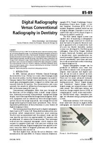
Fig. 1—40-year-old woman with malignant papillary projections in serous cystadenocarcinoma. A, Endovaginal ultrasound image shows 7-cm cystic ovarian mass containing endocystic papillary projections, largest one > 2 cm, with broad base of implantation forming obtuse angle with inner wall and irregular surface. Cystic content is uniformly low echoic. B, Color Doppler ultrasound image shows large vessels at base of papillary projection. C, Photograph of surgical specimen shows multiple papillary projections, some of which are confluent. Scale is cm.
Among the 76 tumors with papillary projections at pathology, papillary projections were not detected on ultrasound in 17 (22%) of 76 tumors: four benign, 12 malignant, and one borderline. In the four benign tumors, the size of papillary projections was ≤ 2 mm. These four tumors were unilocular cysts with a thin wall and without solid tissue, so that these tumors were preoperatively considered benign tumors. In the one borderline and 12 malignant tumors, endocystic papillary projections could not be visualized in 12 tumors in which the size of papillary projections was ≤ 2 mm and in one tumor in which the size was 5 mm; moreover, in these 13 tumors, exocystic papillary projections with size ≤ 5 mm were invisible in six tumors. It must be noted that in all these 13 cases, malignant irregular solid tissue was present, and therefore these cases were considered malignant preoperatively. Among the 267 ovarian tumors without papillary projection at pathology, papillary projections on ultrasound were falsely identified in 11 (4%) of 267 cases. These cases were related to a clot in six endometriomas
A
and one functional cyst, accumulation of mucin in one mucinous cystadenoma, a small portion of normal parenchyma protruding in a serous cyst, a small echogenic portion in a dermoid cyst, and a tubal fold in a hydrosalpinx falsely considered as an ovarian cyst. Color flow was absent in all these 11 echoic structures falsely considered as papillary projection. Morphologic Findings of Papillary Projections Morphologic findings and topography of the largest papillary projection assessed in 59 tumors (22 benign, 10 borderline, 27 malignant) (Figs. 1–3) showed that dissemination, size, wide angle, and irregular surface were indicative of malignancy (p values in Table 3). Calcification was indicative of benign lesions, but we did not have sufficient data to compute the chi-square test. Color Doppler Findings in Papillary Projections Color Doppler findings were present in 30 of 37 borderline and malignant tumors and absent in all 22 benign tumors, with sensitivity of 81% (95% CI, 64.8–92.0%), specificity of 100% (95% CI, 84.6–100%), PPV
B
of 100%, NPV of 76%, and an accuracy of 88%. Color flow was absent in all papillary projections < 10 mm whether benign, borderline, or malignant (Figs. 1–3). For the assessment of tumors containing papillary projections ≥ 10 mm, color Doppler findings were absent in nine of nine (100%) benign tumors. Conversely, color Doppler findings were present in 24 of 24 malignant tumors and in six of seven borderline tumors, with sensitivity of 97% (95% CI, 83.3– 99.9%), specificity of 100% (95% CI, 66.4– 100%), PPV of 100%, NPV of 90%, and an accuracy of 97%. Multivariate Analysis Three variables were dropped from the multivariate analysis because they were fully predictive of the outcome of interest—that is, malignancy (presence of color Doppler flow in the papillary projection and dissemination of papillary projections ≥ 50% of the surface) or benignity (presence of calcifications in the papillary projection). Therefore, we performed a logistic regression test with malignancy as the binary dependent variable,
Fig. 2—20-year-old woman with serous cystadenoma. A and B, Endovaginal color Doppler ultrasound image (A) and photograph of surgical specimen (B) show 4-cm ovarian cyst with small group of four papillary projections, measuring less than 5 mm at base of implantation. Papillary projections form acute angle with inner wall of cyst. No color flow is detected in papillary projections. Scale is cm.
AJR:196, June 2011 1447
Downloaded from www.ajronline.org by 37.44.207.116 on 01/23/17 from IP address 37.44.207.116. Copyright ARRS. For personal use only; all rights reserved
Hassen et al.
A
B
C
Fig. 3—58-year-old woman with serous cystadenoma. A, Endovaginal ultrasound image discloses right cystic ovarian mass of 6 cm containing 2.5-cm endocystic papillary projection with broad base of implantation and obtuse angle. B, Color Doppler ultrasound image shows no vessel is detected in this papillary projection, which measured greater than 1 cm. C, Photograph of surgical specimen shows that papillary projection seen on ultrasound corresponds to group of gathered small endocystic benign papillary projections. Scale is cm.
and three independent binary variables significantly associated with malignancy in univariate analysis were included in the model: size of the papillary projection (using a threshold of 10 mm or more), angle within the cyst (obtuse or indeterminate versus acute), and aspect of the surface of the papillary projection (irregular or indeterminate versus regular). Using a backward approach, only size ≥ 10 mm and irregular or indeterminate surface remained significant predictors of malignancy, with odds ratios (95% CI) as 8.35 (1.85– 37.61) and 13.46 (3.15–57.52), respectively. Because color Doppler flow was negative in all 19 subjects with size of papillary projections < 10 mm, we looked more specifically at this subgroup. None had a dissemination ≥ 50% of the surface. The presence of one of the two remaining characteristics, i.e., irregular surface and an obtuse angle, was more common in the presence of malignancy, with odds ratios of 4.15 (0.42–40.79) and 1.21 (0.12–11.99), respectively, although not statistically significant. Discussion As has already been reported, papillary projections can be missed on ultrasound [10] and MRI [2], mainly because of their small size (< 5 mm). Papillary projections can also be missed because of their exocystic location, as in six (8%) of the 76 cases in our study; however, in these cases, these exocystic papillary projections were associated with endocystic papillary projections. On the contrary, echogenic structures against the inner wall of the cyst can be falsely considered as papillary projections [11]. In our study, the main source of false-positive findings was amorphous material, essentially clots; however, vascularized structures, es-
1448
pecially tubal fold in a hydrosalpinx, can also be confused with an endocystic papillary projection. No color flow was detected in these false-positive cases, but this is not sufficient to exclude papillary projections because color flow may be absent in benign, borderline, and even small malignant papillary projections. On the other hand, although not present in this series, tubal folds in a hydrosalpinx or protrusion of normal ovarian parenchyma may show exceptional color flow. Although all patients with a cyst containing papillary projections will likely undergo surgical removal of the cyst [2], preoperative assessment of a tumor or a papillary projection as benign, borderline, or malignant is fundamental, particularly in young women. This will help the surgeon to decide the type of surgery (cystectomy, ovariectomy, or adnexectomy) and the surgical approach (laparoscopy or laparotomy through Pfannenstiel or midline incision). Although several scoring systems and models for masses containing papillary projections have been suggested to characterize papillary projections [12, 13], this study shows that on conventional ultrasound, topography, size, and morphologic findings correlated with macroscopic findings, and presence or absence of color flow can be used to characterize papillary projections. Benign epithelial tumors have fewer papillary projections than borderline or malignant masses [1, 3, 4, 14]. In our study, disseminated papillary projections (covering ≥ 50% of the surface of the tumor) were present in 0% of benign, 25% of borderline, and 50% of malignant tumors. At pathology, benign epithelial tumors show smaller papillary projections than borderline
or malignant masses [1, 3, 4]. However, there is an overlap of sizes, and our data outline the problem of choosing a threshold. If a threshold < 10 mm was chosen for benignity, there would be 13 (59%) of 22 benign, three (30%) of 10 borderline, and three (11%) of 27 carcinomas. However, in these three cases of malignant tumors, the small papillary projections were associated with solid irregular tissue, and these tumors would be correctly classified as malignant on the basis of the solid irregular tissue. The problem would be with the three borderline tumors. If a threshold ≤ 5 mm was chosen, there would be still a problem with two borderline tumors. If a threshold > 20 mm was chosen for malignancy, papillary projections would be found in 23 (85%) of 27 malignant tumors, in three (30%) of 10 borderline tumors, and in only one (5%) of 22 benign tumors. The angle of the papillary projection with the wall could not be assessed as acute or obtuse in seven (12%) of 59 tumors. In the other cases, an acute angle was more common in benign tumors and an obtuse angle in malignant tumors, whereas no significant difference was found in borderline tumors (Table 3). As reported by Krigman et al. [1], the lining of the surface of benign papillary projections is usually smooth, whereas papillary projections of malignant tumors have highly irregular angular margins. These findings could not be clearly defined in eight (14%) of 59 tumors in our series. As for the angle in the other cases, there was a difference for benign and malignant tumors but not for borderline (Table 3). Although calcifications have been reported in carcinoma at pathology [1], calcifications of a sufficient size to be detected on ultrasound
AJR:196, June 2011
Downloaded from www.ajronline.org by 37.44.207.116 on 01/23/17 from IP address 37.44.207.116. Copyright ARRS. For personal use only; all rights reserved
Ultrasound of Ovarian Masses were only encountered in four of 22 benign tumors and never in borderline and malignant tumors in our series. Although the sensitivity of this finding was low (18%), its specificity was high (100%) for prediction of a benign lesion. Although papillary projections in benign tumors have a loose stromal fibromatous core, malignant tumors have intricate papillary projections with a central fibrovascular core [1]. Because of the presence or absence of angiogenesis, size and number of vessels are substantially more common in malignant than in borderline or benign tumors [5]. The size of a structure is also an important factor that can influence the detection of Doppler flow [15]. This study and a previous study [5] show the absence of color flow in all papillary projections < 10 mm and in all benign papillary projections, even in those ≥ 10 mm. Conversely, color flow was present in all malignant and borderline papillary projections ≥ 10 mm except in one borderline tumor in this study. Association of morphologic findings and color Doppler findings seems to be the best method to characterize the type of papillary projections. When size of the papillary projection is ≥ 10 mm, the PPV for malignancy is 77.5%. If, in addition, color Doppler flow is detected or dissemination ≥ 50% is present, then the PPV is 100%. If size of the papillary projection is ≥ 10 mm but neither Doppler flow nor dissemination ≥ 50% is present, an irregular surface significantly increases the likelihood of malignancy. When the size of the papillary projection is < 10 mm, the corresponding NPV is 68.4% regardless of the other determinants of malignancy defined in our method, i.e., an irregular thickened septa or wall and an irregular solid component. Excluding subjects with such determinants of malignancy, the NPV is 86.7%. It is worth noting that among subjects with papillary projections < 10 mm, dissemination ≥ 50% was always absent. In this group, an irregular surface or obtuseindeterminate angle favored malignancy, although the association was not statistically significant because of the small sample size. Our study has limitations. The study is retrospective and purely descriptive. The use of
still images may limit the descriptive accuracy of some findings, such as angle and dissemination, but this is tempered by the use of contiguous images obtained by an experienced radiologist aware of the importance of these features. A large prospective study combining the different morphologic and vascular findings defined as suggestive of benign versus malignant tumor is needed to provide more elaborate statistical results in the characterization of papillary projections. Only surgically proven masses were included in this series. Thus, all functional hemorrhagic cysts that disappeared on follow-up were excluded, limiting the falsepositive diagnosis of papillary projections because of clots that can occur in practice. Exocystic papillary projections are present in as many as 70% of cases [3, 4] in borderline serous tumors. However, these papillary projections, which were present in six borderline serous tumors of our series, were overlooked on ultrasound. In conclusion, this study shows that a certain number of morphologic and Doppler findings can highly suggest the diagnosis of benign versus malignant papillary projection, although an overlap exists, especially with borderline tumors. Large size, acute angle, irregular surface, and disseminated pattern are suggestive of malignancy whereas calcification is suggestive of benignity. Absence of color flow in a papillary projection ≥ 10 mm is suggestive of benignity. These findings will help the surgeon decide the type of surgery and the surgical approach. References 1. Krigman H, Bentley R, Robboy SJ. Pathology of epithelial ovarian tumors. Clin Obstet Gynecol 1994; 37:475–491 2. Outwater EK, Huang AB, Dunton CJ, Talerman A, Capuzzi DM. Papillary projections in ovarian neoplasms: appearance on MRI. J Magn Reson Imaging 1997; 7:689–695 3. Scully R, Young R, Clement P. Tumors of the ovary, maldeveloped gonads, fallopian tubes and broad ligament, 3rd ed. Washington, DC: Armed Forces Institute of Pathology, 1998:51–168 4. Seidman J, Russell P, Kurman R. Surface epithelial tumors of the ovary. In: Kurman RJ, ed.
Blaustein’s pathology of the female genital tract. New York, NY: Springer-Verlag, 2002:791–904 5. Buy JN, Ghossain MA, Hugol D, et al. Characterization of adnexal masses: combination of color Doppler and conventional sonography compared with spectral Doppler analysis alone and conventional sonography alone. AJR 1996; 166:385–393 6. Saini A, Dina R, McIndoe GA, Soutter WP, Gishen P, deSouza NM. Characterization of adnexal masses with MRI. AJR 2005; 184:1004–1009 7. Guerriero S, Alcazar JL, Coccia ME, et al. Complex pelvic mass as a target of evaluation of vessel distribution by color Doppler sonography for the diagnosis of adnexal malignancies: results of a multicenter European study. J Ultrasound Med 2002; 21:1105–1111 8. Timmerman D, Testa AC, Bourne T, et al. Simple ultrasound-based rules for the diagnosis of ovarian cancer. Ultrasound Obstet Gynecol 2008; 31: 681–690 9. Buy JN, Ghossain MA, Sciot C, et al. Epithelial tumors of the ovary: CT findings and correlation with US. Radiology 1991; 178:811–818 10. Ekerhovd E, Wienerroith H, Staudach A, Granberg S. Preoperative assessment of unilocular adnexal cysts by transvaginal ultrasonography: a comparison between ultrasonographic morphologic imaging and histopathologic diagnosis. Am J Obstet Gynecol 2001; 184:48–54 11. Laing FC. US analysis of adnexal masses: the art of making the correct diagnosis. Radiology 1994; 191:21–22 12. Ameye L, Valentin L, Testa AC, et al. A scoring system to differentiate malignant from benign masses in specific ultrasound-based subgroups of adnexal tumors. Ultrasound Obstet Gynecol 2009; 33:92–101 13. Valentin L, Ameye L, Jurkovic D, et al. Which extrauterine pelvic masses are difficult to correctly classify as benign or malignant on the basis of ultrasound findings and is there a way of making a correct diagnosis? Ultrasound Obstet Gynecol 2006; 27:438–444 14. Thurnher SA. MR imaging of pelvic masses in women: contrast-enhanced vs unenhanced images. AJR 1992; 159:1243–1250 15. Ramos I, Fernandez LA, Morse SS, Fortune KL, Taylor KJ. Detection of neovascular signals in a 3 day Walker 256 rat carcinoma by CW Doppler ultrasound. Ultrasound Med Biol 1988; 14:123–126
AJR:196, June 2011 1449