50 Celiac Disease Dascha C. Weir, MD Ciaran Kelly, MD
Celiac disease (CD) is an immune-mediated enteropathy secondary to permanent sensitivity to wheat gluten and related proteins in rye and barley. It results in characteristic histologic changes consisting of inflammation, crypt hyperplasia, and villous atrophy of the small intestine in genetically susceptible individuals. Significant variability in the clinical presentation of CD in the pediatric population complicates recognition of the disease in many patients. Treatment for CD consists of a lifelong strict gluten-free diet (GFD). Adherence to this diet is associated with resolution of most related signs and symptoms and a decreased risk of related complications. With an explosion of new knowledge over the span of two decades, our understanding of CD has changed dramatically. CD has gone from a rare condition causing gastrointestinal symptoms in children of European origin to a common disorder causing symptoms that affect multiple organ systems in all ages virtually worldwide.1 The overall prevalence of CD is similar in Europe and North America affecting up to 1% of the population.2,3 A large multicenter study in the United States, using serologic screening with biopsy confirmation to identify cases of CD, showed a prevalence of CD of 1:133 in individuals with no evident risk factors. Prevalence of CD in symptomatic patients was 1:56. The prevalence of diagnosed CD is much lower, especially in the United States. This reflects underrecognition and underdiagnosis related to the wide clinical spectrum of disease and the presence of silent disease.4 In the United States, the prevalence of CD in people of non-European descent is not known. Limited studies suggest that, in the United States, Hispanic and AfricanAmerican populations have a lower prevalence of CD than non-Hispanic Caucasians. For example, a study in Colorado showed that the incidence of positive serology in Hispanic children was three times lower than in non-Hispanic Whites.5 African-Americans also represent a small number of patients currently diagnosed with CD. A large center in New York City reported that African-Americans comprised 1.3% of their patients with biopsy-proven CD.6 This is likely partially because of genetic differences in these populations. However, given the increasing recognition of CD in non-Caucasian populations internationally, serious consideration should be Compliments of AbbottNutritionHealthInstitute.org
given to the possibility of underdiagnosis in minority populations in the United States. CD is being increasingly diagnosed in other parts of the world, involving populations that were not traditionally thought to be affected by the disease. Reports from Mexico, South America, North India, the Middle East, Turkey, and North Africa have changed previous assumptions that CD is a European affliction.7–15 CD is rare in people of Japanese, Chinese, and purely AfricanCaribbean descent.16 CD is associated with several autoimmune conditions and genetic disorders and often presents atypically or silently in these populations. In children, it is associated with autoimmune thyroid disease and with type I diabetes mellitus with a 7.8 and 4.5% prevalence of CD in affected children.17,18 Up to 7% of patients with a selective IgA deficiency also have CD.19 Patients with Trisomy 21, Turner syndrome, and Williams syndrome have also been found to be at higher risk of CD than the general population.19–22 PATHOGENESIS Environmental, immunologic, and genetic factors are all important contributors to the pathogenesis of CD. Clearly, enteric exposure to specific proteins in wheat, rye, and barley are essential to disease activation in a genetically susceptible person. Wheat, rye, and barley have common ancestry and are all derived from the Triticeae tribe of the grass family. Broadly termed “gluten,” the specific
proline- and glutamine-rich proteins that activate disease are gliadins in wheat, secalins in rye, and hordeins in barley. Gluten, the second major protein fraction of wheat gluten, is likely involved to a lesser degree. Multiple toxic antigenic epitopes have been described (Figure 1). These peptides activate immune cells causing a chain of events that result in tissue damage. The mechanisms for this activation are incompletely understood and are likely a combination of adaptive and innate immunity. Partially because of their high proline content, these large cereal peptides are resistant to digestion by intestinal proteases and pass through the epithelium and into the lamina propria in an intact state. The mechanisms of this passage through the intestinal barrier are not well understood. Tissue transglutaminase deamidates these peptides in the lamina propria causing them to become negatively charged as the glutamine residues are converted to glutamic acid. Deamidated peptides bind with far greater affinity to specific positively charged residues on the peptide-binding groove of HLA-DQ2 or HLA-DQ8 major histocompatibility complex (MHC) Class II molecules. These peptides are then presented by HLA-DQ2 or HLA-DQ8 to activate gliadin-specific CD4� T cells thereby activating an intestinal inflammatory response leading to the characteristic histopathologic changes of CD. Intraepithelial lymphocytes, T cells that are markedly increased in the intestinal biopsies of patients with active CD, are also likely involved. It is now evident that gluten can
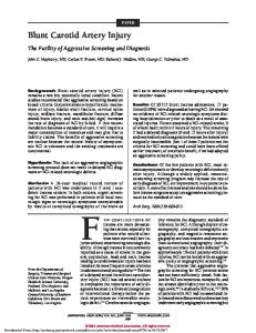
Gramineae (Poaceae) The grass family
Subfamily
Tribe
Festucoideae
Zizaneae
Wild rice
Oryzeae
Rice
Hordeae Aveneae (Triticeae)
Wheat Rye Barley
Oat ?
Festuceae Chlorideae
Finger millet (Ragi)
Teff
Figure 1 The grass family: Grains in the tribe Triticeae mediate celiac disease. Species of interest in the Festucoideae subfamily of the grass family are depicted. It seems likely that, for most patients with celiac disease, only grains in the tribe Triticeae mediate celiac disease. (Adapted from reference 23.)
With permission from Duggan C, et al. Nutrition in Pediatrics. 4th ed. Hamilton, Ontario, Canada: BC Decker Inc; 2008.
562
PART V / Nutritional Aspects of Specific Disease States Dietary gluten peptides Permeability increase
Intestinal epithelium
Basement membrane Recognition by PRR and release of IL-15
Gluten peptide deamidation and crosslinking by tTG
Release and activation of tTG
Antigen presentation via HLA-DQ2/8 CD4+ T cell
APC
Mature dendritic cell
Gluten peptide Deamidated gluten peptide Tissue transglutaminase (tTG) Pattern-recognition receptors (PRR)
Lamina propria
IL-15
Antibodies to gluten and tTG
TH1 cytokines
Mucosal destruction and epithelial apoptosis
TH2 cytokines
B cell
Figure 2 Adaptive and innate immune system responses in celiac disease. Cross-linking of gluten by tTG potentiates presentation of peptides by antigen-presenting cells (APC); deamidation of gluten improves binding to HLA-DQ2 or HLADQ8, triggering adaptive T-cell responses (inflammation and tissue remodeling (Th1) or antibody production (Th2)). Cells of the innate system recognize gluten peptides directly via pattern recognition receptors, and release cytokines (IL-15) that drive adaptive responses. Dendritic and T cells circulate to mesenteric lymph nodes (where they encounter T cells) and back to lamina propria. (Adapted from reference 26.)
stimulate interleukin-15 production, leading to the transformation of some T cells into natural killer cells and ultimately to enterocyte destruction (Figure 2).1,24,25 Genetics are clearly a factor in the development of CD. Among monozygous twins, there is a 70 to 75% CD concordance rate and firstdegree relatives have a 10% risk of disease.27,28 CD is strongly associated with HLA class II genes that map to the DQ locus. The HLA-DQ2 heterodimer is expressed in up to 95% of celiac patients with most of the remaining 5% expressing HLA-DQ8 heterodimer. These HLA types are present in 30% of the general population. Additionally, CD is concordant in only 30 to 40% of HLA-matched siblings. Clearly, these HLA types are necessary but not sufficient for CD to develop and other genes are likely to be involved. Candidate susceptibility genes include MY09B (myosin IXB) and regions of chromosome 5, 6, 11, and 19.1,29 Investigation in this area is ongoing. CLINICAL PRESENTATION Traditionally, CD was thought to present primarily with gastrointestinal symptoms in the young. This “classical” presentation consists of poor growth, chronic diarrhea, abdominal distention, muscle wasting, poor appetite, and irritability typically seen in patients 6 to 24 months of age. Edema, pallor, and emesis are also seen. Rarely, children can present in celiac crisis with copious watery diarrhea, marked abdominal distension, dehydration, electrolyte imbalances, hypotension, and lethargy.30 As serologic testing and our understanding of CD have improved, “nonclassical” or “atypical” presentations of CD are being increasingly recognized. In pediatric patients, this Compliments of AbbottNutritionHealthInstitute.org
form of presentation is characterized by delayed onset with gastrointestinal complaints, such as nausea, bloating, recurrent abdominal pain, or constipation.19 Many patients have no gastrointestinal complaints but have extraintestinal manifestations such as short stature, osteopenia, joint pains, pubertal delay, iron-deficiency anemia, dental enamel defects, neurologic disorders, or elevated transaminases. Some patients, with classic histologic changes on intestinal biopsy, are entirely asymptomatic and are described as having “silent” CD.16,30 CD is being increasingly diagnosed in adults. In fact, the mean age of diagnosis is now in
midadult life (circa 45 years of age).1 Many of these patients have likely had CD since childhood. Adults can present with gastrointestinal symptoms such as diarrhea, flatulence, abdominal bloating, and discomfort but often present with extraintestinal manifestations such as iron-deficiency anemia, macrocytic anemia, osteopenia, malaise, infertility, obstetrical complications, and neurologic conditions. Dermatitis herpetiformis, a pruritic blistering skin disorder, is also seen with CD but rarely occurs in pediatric patients (Table 1 and Figure 3).16,30 DIAGNOSIS While the pathogenesis of CD and the genetic factors involved are being still elucidated, the resulting inflammation and tissue damage causes characteristic and well-recognized histopathologic changes. Small intestinal biopsy histology remains the gold standard in the diagnosis of CD. The spectrum of histopathologic changes in CD have been classified by Marsh with additional more recent modifications.31 Marsh I lesions, an early but often nonspecific finding, are characterized by increased intraepithelial lymphocytes (�30 IEL per 100 enterocytes) and normal villous architecture. Marsh II lesions consist of crypt hyperplasia in the setting of increased numbers of intraepithelial lymphocytes (�30 per 100 epithelial cells). Marsh III lesions, type a–c, are characterized by partial to total villous atrophy in addition to crypt hyperplasia and increased numbers of intraepithelial lymphocytes. To avoid misinterpretation, biopsies are best reviewed by an experienced gastrointestinal pathologist. Because of patchy distribution of these histopathologic changes, multiple biopsies taken from the duodenal mucosa are indicated (Figure 4).32
Table 1 Clinical Manifestation of Celiac Disease in Children Manifestations Secondary to Untreated CD
Associated Diseases (or Diseases Secondary to Untreated CD)
CD with classic symptoms –Abdominal distension –Anorexia –Chronic or recurrent diarrhea –Failure to thrive/weight loss –Irritability –Muscle wasting –Celiac crisis (rare)
Autoimmune diseases –Type 1 diabetes –Thyroiditis –Sjogren’s syndrome
CD with nonclassic symptoms –Arthritis –Aphthous stomatitis –Constipation –Dental enamel defects –Dermatitis herpetiformis –Hepatitis –Iron-deficient anemia –Pubertal delay –Recurrent abdominal pain –Short stature –Vomiting
Neurologic and psychologic disturbances –Ataxia –Depression –Epilepsy with intracranial calcifications Other disorders –IgA nephropathy –Osteopenia/osteoporosis Genetic disorders –Down syndrome –Turner syndrome –Williams syndrome –IgA deficiency
Adapted from reference 30. With permission from Duggan C, et al. Nutrition in Pediatrics. 4th ed. Hamilton, Ontario, Canada: BC Decker Inc; 2008.
CHAPTER 50 / Celiac Disease Classical CD IgA TTG positive
Mucosal abnormality
“Atypical” CD Silent CD Latent CD
Genetic susceptibility (HLA DQ2 or DQ8)
Healthy individual
The celiac “iceberg”
Figure 3 Clinical manifestations of celiac disease—the celiac iceberg. Celiac disease (CD) presents with a wide range of symptoms. Classical CD with prominent diarrhea and malabsorption is now less common than atypical CD presenting with iron-deficiency anemia or other disease manifestations with mild or absent gastrointestinal symptoms. Many individuals have silent CD with no symptoms or disease manifestations despite positive tTG serology and abnormal small bowel histology. Only a subset of individuals with CD has been diagnosed. This subgroup is often referred to as the “tip of the celiac iceberg.” Most individuals with genetic susceptibility (HLA DQ2 or DQ8 positive) do not develop CD.
The development of accurate serologic markers has markedly improved our ability to identify patients with untreated CD and to monitor them after diagnosis. Serologic testing, coupled with clinical assessment and histopathologic confirmation, has become a cornerstone of diagnosis. IgA endomysial antibodies (EMA IgA) can be detected by indirect immunofluorescence using human umbilical cord or monkey esophagus. In pediatric patients, the sensitivity ranges from 89 to 94% and the specificity ranges from 99 to 100%.33,34 However, this test is expensive and heavily operator dependent. IgA tissue transglutaminase (TTG IgA) is a less expensive, less operator-dependent assay. Its sensitivity ranges from 84.6 to 100% and specificity ranges from 76 to 97.4% in the pediatric population.33,35,36 These tests perform less well in pediatric patients compared to adults, especially in patients under the age of 2 years. False negatives can also occur in the setting of mild
(A)
enteropathy and in patients with IgA deficiency. TTG IgA and EMA IgA have largely replaced the older IgA and IgG antigliadin antibody tests. Antigliadin antibodies have poorer test performance, with low specificity in all but the youngest patients. However, in patients under 2 years of age, they may still be useful. Serologic markers also have a role in monitoring disease control once a GFD has been initiated. TTG IgA gradually normalizes within 3 to 12 months on a GFD depending upon the initial antibody concentration. TTG IgA has greater sensitivity than EMA IgA in monitoring for continued gluten intake but may not be sensitive enough to detect occasional or minor dietary errors.33,37 Positive TTG or EMA serology and/or resolution of symptoms on a GFD are not sufficient to make a definitive diagnosis of CD and do not replace the need for duodenal biopsies to make the initial diagnosis. However, repeat biopsies are no longer routinely recommended. Historically, repeat intestinal biopsies were used to confirm diagnosis and to monitor disease control. However, the availability of more accurate serologic markers has greatly reduced the use of repeat biopsies in patients with CD. A second biopsy is now limited to patients with poor clinical response to a strict GFD. Gluten rechallenge, once a mainstay of diagnosis, has also largely fallen by the wayside, especially in the pediatric population. Rechallenge is now indicated only in patients on a GFD in whom the diagnosis remains in doubt (Figure 5).16 First-degree relatives of people with CD have a 10% risk of disease. In family members, symptoms may be subtle or differ significantly from those in the affected child. It is controversial whether or not to perform serologic screening of all symptomless family members. However, case finding by a careful review of symptoms or signs of possible CD in first-degree relatives should be sought routinely when a child or adolescent is diagnosed with CD.
563
EFFECT OF CD ON GROWTH AND BODY COMPOSITION Short stature is a common presenting sign of CD, especially in the pediatric population. It may be the only manifestation.39–43 The pathophysiology of CD growth retardation is not fully defined but is traditionally thought to be secondary to associated malnutrition. With the initiation of a GFD and nutritional rehabilitation, children with CD undergo signifi cant growth acceleration. This catch-up growth appears to be most evident during the first year on a GFD.44 There is some evidence that, even when diagnosed early, monitored closely, and kept on a strict GFD, many children do not catch up completely and remain slightly below average height for age and skeletal age into adolescence.45,46 New evidence suggests overlap of CD with growth hormone deficiency.47,48 When catch-up growth on a GFD is not observed, further evaluation and possible growth hormone replacement is warranted. In addition to linear growth, body composition is also significantly affected by CD. Longitudinal study of children and adolescents with newly diagnosed CD has shown significantly decreased weight, fat mass, and muscle mass compared to controls.49 Decreased fat mass has also been described in children presenting with asymptomatic disease.50 Similar findings have also been described in the adult population.51 After 1 year on a GFD, affected children have been found to have complete restoration of body composition with no significant differences in weight, fat mass, or lean mass of the limbs compared to healthy controls. This restoration appears to occur faster and more completely when the GFD is initiated in childhood or adolescence compared to adulthood. It also appears to be a lasting effect, as body composition in young adults who had initiated a GFD in childhood or adolescence was entirely normal.49 Even without strict adherence, long-term
(B)
(C)
(D) Compliments of AbbottNutritionHealthInstitute.org
(E)
Figure 4 The spectrum of intestinal damage in celiac disease, Marsh classification. (A) Marsh I: Increased intraepithelial lymphocytes. (B) Marsh II: Increased intraepithelial lymphocytes and crypt hyperplasia. (C) Marsh IIIA: Partial villious atrophy. (D) Marsh IIIB: Subtotal villious atrophy. (E) Marsh IIIC: Total villous atrophy. (Adapted from reference 119.) With permission from Duggan C, et al. Nutrition in Pediatrics. 4th ed. Hamilton, Ontario, Canada: BC Decker Inc; 2008.
564
PART V / Nutritional Aspects of Specific Disease States Probability >2 to 5% (eg, family history of celiac disease, otherwise unexplained irondeficiency anemia or steatorrhea, failure to thrive in children, type 1 daibetes mellitus (see text))
Probability >2 to 5%
IgA EMA or tTG ± serum IgA level Positive
Negative
Small bowel biopsy and IgA EMA or tTG serology Small bowel biopsy
Histology negative IgA EMA or tTG positive
Both positive
Histology negative IgA EMA or tTG negative
Diagnosis adequately excluded
Both negative
Review OR Positive Treat repeat biopsy
These diagnoses excluded or unlikely
Consider other causes of villous atrophy - Cow’s milk protein intolerance (children) - Postgastroenterititis - Glardiasis - Peptic duodenitis (including Zollinger–Ellison syndrome) - Tropical sprue - Small intestinal bacterial overgrowth - Crohn’s disease - Common variable immunodeficiency - Other immunodeficiency states (usually apparent clinically)
Figure 5 Making the diagnosis of celiac disease. (Adapted from reference 38.)
maintenance of the diet through adolescence was associated with improvements in body composition.52 As nonclassical forms of CD are being increasingly recognized, these characteristic changes in body composition may not be found consistently. A recent analysis of adults with CD in the United States showed that only 18% were underweight at the time of diagnosis with 21% meeting criteria of being either overweight or obese (specifically defined as a BMI � 25).53 This has not yet been demonstrated in the pediatric population (Figures 6 and 7).
(A)
(B)
Osteoporosis, rickets, and osteomalacia are all seen with CD. The specific mechanism of bone disease associated with CD is unclear but is likely related to malabsorption of calcium and vitamin D, and a possible effect of increased interleukins and other inflammatory mediators. Decreased bone mineral density is frequently seen in patients with CD, even in patients with clinically silent CD.54 In untreated adult celiac patients, the reported prevalence of decreased bone mineral content is 22 to 80%. In children and adolescents with CD, bone mineral density
(C)
Figure 6 A toddler with “classic” presentation of celiac disease: (A) at the time of diagnosis (1); (B) at the time of diagnosis (2); (C) 1 year on a gluten-free diet. Compliments of AbbottNutritionHealthInstitute.org
is often decreased at the time of diagnosis (3 to 39% prevalence).55 However, after 1 year on a strict GFD, the bone mineral density of children and adolescents with CD appears to normalize and reach the level of healthy controls. After 4 years of a GFD, these patients continue to have normal values.54,56 Again, this restoration appears to be most consistent in the setting of early recognition and treatment.57 Interestingly, patients who are poorly compliant did not appear to achieve peak bone mineralization.56 The effect of the GFD on bone mineral density in adult studies has shown confl icting results. Some studies report improvement of bone mineral density on the diet and others show no such effect.51,58–60 The age at diagnosis and initiation of the GFD seems to be a determining factor in bone restoration perhaps because of the increased activity of bone metabolism in childhood and adolescence.61 There are conflicting data, however, on whether fracture risk is increased in adult patients with CD so the clinical significance of this osteoporosis is not clear.62,63 Another characteristic change associated with celiac disease involves dentition. Dental enamel defects of permanent teeth involving symmetric and chronologic distribution are well recognized in CD affecting up to 96% of children with CD and 83% of adults.64 NUTRITIONAL DEFICIENCIES Nutritional deficiencies associated with CD are well recognized and should be an important consideration at the time of diagnosis and in continued management. Abnormal folate, iron, and vitamin B12 levels are found in children, adolescents, and adults with newly diagnosed or untreated CD. This appears to be true in patients with clinically silent disease as well as those with more “classic” manifestations. A study of screening-detected adolescent cases of CD in Finland (30% asymptomatic) found that 1 of 3 had abnormal folate or iron status.65 European studies in adults have shown decreased levels of vitamin B12 in 11 to 41% of patients with CD.66,67 Additionally, there is some evidence that untreated adult patients with CD have reduced endogenous pools of zinc.68 Children with active gastrointestinal CD have also been shown to have lower serum zinc levels than healthy controls and children with celiac on the GFD.69 Fat-soluble vitamins such as vitamin E, vitamin D, and vitamin K levels may also be low. Iron-deficiency anemia is the most common clinical presentation of CD in adults but is also seen in children.70,71 This is likely secondary to decreased absorption of iron in the proximal small bowel but may also be related to inadequate dietary intake as a result of CD-associated anorexia or chronic inflammation. Megoblastic anemia due to folate or vitamin B12 deficiency is also seen. Thrombocytopenia, leukopenia, and prolonged coagulation have also been reported in the pediatric population.72 Notably, these abnormalities appear
With permission from Duggan C, et al. Nutrition in Pediatrics. 4th ed. Hamilton, Ontario, Canada: BC Decker Inc; 2008.
CHAPTER 50 / Celiac Disease
565
82–84
The syndrome to a third of patients with CD. of bilateral occipital cerebral calcifications and seizures, also known as CD, epilepsy, and cerebral calcifications (CEC syndrome), has been well described in association with CD. It presents primarily in childhood.85 There are limited studies on other neurologic complications of CD in pediatrics. Headache appears to be the most common neurologic complaint in children affecting almost a third of patients. Neurologic disorders such as hypotonia, developmental delay, learning disabilities, attention deficit hyperactivity disorder, headache, and ataxia have been described in up to 52% of patients with CD diagnosed in childhood which was significantly higher than a healthy control group. Epilepsy unrelated to CEC syndrome was not strongly associated with CD in children. There is no increase in the incidence of tics in patients with CD.86 Despite previous reports to the contrary an association between CD and autism has not been confirmed.30,84,87 The spectrum of neurologic complications responds variably to a GFD. Up to half of children experiencing headaches improved on the diet. High rates of response in transient infantile hypotonia associated with classical early onset CD have also been reported, perhaps because of resolution of nutritional deficiencies.86 In cases of epilepsy, improved seizure control has been reported but not seizure resolution. Depressive symptoms and anxiety may improve with a GFD in some patients but depression remains common in the treated celiac population. A few reports describe improvement of neuropathy and ataxia on a GFD but for most patients these neurologic problems persist or progress despite gluten avoidance.84 NATURAL HISTORY Figure 7 Growth chart of a classically presenting toddler with celiac disease. The arrow indicates time of diagnosis and initiation of a gluten-free diet.
to resolve with treatment. For example, up to 95% of patients have correction of their anemia after 1 year on the GFD.73 OTHER EXTRAINTESTINAL MANIFESTATIONS Hepatic involvement is seen in adult and pediatric patients with CD. In a European study of 114 consecutive pediatric patients with CD, mild hypertransaminasemia was observed in 32% of patients at the time of diagnosis. In 5 patients (4.3%), it was the only manifestation of CD. In these children, aminotransferases normalized within 1 year of initiating a GFD either before or at the same time as serologic marker normalization. Hypertransaminasemia does not seem to be related to malnutrition in these patients and the mechanism of hepatic injury is not clear.74 Similar findings have been reported in the adult celiac population.75 Severe liver disease that Compliments of AbbottNutritionHealthInstitute.org
resolved with a GFD has also been reported in adults with CD.76 Pubertal delay can occur in both boys and girls with CD. Hypogonadism, infertility, and sexual dysfunction have all been described in the literature. Boys have been found to have an abnormal pattern of androgen resistance. Girls with untreated CD have been shown to have delayed menarche compared to their mothers and to girls with CD who are on a GFD.77,78 Increased risk for recurrent miscarriage, premature delivery, and lower birth weight babies have each been reported in women with untreated CD.79–81 Neurologic complications are a rare but recognized manifestation of CD. In fact, there are a growing number of reports linking neurologic symptoms with CD, sometimes as the presenting manifestation. Cerebellar ataxia, peripheral neuropathy, and epilepsy have been described primarily in the adult celiac population. Depression and anxiety have also been described to affect up
Overall mortality in patients with CD has been described as two times that of the general population. The risk of increased mortality appears higher in patients with malabsorptive symptoms with no increased risk in patients with minor or no symptoms. Increased mortality has also been observed in association with delayed diagnosis and with poor adherence to the GFD.88,89 Specifically, an increased risk of malignancy has been associated with CD since the 1960s. Patients with CD appear to be at increased risk for non-Hodgkin lymphoma, especially small-intestinal T-cell lymphoma, small-intestinal adenocarcinoma, squamous cell carcinomas of the esophagus, mouth, and pharynx, and other gastrointestinal tract tumors. However, the absolute risk for malignancy remains small and recent data suggest that the risk while present is less than previously estimated and is low in patients with clinically silent CD.89,90–93 The mechanism for this increased risk of cancer is not well understood but is perhaps related to chronic inflammation, increased exposure to environmental carcinogens secondary to intestinal permeability, immune surveillance abnormalities, or nutritional deficits.
With permission from Duggan C, et al. Nutrition in Pediatrics. 4th ed. Hamilton, Ontario, Canada: BC Decker Inc; 2008.
566
PART V / Nutritional Aspects of Specific Disease States
Increased mortality and malignancy appear to be issues that primarily effect adult patients with CD. Few cases of cancer in children and adolescents with CD have been reported and increased risk of malignancy in this population has not been shown clearly.91,93–95 Nonetheless, prompt identification of CD and appropriate treatment likely has long-term effects into adulthood. Adult patients with CD who were diagnosed in childhood appear to have a normal mortality rate presumably a benefit of long-term adherence to a GFD.89 Additionally, patients who adhere to a GFD appear to have decreased malignancy risk that approaches that of the general population.93,94 The prevalence of autoimmune disorders is higher in adolescents and adults with CD and in their relatives as compared to the general population.97–99 This association is likely related to the common genetic background including HLA type. It has been suggested that the risk of autoimmunity is related to the duration of gluten exposure and the age of diagnosis.97 However, other studies have disputed this theory.100,101 TREATMENT Gluten and related proteins in rye and barley are clearly implicated in the pathogenesis of CD and eliminating these proteins from the diet is currently the mainstay of treatment. Adherence to a strict GFD leads to histopathologic remission, serologic normalization, resolution of most of the related symptoms, and likely prevention of longterm complications. However, the GFD is often challenging and sometimes overwhelming to people with CD. Wheat-based foods are a major staple of the American diet and finding palatable gluten-free alternatives can be diffi cult and expensive. Additionally, gluten is a hidden ingredient in many foods and medications and labeling can be confusing. The initiation of a strict GFD can have significant implications on quality of life. In fact, both adults and children with CD report negative feelings toward the diet and report difficulties with adherence especially related to eating out of the home, traveling, and social events.102,103 Thus it is not surprising that adherence to the GFD is a major issue in CD management. This is true across all ages but especially in adolescents and young adults. Strict adherence to a GFD in this age group has been reported to be between 45 to 65%.104,105 A very young age of diagnosis is associated with higher rates of adherence.106 Screening detected disease, with either atypical or silent features, is associated with poorer compliance.107 Patient and family education and support help the transition to a GFD and increase adherence. Intensive support is best given via a team approach with involvement of both a physician and a dietitian experienced in CD. Involvement in support groups is also helpful.102,103 The role of oats in the GFD has been somewhat controversial. In Europe, they are often Compliments of AbbottNutritionHealthInstitute.org
included as part of a GFD. Various studies show that the inclusion of oats is safe for the majority of children and adults with CD. In some individuals, the avenin proteins in oats can trigger a response similar to gluten.108–110 However, because of concerns about high levels of cross-contamination with wheat in the North American oats supply, it is not yet widely recommended in the United States. As uncontaminated sources of oats become more available, gradual introduction of oats with close clinical follow-up may be attempted in patients with well-treated CD. Lactose intolerance is common in both children and adults with untreated CD. In fact, up to half of newly diagnosed patients are affected. Lactose intolerance in the setting of active CD is likely secondary to low lactase enzyme activity in the damaged small intestinal mucosa. Lactose-containing foods may need to be avoided initially in newly diagnosed patients with significant symptoms. Fortunately, most patients can tolerate dairy intake within 3 to 6 months after initiation of a GFD.111,112 While general nutritional status improves with treatment, a lifelong GFD may not be nutritionally well balanced. Few gluten-free products in the United States are enriched and most are lower in fiber than their wheat-containing counterparts.108 In fact, adult and adolescent patients on a GFD have been shown to have decreased fiber intake.113,114 Mean daily intakes of folate and vitamin B12 were significantly lower in adults with CD on a GFD as compared to healthy controls.115 Children on a GFD were also found to consume only 60% of the recommended daily allowance of iron.116 Another nutritional study of children with CD found that those children on a strict diet were more likely to have a nutritionally unbalanced diet and were more likely to be overweight than children who were not adherent to the diet.117 A study in adult celiac patients after 8 to 12 years of GFD with biopsy-proven remission showed that 56% showed signs of poor vitamin status, specifically with lower plasma levels of folate, vitamin B6, and elevated levels of homocysteine (a metabolic marker of folate, vitamin B6, and vitamin B12 deficiency).115 Thus the use of a daily gluten-free multivitamin supplement is often recommended.108 Please refer to Chapter 71, “Special Diets,” for additional information about a gluten-restricted diet and celiac resources and support groups. BEYOND THE GFD While highly effective, a GFD is not an ideal therapeutic option for many individuals with CD because of the challenges associated with diet adherence and the negative impact on quality of life. Developing grains, through selective breeding and transgenic technology, that have low or nontoxic peptide sequences and acceptable baking qualities is technically challenging. Fortunately, as our understanding of the pathophysiology of CD has grown, several potential targets for new
treatments have been identified. Alternatives to the GFD are currently being investigated. Intraluminal therapies, such as endoproteolytic enzymes, may help degrade gluten peptides in the lumen thereby rendering them nontoxic. Blocking zonulin, a protein that regulates intestinal permeability, is also being explored and may act by preventing disruption of the intestinal epithelial barrier and the passage of toxic gluten peptides into the lamina propria. Other strategies target recognition and presentation of the offending gluten antigens via blockade of dendritic cells and other antigen-presenting cells, specific antagonization of HLA-DQ peptide binding or by inhibition of TTG. Interfering with the subsequent inflammatory response by cytokine therapy and selective adhesion molecule inhibition is another potential intervention that is currently being explored in other disease processes.118 Many of these potential new treatments, while promising, are in the early phases of investigation. Ideally, a safe and effective treatment will be developed that can replace the GFD and allow patients with CD to eat freely. However, a more realistic goal may be to develop therapies that allow patients with CD increased flexibility with their diet. A treatment that allows individuals to tolerate small or moderate amounts of gluten in their diets would clearly have a positive impact on the lives of people with CD. REFERENCES 1. Treem WR. Emerging concepts in celiac disease. Curr Opin Pediatr 2004;16:552–9. 2. Maki M, Mustalahti K, Kokkonen J, et al. Prevalence of celiac disease among children in Finland. N Engl J Med 2003;348:2517–24. 3. Carlsson AK, Axelsson IE, Borulf SK, et al. Serological screening for celiac disease in healthy 2.5-year-old children in Sweden. Pediatrics 2001;107:42–5. 4. Fasano A, Berti I, Gerarduzzi T, et al. Prevalence of celiac disease in at-risk and not-at-risk groups in the United States: A large multicenter study. Arch Intern Med 2003;163:286–92. 5. Hoffenberg EJ, MacKenzie T, Barriga KJ, et al. A prospective study of the incidence of childhood celiac disease. J Pediatr 2003;143:308–14. 6. Brar P, Lee AR, Lewis SK, et al. Celiac disease in AfricanAmericans. Dig Dis Sci 2006;51:1012–5. 7. Remes-Troche JM, Ramirez-Iglesias MT, Rubio-Tapia A, et al. Celiac disease could be a frequent disease in Mexico: Prevalence of tissue transglutaminase antibody in healthy blood donors. J Clin Gastroenterol 2006;40:697–700. 8. Melo SB, Fernandes MI, Peres LC, et al. Prevalence and demographic characteristics of celiac disease among blood donors in Ribeirao Preto, State of Sao Paulo, Brazil. Dig Dis Sci 2006;51:1020–5. 9. Sood A, Midha V, Sood N, et al. Prevalence of celiac disease among school children in Punjab, North India. J Gastroenterol Hepatol 2006;21:1622–5. 10. Rawashdeh MO, Khalil B, Raweily E. Celiac disease in Arabs. J Pediatr Gastroenterol Nutr 1996;23:415–8. 11. Masjedizadeh R, Hajiani E, Hashemi J, et al. Celiac disease in South-West of Iran. World J Gastroenterol 2006;12:4416–9. 12. Ertekin V, Selimoglu MA, Kardas F, Aktas E. Prevalence of celiac disease in Turkish children. J Clin Gastroenterol 2005;39:689–91. 13. Catassi C, Ratsch IM, Gandolfi L, et al. Why is coeliac disease endemic in the people of the Sahara? Lancet 1999;354:647–8. 14. Bdioui F, Sakly N, Hassine M, Saffar H. Prevalence of celiac disease in Tunisian blood donors. Gastroenterol Clin Biol 2006;30:33–6. 15. al-Tawaty AI, Elbargathy SM Coeliac disease in northeastern Libya. Ann Trop Paediatr 1998;18:27–30.
With permission from Duggan C, et al. Nutrition in Pediatrics. 4th ed. Hamilton, Ontario, Canada: BC Decker Inc; 2008.
CHAPTER 50 / Celiac Disease 16. Farrell RJ, Kelly CP. Celiac sprue. N Engl J Med 2002;346:180–8. 17. Larizza D, Calcaterra V, De Giacomo C, et al. Celiac disease in children with autoimmune thyroid disease. J Pediatr 2001;139:738–40. 18. Holmes GK. Screening for coeliac disease in type 1 diabetes. Arch Dis Child 2002;87:495–8. 19. Hill ID, Dirks MH, Liptak GS, et al. Guideline for the diagnosis and treatment of celiac disease in children: Recommendations of the North American Society for Pediatric Gastroenterology, Hepatology and Nutrition. J Pediatr Gastroenterol Nutr 2005;40:1–19. 20. Bonamico M, Pasquino AM, Mariani P, et al. Prevalence and clinical picture of celiac disease in Turner syndrome. J Clin Endocrinol Metab 2002;87:5495–8. 21. Ivarsson SA, Carlsson A, Bredberg A, et al. Prevalence of coeliac disease in Turner syndrome. Acta Paediatr 1999;88:933–6. 22. Giannotti A, Tiberio G, Castro M, et al. Coeliac disease in Williams syndrome. J Med Genet 2001;38:767–8. 23. Jabri B, Kasarda DD, Green PH. Innate and adaptive immunity: The yin and yang of celiac disease. Immunol Rev 2005;206:219–31. 24. Kagnoff MF. Overview and pathogenesis of celiac disease. Gastroenterology 2005;128:S10–8. 25. Hourigan CS. The molecular basis of coeliac disease. Clin Exp Med 2006;6:53–9. 26. Schuppan D, Esslinger B, Dieterich W. Innate immunity and coeliac disease. Lancet 2003;362:3–4. 27. Greco L, Romino R, Coto I, et al. The first large population based twin study of coeliac disease. Gut 2002;50:624–8. 28. van Heel DA, Hunt K, Greco L, Wijmenga C Genetics in coeliac disease. Best Pract Res Clin Gastroenterol 2005;19:323–39. 29. Stepniak D, Koning F. Celiac disease–sandwiched between innate and adaptive immunity. Hum Immunol 2006;67:460–8. 30. Fasano A. Clinical presentation of celiac disease in the pediatric population. Gastroenterology 2005;128:S68–73. 31. Marsh MN. Gluten, major histocompatibility complex, and the small intestine. A molecular and immunobiologic approach to the spectrum of gluten sensitivity (‘celiac sprue’). Gastroenterology 1992;102:330–54. 32. Bonamico M, Mariani P, Thanasi E, et al. Patchy villous atrophy of the duodenum in childhood celiac disease. J Pediatr Gastroenterol Nutr 2004;38:204–7. 33. Baudon JJ, Johanet C, Absalon YB, et al. Diagnosing celiac disease: A comparison of human tissue transglutaminase antibodies with antigliadin and antiendomysium antibodies. Arch Pediatr Adolesc Med 2004;158:584–8. 34. Tonutti E, Visentini D, Bizzaro N, et al. The role of antitissue transglutaminase assay for the diagnosis and monitoring of coeliac disease: A French-Italian multicentre study. J Clin Pathol 2003;56:389–93. 35. Barker CC, Mitton C, Jevon G, Mock T. Can tissue transglutaminase antibody titers replace small-bowel biopsy to diagnose celiac disease in select pediatric populations? Pediatrics 2005;115:1341–6. 36. Hansson T, Dahlbom I, Rogberg S, et al. Recombinant human tissue transglutaminase for diagnosis and follow-up of childhood coeliac disease. Pediatr Res 2002;51:700–5. 37. Bazzigaluppi E, Roggero P, Parma B, et al. Antibodies to recombinant human tissue-transglutaminase in coeliac disease: Diagnostic effectiveness and decline pattern after gluten-free diet. Dig Liver Dis 2006;38:98–102. 38. Kelly C. Diagnosis of celiac disease. In: Rose BD, editor. Waltham, MA: Up To Date, Inc; 2006. 39. Bonamico M, Scire G, Mariani P, et al. Short stature as the primary manifestation of monosymptomatic celiac disease. J Pediatr Gastroenterol Nutr 1992;14:12–6. 40. Rosenbach Y, Dinari G, Zahavi I, Nitzan M. Short stature as the major manifestation of celiac disease in older children. Clin Pediatr (Phila) 1986;25:13–6. 41. Knudtzon J, Fluge G, Aksnes L. Routine measurements of gluten antibodies in children of short stature. J Pediatr Gastroenterol Nutr 1991;12:190–4. 42. Groll A, Candy DC, Preece MA, et al. Short stature as the primary manifestation of coeliac disease. Lancet 1980;2:1097–9. 43. Cacciari E, Salardi S, Lazzari R, et al. Short stature and celiac disease: A relationship to consider even in patients with no gastrointestinal tract symptoms. J Pediatr 1983;103:708–11. 44. Bosio L, Barera G, Mistura L, et al. Growth acceleration and final height after treatment for delayed diagnosis of celiac disease. J Pediatr Gastroenterol Nutr 1990;11:324–9. 45. Gemme G, Vignolo M, Naselli A, Garzia P. Linear growth and skeletal maturation in subjects with treated celiac disease. J Pediatr Gastroenterol Nutr 1999;29:339–42. Compliments of AbbottNutritionHealthInstitute.org
46. Patwari AK, Kapur G, Satyanarayana L, et al. Catch-up growth in children with late-diagnosed coeliac disease. Br J Nutr 2005;94:437–42. 47. Giovenale D, Meazza C, Cardinale GM, et al. The prevalence of growth hormone deficiency and celiac disease in short children. Clin Med Res 2006;4:180–3. 48. Salardi S, Cacciari E, Volta U, et al. Growth and adult height in atypical coeliac patients, with or without growth hormone deficiency. J Pediatr Endocrinol Metab 2005;18:769–75. 49. Barera G, Mora S, Brambilla P, et al. Body composition in children with celiac disease and the effects of a gluten-free diet: A prospective case-control study. Am J Clin Nutr 2000;72:71–5. 50. Mazure RM, Vazquez H, Gonzalez D, et al. Early changes of body composition in asymptomatic celiac disease patients. Am J Gastroenterol 1996;91:726–30. 51. Bode S, Hassager C, Gudmand-Hoyer E, Christiansen C. Body composition and calcium metabolism in adult treated coeliac disease. Gut 1991;32:1342–5. 52. Carbone MC, Pitzalis G, Ferri M, et al. Body composition in coeliac disease adolescents on a gluten-free diet: A longitudinal study. Acta Diabetol 2003;40:S171–3. 53. Brar P, Egbuna I, Ramakrishnan S, et al. BMI in the celiac disease patients of United States—Abstract. XII International Celiac Disease Symposium; 2006. p. D-325. 54. Kavak US, Yuce A, Kocak N, et al. Bone mineral density in children with untreated and treated celiac disease. J Pediatr Gastroenterol Nutr 2003;37:434–6. 55. Bernstein CN, Leslie WD, Leboff MS. AGA technical review on osteoporosis in gastrointestinal diseases. Gastroenterology 2003;124:795–841. 56. Mora S, Barera G, Beccio S, et al. A prospective, longitudinal study of the long-term effect of treatment on bone density in children with celiac disease. J Pediatr 2001;139:516–21. 57. Scotta MS, Salvatore S, Salvatoni A, et al. Bone mineralization and body composition in young patients with celiac disease. Am J Gastroenterol 1997;92:1331–4. 58. Caraceni MP, Molteni N, Bardella MT, et al. Bone and mineral metabolism in adult celiac disease. Am J Gastroenterol 1988;83:274–7. 59. Molteni N, Caraceni MP, Bardella MT, et al. Bone mineral density in adult celiac patients and the effect of gluten-free diet from childhood. Am J Gastroenterol 1990;85:51–3. 60. McFarlane XA, Bhalla AK, Robertson DA. Effect of a gluten free diet on osteopenia in adults with newly diagnosed coeliac disease. Gut 1996;39:180–4. 61. Ciacci C, Maurelli L, Klain M, et al. Effects of dietary treatment on bone mineral density in adults with celiac disease: Factors predicting response. Am J Gastroenterol 1997;92:992–6. 62. Moreno ML, Vazquez H, Mazure R, et al. Stratification of bone fracture risk in patients with celiac disease. Clin Gastroenterol Hepatol 2004;2:127–34. 63. Thomason K, West J, Logan RF, et al. Fracture experience of patients with coeliac disease: A population based survey. Gut 2003;52:518–22. 64. Aine L, Maki M, Collin P, Keyrilainen O. Dental enamel defects in celiac disease. J Oral Pathol Med 1990;19:241–5. 65. Haapalahti M, Kulmala P, Karttunen TJ, et al. Nutritional status in adolescents and young adults with screendetected celiac disease. J Pediatr Gastroenterol Nutr 2005;40:566–70. 66. Dickey W. Low serum vitamin B12 is common in coeliac disease and is not due to autoimmune gastritis. Eur J Gastroenterol Hepatol 2002;14:425–7. 67. Dahele A, Ghosh S. Vitamin B12 deficiency in untreated celiac disease. Am J Gastroenterol 2001;96:745–50. 68. Crofton RW, Aggett PJ, Gvozdanovic S, et al. Zinc metabolism in celiac disease. Am J Clin Nutr 1990;52:379–82. 69. Naveh Y, Lightman A, Zinder O. A prospective study of serum zinc concentration in children with celiac disease. J Pediatr 1983;102:734–6. 70. Bonamico M, Vania A, Monti S, et al. Iron deficiency in children with celiac disease. J Pediatr Gastroenterol Nutr 1987;6:702–6. 71. Kalayci AG, Kanber Y, Birinci A, et al. The prevalence of coeliac disease as detected by screening in children with iron deficiency anaemia. Acta Paediatr 2005;94:678–81. 72. Fisgin T, Yarali N, Duru F, et al. Hematologic manifestation of childhood celiac disease. Acta Haematol 2004;111:211–4. 73. Green PHR, Stavropoulos SN, Panagi SG, et al. Characteristics of adult celiac disease in the USA: Results of a national survey. Am J Gastroenterol 2001;96:126–31. 74. Farre C, Esteve M, Curcoy A, et al. Hypertransaminasemia in pediatric celiac disease patients and its prevalence as a diagnostic clue. Am J Gastroenterol 2002;97:3176–81. 75. Bardella MT, Fraquelli M, Quatrini M, et al. Prevalence of hypertransaminasemia in adult celiac patients and effect of gluten-free diet. Hepatology 1995;22:833–6.
567
76. Kaukinen K, Halme L, Collin P, et al. Celiac disease in patients with severe liver disease: Gluten-free diet may reverse hepatic failure. Gastroenterology 2002;122:881–8. 77. Rujner J. Age at menarche in girls with celiac disease. Ginekol Pol 1999;70:359–62. 78. Bona G, Marinello D, Oderda G. Mechanisms of abnormal puberty in coeliac disease. Horm Res 2002;57:63–5. 79. Sher KS, Mayberry JF. Female fertility, obstetric and gynaecological history in coeliac disease: A case control study. Acta Paediatr Suppl 1996;412:76–7. 80. Smecuol E, Maurino E, Vazquez H, et al. Gynaecological and obstetric disorders in coeliac disease: Frequent clinical onset during pregnancy or the puerperium. Eur J Gastroenterol Hepatol 1996;8:63–89. 81. Ludvigsson JF, Montgomery SM, Ekbom A. Celiac disease and risk of adverse fetal outcome: A population-based cohort study. Gastroenterology 2005;129:454–63. 82. Hadjivassiliou M, Grunewald RA, Kandler RH, et al. Neuropathy associated with gluten sensitivity. J Neurol Neurosurg Psychiatry 2006;77:1262–6. 83. Muller AF, Donnelly MT, Smith CM, et al. Neurological complications of celiac disease: A rare but continuing problem. Am J Gastroenterol 1996;91:1430–5. 84. Bushara KO. Neurologic presentation of celiac disease. Gastroenterology 2005;128:S92–7. 85. Gobbi G, Bouquet F, Greco L, et al. Coeliac disease, epilepsy, and cerebral calcifications. The Italian Working Group on Coeliac Disease and Epilepsy. Lancet 1992;340: 439–43. 86. Zelnik N, Pacht A, Obeid R, Lerner A. Range of neurologic disorders in patients with celiac disease. Pediatrics 2004;113:1672–6. 87. Pavone L, Fiumara A, Bottaro G, et al. Autism and celiac disease: Failure to validate the hypothesis that a link might exist. Biol Psychiatry 1997;42:72–5. 88. Corrao G, Corazza GR, Bagnardi V, et al. Mortality in patients with coeliac disease and their relatives: A cohort study. Lancet 2001;358:356–61. 89. Logan RF, Rifkind EA, Turner ID, Ferguson A. Mortality in celiac disease. Gastroenterology 1989;97:265–71. 90. Swinson CM, Slavin G, Coles EC, Booth CC. Coeliac disease and malignancy. Lancet 1983;1:111–5. 91. Askling J, Linet M, Gridley G, et al. Cancer incidence in a population-based cohort of individuals hospitalized with celiac disease or dermatitis herpetiformis. Gastroenterology 2002;123:1428–35. 92. Mearin ML, Catassi C, Brousse N, et al. European multicentre study on coeliac disease and non-Hodgkin lymphoma. Eur J Gastroenterol Hepatol 2006;18:187–94. 93. Catassi C, Bearzi I, Holmes GK. Association of celiac disease and intestinal lymphomas and other cancers. Gastroenterology 2005;128:S79–86. 94. Cereda S, Cefalo G, Spreafico F, et al. Celiac disease and childhood cancer. J Pediatr Hematol Oncol 2006;28:346–9. 95. Schweizer JJ, Oren A, Mearin ML. Cancer in children with celiac disease: A survey of the European Society of Paediatric Gastroenterology, Hepatology and Nutrition. J Pediatr Gastroenterol Nutr 2001;33:97–100. 96. Holmes GK, Prior P, Lane MR, et al. Malignancy in coeliac disease—effect of a gluten free diet. Gut 1989;30:333–8. 97. Ventura A, Magazzu G, Greco L. Duration of exposure to gluten and risk for autoimmune disorders in patients with celiac disease. SIGEP Study Group for Autoimmune Disorders in Celiac Disease. Gastroenterology 1999;117:297–303. 98. Petaros P, Martelossi S, Tommasini A, et al. Prevalence of autoimmune disorders in relatives of patients with celiac disease. Dig Dis Sci 2002;47:1427–31. 99. Cataldo F, Marino V. Increased prevalence of autoimmune diseases in first-degree relatives of patients with celiac disease. J Pediatr Gastroenterol Nutr 2003;36:470–3. 100. Sategna Guidetti C, Solerio E, Scaglione N, et al. Duration of gluten exposure in adult coeliac disease does not correlate with the risk for autoimmune disorders. Gut 2001;49:502–5. 101. Viljamaa M, Kaukinen K, Huhtala H, et al. Coeliac disease, autoimmune diseases and gluten exposure. Scand J Gastroenterol 2005;40:437–43. 102. Case S. The gluten-free diet: How to provide effective education and resources. Gastroenterology 2005;128: S128–34. 103. Pietzak MM. Follow-up of patients with celiac disease: Achieving compliance with treatment. Gastroenterology 2005;128:S135–41. 104. Mayer M, Greco L, Troncone R, et al. Compliance of adolescents with coeliac disease with a gluten free diet. Gut 1991;32:881–5. 105. Bardella MT, Molteni N, Prampolini L, et al. Need for follow up in coeliac disease. Arch Dis Child 1994;70:211–3. 106. Hogberg L, Grodzinsky E, Stenhammar L. Better dietary compliance in patients with coeliac disease diagnosed in early childhood. Scand J Gastroenterol 2003;38:751–4.
With permission from Duggan C, et al. Nutrition in Pediatrics. 4th ed. Hamilton, Ontario, Canada: BC Decker Inc; 2008.
568
PART V / Nutritional Aspects of Specific Disease States
107. Fabiani E, Catassi C, Villari A, et al. Dietary compliance in screening-detected coeliac disease adolescents. Acta Paediatr Suppl 1996;412:65–7. 108. Kupper C. Dietary guidelines and implementation for celiac disease. Gastroenterology 2005;128:S121–7. 109. Janatuinen EK, Kemppainen TA, Julkunen RJ, et al. No harm from five year ingestion of oats in coeliac disease. Gut 2002;50:332–5. 110. Janatuinen EK, Pikkarainen PH, Kemppainen TA, et al. A comparison of diets with and without oats in adults with celiac disease. N Engl J Med 1995;333:1033–7. 111. Bode S, Gudmand-Hoyer E. Incidence and clinical significance of lactose malabsorption in adult coeliac disease. Scand J Gastroenterol 1988;23:484–8.
Compliments of AbbottNutritionHealthInstitute.org
112. Roggero P, Ceccatelli MP, Volpe C, et al. Extent of lactose absorption in children with active celiac disease. J Pediatr Gastroenterol Nutr 1989;9:290–4. 113. Kemppainen T, Uusitupa M, Janatuinen E, et al. Intakes of nutrients and nutritional status in coeliac patients. Scand J Gastroenterol 1995;30:575–9. 114. Hopman EG, le Cessie S, von Blomberg BM, Meari n ML Nutritional management of the gluten-free diet in young people with celiac disease in The Netherlands. J Pediatr Gastroenterol Nutr 2006;43:102–8. 115. Hallert C, Grant C, Grehn S, et al. Evidence of poor vitamin status in coeliac patients on a gluten-free diet for 10 years. Aliment Pharmacol Ther 2002;16:1333–9.
116. Hjelt K, Krasilnikoff PA. The impact of gluten on haematological status, dietary intakes of haemopoietic nutrients and vitamin B12 and folic acid absorption in children with coeliac disease. Acta Paediatr Scand 1990;79:911–9. 117. Mariani P, Viti MG, Montuori M, et al. The gluten-free diet: A nutritional risk factor for adolescents with celiac disease? J Pediatr Gastroenterol Nutr 1998;27:519–23. 118. Sollid LM, Khosla C. Future therapeutic options for celiac disease. Nat Clin Pract Gastroenterol Hepatol 2005;2:140–7. 119. Green PH, Rostami K, Marsh MN. Diagnosis of coeliac disease. Best Pract Res Clin Gastroenterol 2005;19:389–400.
With permission from Duggan C, et al. Nutrition in Pediatrics. 4th ed. Hamilton, Ontario, Canada: BC Decker Inc; 2008.