Directed differentiation of umbilical cord blood stem cells into cortical GABAergic neurons Hamad Ali1*, Nadhim Bayatti2, Susan Lindsay3, Ali A. Dashti1, and Fahd Al-Mulla4 Department of Medical Laboratory Sciences (MLS), Faculty of Allied Health Sciences, Health Sciences Center, Kuwait Universiy, The State of Kuwait, *Email: [email protected]; 2Academic Unit of Neurology, Sheffield Institute for Translational Neuroscience, University of Sheffield, Sheffield, United Kingdom; 3Institute of Genetic Medicine, International Centre for Life, Newcastle University, Newcastle-upon-Tyne, United Kingdom; 4Department of Pathology, Molecular Pathology Unit, Faculty of Medicine, Health Sciences Center, Kuwait University, The State of Kuwait
1
Umbilical cord blood contains a population of non-hematopoietic multipotent stem cells that are capable of neuronal differentiation in-vitro. These cells have shown great potential as a therapeutic tool for central nervous system diseases and disorders. However whether these cells are able to produce neurons with similar developmental and functional characteristics to indigenous neurons within the brain remains poorly investigated. In this study, we used purified umbilical cord blood non-hematopoietic stem cells to produced GABAergic neurons with similar developmental and functional characteristics to cortical GABAergic neurons. We analyzed the expression of transcription factors MASH1, DLX1 and DLX2 throughout the 24 days of a sequential neuronal induction protocol and found that their expression patterns resembled those reported in the developing human cortex. The derived neurons also expressed components of GABAergic neurotransmission including GABA regulatory enzymes, GABA receptor subunits and GABA transporters. Thus we have demonstrated that umbilical cord blood stem cells are capable of producing cortical-like GABAergic neurons in vitro. This highlights the potential of umbilical cord blood stem cells as a therapeutic tool for neural injuries and disorders. Key words: stem cell, umbilical cord blood, cortex, brain, GABA, therapy
INTRODUCTION The human cortex is composed of two main types of neurons; Pyramidal neurons which are glutamatergic excitatory neurons representing approximately 80% of the cortical neuronal content and interneurons which are GABAergic inhibitory neurons that account for about 20% of the total neuronal cortical content. The excitatory and inhibitory systems, represented by both types of cortical neurons, function in a collaborative and coordinated manner regulating neuronal firing patterns, network activity and synaptic plasticity which contribute to the diverse and complex CNS functions (Foster and Kemp 2006). We have previously shown that directed neuronal differentiation of umbilical cord blood non-hematopoietic stem cells Correspondence should be addressed to H. Ali, Email: [email protected] Received 27 February 2013, accepted 07 May 2013
recapitulated key events taking place in human neocortex regulating cortical neurogenesis of glutamatergic neurons (Ali et al. 2012). The ability to additionally model GABAergic neurogenesis would provide a more accurate and relevant model of human cortical neurogenesis. GABAergic neurons in human central nervous system are derived from two regions; progenitors in the cortex itself which account for around 65% of all the GABAergic neuronal content, and from progenitors in the ganglionic eminences which account for the remaining 35% (Meyer et al. 2000, Letinic et al. 2002, Jones 2009). Expression patterns of certain transcription factors have been shown to control the GABAergic fate specification in human CNS (Letinic et al. 2002). DLX1 and DLX2, which are members of the DLX homeobox family of transcription factors, have been shown to specify the neuronal GABAergic neuronal fate in the human forebrain and repress neuronal glutamatergic and glial oligodendrocytic fates (Letinic et
GABAergic neurons from cord blood stem cells 251 al. 2002, Petryniak et al. 2007). Pro-neural b-HLH transcription factors are also involved in regulation of GABAergic differentiation in the forebrain (Poitras et al. 2007). It has been shown that MASH1 expression in progenitor cells of the ventricular (VZ) and sub-ventricular zones (SVZ) promotes a GABAergic fate (Campbell 2005, Mattar et al. 2008, Roybon et al. 2010, Schuurmans et al. 2004). The restricted expression patterns of these transcription factors in human CNS highlights their important regulatory role in regulating GABAergic neurogenesis. Although advances in understanding neurogenesis have been achieved, the exact mechanisms determining neuronal cortical subtyping are poorly understood in the human CNS as a result of the histological and cellular complexity of the neocortex (Gaspard et al. 2009). Many animal models have been used in attempts to understand these complex molecular mechanisms, nevertheless major molecular, anatomical and physiological differences between animal models and human CNS represent a major limitation for those models and highlight the need of in vitro models of human corticogensis (Bystron et al. 2008, Clowry et al. 2010). Such in vitro models need to be able to recapitulate main molecular events that govern the specification of neuronal subtypes in the human cortex to allow a more detailed analysis of such complex processes. It has been shown previously that umbilical cord blood contains a distinct population of non-hematopoietic stem cells, reviewed in (Ali and Al-Mulla 2012), that are capable of neuronal differentiation in vitro (Domanska-Janik et al. 2006, Ali et al. 2009, Ali and Bahbahani 2010, Janowski et al. 2011). We previously established an in-vitro model of human neocortical neurogenesis of glutamatergic neurons by sequential induction protocol of umbilical cord blood non-hematopoietic stem cells (Ali et al. 2012). Being able to model the GABAergic fate commitment using a similar in vitro system would provide a more accurate and complete model of human cortical neurogenesis. As stem cells are being proposed as a potential therapeutic tool for neurological injuries and disorders (Gornicka-Pawlak et al. 2011), it is important to understand the molecular mechanisms that regulate the differentiation of cortical endogenous neurons and see if stem cells can produce neurons with similar characteristics to the ones targeted for therapy. In this study, we show that purified umbilical cord blood non-hematopoietic multipotent stem cells can dif-
ferentiate into GABAergic neurons with similar functional and developmental characteristics to endogenous cortical GABAergic neurons. The differentiating cells express certain transcription factors known to be crucially involved in the developmental regulation of GABAergic neurons in human cortex. In addition, the derived neurons expressed proteins associated with GABA synthesis, GABAergic neurotransmission and action potential propagation. Such stem cell in vitro system can provide a tool to further analyze the developmental mechanisms regulating the subtyping of human GABAergic cortical neurons which would allow better understanding of the complex molecular regulating systems. This could also aid in developing better stem cells therapeutic interventions for neural injuries and disorders. METHODS Umbilical cord blood collection Umbilical cord blood was collected in the caesarean sections delivery suite of the Maternity Hospital. A negative viral profile and infection status was required. Informed consent from parents was obtained prior to delivery. All the ethical requirements for collection of samples and experimental procedures required by Hospital, University and health authorities have been fulfilled. Cord blood processing and stem cells purification Mononuclear cells were separated from whole umbilical cord blood using Ficoll-Paque PREMIUM [GE healthcare] on the same day as cord blood collection. Cord blood diluted with PBS was layered over the Ficoll and centrifuged at 400 g for 30 minutes at room temperature with minimum acceleration and no deceleration to isolate the mononuclear cells fraction (Ali et al. 2009). Targeted stem cells were purified from mononuclear cells fraction using StemSep® Primitive hematopoietic Progenitor cell enrichment kit (Stemcell technologies) following the manufacturer’s protocol (Ali et al. 2012). FACS analysis The analysis was performed on 100 000 cells of cord blood, mononuclear fraction and purified stem cells.
252
H. Ali et al.
BD LSRII machine was used and two markers were analyzed SSEA-4 (R & D systems, FAB1435P) and CD45 (BD Pharmingen, 557833). A minimum of 3 trials were performed per each test. Results were analyzed using BD FACSDiva™ 6.0 software. Culturing and differentiation protocol Purified cells were cultured initially at a concentration of 1 million cells per ml. Cells were differentiated using our previously published 3-steps neuronal induction (Ali et al. 2012). In the first step, cells were cultured in early commitment media containing 20 ng/ml EGF (Immunotools) and 20 ng/ml bFGF (Immunotools) for 10 days (referred to in text as Day 10). Then cells were switched to differentiation media containing 10 ng/ml EGF (Immunotools), 10 ng/ml brain derived neurotrophic factor (Immunotools) and 1 μM Retinoic acid (Sigma Aldrich) for 7 days (referred to in text as Day 17). Finally cells were transferred into maturation media containing 10 ng/ml EGF (Immunotools), 5 ng/
ml Brain Derived Neurotrophic Factor (Immunotools), 10 ng/ml Nerve growth factor (Immunotools) and 1 μM Di-buthyryl cyclic AMP (Sigma Aldrich) for 7 days (referred to in text as Day 24). Immunocytochemistry Cells were fixed with Accustain for 30 minutes. Blocking of nonspecific binding was achieved with 8% fetal calf serum, 8% goat serum and 0.1% tritonX100 in PBS for 60 minutes. Primary antibodies were added after being properly diluted in PBS and incubated overnight. Antibodies used including VGAT (Synaptic biosystems, 131011), GAD1 (Synaptic biosystems, 198011) and β-Tubulin (Abcam, Ab18207). Cells were incubated with appropriate secondary antibody, Alexa 488 (Invitrogen A21121) and Alexa 594 (Invitrogen A11012) for 30 minutes. Cover slips were mounted and cells were visualized using Axio Imager by Zeiss.
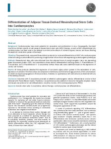
Fig. 1. Purification of non-hematopoietic umbilical cord blood stem cells. FACS analysis of SSEA-4 expression reveals that cells expressing the marker were enriched after the purification process. The upper panel shows the analysis of SSEA-4 expression (X-axis) against the forward scattered beam (Y-axis) in unprocessed samples (Cord blood), after Ficoll treatment (Mononuclear Cells) and after negative depletion (purified stem cells). The lower panel shows a significant increase in signal of SSEA-4 positive cells in the “purified stem cells” sample which reflects the relative number of cells in comparison with samples before the depletion step.
GABAergic neurons from cord blood stem cells 253 Total RNA isolation and reverse transcription Total RNA was isolated from the samples using Qiagen RNeasy Plus mini kit following the manufacturer’s protocol. For optimal RNA purification DNase digestion step was added. cDNA was produced using script VILO kit (Invitrogen) following manufacturer’s protocol. Quantitative polymerase chain reaction (PCR) For Q-PCR reactions, we used Profiler™ PCR arrays purchased from Sabiosciences [PAHS-060E and PAHS-036E] in addition to conventional reactions. Primers sets were obtained from the PrimerBank website (http://pga.mgh.harvard.edu/ primerbank) (Wang and Seed 2003, Spandidos et al. 2008) and purchased from Sigma, GAPDH 5-TGTTGCCATCAATGACCCCTT-3 and 5-CTCCACGACGTACTCAGCG-3, DLX1 5-CCATGCCAGAAAGTCTCAACA-3 and 5-GGCCCAAACTCCATAAACACC-3, DLX2 5-GCCTCAACAACGTC-
CCTTACT-3 and 5-TCACTATCCGAATTTCAGGCTCA-3, OLIG1 5-AAAAGACCAGTTAGGCGGTG-3 and 5-AAGAGCGAAAACTCTCTGCG-3, S100B 5-CCACCAATATTCTGGAAGGG-3 and 5-GTGGCAGGCAGTAGTAACCA-3, DLG4 5-GGACCAGATCCTGTCGGTCA-3 and 5-CCTCGAATCGGCTGTACTCTT-3, MASH1 5-CGCGGCCAACAAGAAGATG-3 and 5-CGACGAGTAGGATGAGACCG-3. Q-PCR reactions were carried out using RT2 SYBR® Green/ROX™ qPCR master mix (Sabiosciences, PA-012-8) and ABI7900HT machine according to a two steps cycling program published previously (Ali et al. 2012). PCR results for each gene were normalized to GAPDH. A minimum of triplicates for each reaction has been performed. Statistical analysis For two groups of data, the student’s paired t-test was used to obtain probability (P) values. For three or more groups of univariate data, single-factor analysis
Fig. 2. Q-PCR expression analysis reveals an induction profile of GABAergic-specific markers during the differentiation process. (A) Analysis of neuronal (DLG4), astrocytic (S100B) and oligodendrocytic (OLIG1) markers between undifferentiated cells (Pre or Day 0) and cells after neuronal maturation (post or Day 24). Expression profiles of (B) transcription factors MASH1, DLX1 and DLX2 in samples isolated from undifferentiated cells (day 0), cells after neuronal commitment (day10), cells after neuronal induction (day 17) and cells after neuronal maturation (day 24), (C) genes encoding enzymes involved in neurotransmitter synthesis and regulation, (D) genes encoding GABA receptors and one transporter and (E) genes encoding subunits of ion channels involved in action potential generation and propagation.
254
H. Ali et al.
of variation (ANOVA) was used to obtain P values. Results with P values of less than 0.05 were considered statistically significant. RESULTS Effective enrichment of non-hematopoietic umbilical cord blood stem cells FACS analysis of SSEA-4 expression revealed that cells expressing SSEA-4 were enriched by 7.62 fold (SD ± 1.43, n=6, P