J Clin Exp Hematop Vol. 53, No. 3, Dec. 2013
Case Study
Diffuse Large B-Cell Lymphoma with Mass Lesions of Skull Vault and Ileocecum Shigeki Kosugi,1) Masaaki Kume,2) Joji Sato,3) Ikuo Sakuma,4) Junta Moroi,5) Keiichi Izumi,6) Yasukazu Sato,6) Naoya Nakamura,7) Masatomo Takahashi,1) and Ikuo Miura1) We report a rare case of non-Hodgkin lymphoma with mass lesions of skull vault and ileocecum. The patient was an 82year-old Japanese woman who exhibited a painless subcutaneous scalp tumor in the right parietal region associated with no neurological abnormalities. Magnetic resonance imaging of the head demonstrated a mass in the skull vault with iso- to hypointense signals on both T1- and T2-weighted imaging. Biopsy of the mass revealed that the tumor comprised large cells that were immunoreactive for CD20 (L-26) and CD79a. Diffuse large B-cell lymphoma (DLBCL) was therefore diagnosed. Further investigation could not identify any other evidence of systemic lymphoma other than ileocecal lesions. She was treated by irradiation (45 Gy) of the mass on the parietal bone and with rituximab, pirarubicin, cyclophosphamide, and vincristine. The patient achieved complete remission after 3 cycles of systemic chemotherapy. As of 30 months after presentation, no signs of lymphoma have been found. 〔J Clin Exp Hematop 53(3) : 215-219, 2013〕 Keywords: cranial vault lymphoma, colonic lymphoma, diffuse large B-cell lymphoma, radiation, chemotherapy We here discuss the biological characteristics and treatment of our case compared with previously reported cases of solitary skull vault lymphoma.
INTRODUCTION The involvement of bone in disseminated non-Hodgkin lymphoma (NHL) is not uncommon, occurring in up to 25% of patients.1,2 Primary involvement of the bone is extremely rare in Hodgkin lymphoma, whereas NHL arises from a skeletal location in up to 4% of cases, particularly in the long bones of the upper and lower extremities, the pelvis, and the spine.1,2 Initial involvement of the skull at presentation is extremely rare and primary cranial vault lymphoma constitutes only 0.2% of lymphoma cases.3 No consensus has been reached regarding the treatment for lymphoma of the skull.
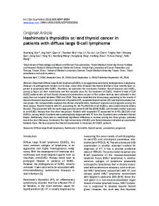
CASE REPORT An 82-year-old Japanese woman visited hospital with a painless subcutaneous scalp tumor on the parietal region. Performance status was 0 according to the Eastern Cooperative Oncology Group.4 The patient had a past history of chronic hepatitis C, but anti-human immunodeficiency virus antibody was negative. There were no episodes of head injury before the emergence of the subcutaneous scalp mass. Physical examination revealed a firm nonpulsatile and immovable subcutaneous mass measuring 3.0 × 3.0 cm in the right parietal area. She was afebrile, with no lymphadenopathy or hepatosplenomegaly. Magnetic resonance imaging (MRI) of the head demonstrated a solitary mass of the skull vault with iso- to hypointense signals on both T1- and T2weighted imaging (Fig. 1). The center of the mass was located within diploe and projected into inner and outer tables. The skull was completely destroyed and the tumor was adjacent to the dura. Biopsy of the subcutaneous scalp mass showed that the tumor comprised large cells that were immunoreactive for CD20 (L-26) (Figs. 2 & 3) and CD79a, but not CD3, CD5, bcl-2, or in situ hybridization for Epstein-Barr virus-encoded RNA (EBER)-1, indicating diffuse large B-cell lymphoma
Received : May 12, 2013 Revised : July 6, 2013 Accepted : July 29, 2013 1)
Department of Internal Medicine, Division of Hematology and Oncology, St. Marianna University School of Medicine, Kawasaki, Japan 2) The Second Department of Internal Medicine, Hiraka General Hospital, Yokote, Japan 3) 6) Department of Internal Medicine and Department of Surgery, Sato Hospital, Yurihonjo, Japan 4) Division of Radiology, Department of Medicine, Akita University School of Medicine, Akita, Japan 5) Department of Neurosurgery, Research Institute for Brain and Blood Vessels-Akita, Akita, Japan 7) Department of Pathology, Tokai University School of Medicine, Isehara, Japan Corresponding author: Dr. Shigeki Kosugi, Department of Internal Medicine, Division of Hematology and Oncology, St. Marianna University School of Medicine, 2-16-1 Sugao, Miyamae-Ku, Kawasaki, Kanagawa 218-8511, Japan E-mail:
[email protected]
215
05-13-018.mcd
13/12/27 13:02
v4.21
Kosugi S, et al.
Fig. 1. Magnetic resonance imaging of the head demonstrating a solitary mass of the cranial vault with signal hypointensity on T1weighted imaging.
Fig. 2. Histological features showing that the tumor comprises large cells. H&E stain, ×200.
Fig. 3. Histological examination showing that the large tumor cells are immunoreactive for CD20 (L-26) (× 200).
(DLBCL). MIB-1 index was 72.8%. The patient was admitted to our hospital. Hematological examination on admission showed a white blood cell count of 5,460/mm3 with a normal differential, hemoglobin level of 10.0 g/dL, and platelet count of 139,000/mm3 . Blood biochemistry was unremarkable. Serological examination showed elevated soluble interleukin-2 receptor (1,093 U/mL). Bone marrow aspiration and biopsy from the posterior iliac spine revealed no marrow involvement. Cerebrospinal fluid cytology was negative for lymphoma cells. Contrast-
Fig. 4. Total colonoscopy demonstrating mass lesions in the ileocecum.
enhanced computed tomography of the neck, chest, abdomen, and pelvis showed no evidence of lymphoma lesions. Bone scintigraphy revealed no abnormal uptake other than in the parietal bone lesion. However, while upper gastrointestinal endoscopy revealed no lymphoma lesion, total colonoscopy 216
05-13-018.mcd
13/12/27 13:02
v4.21
Skull vault and ileocecum lymphoma
molecular analyses, we considered that it was difficult to discuss the possibility of systemic lymphoma in this case. Primary cranial vault lymphomas have been reported in immunocompromised or trauma patients.8,9 However, primary NHL of the skull with extra- and intracranial extension without systemic or skeletal manifestation in a nonimmunocompromised and non-trauma patient is extremely rare. We found only 19 cases in the literature (Table 1).10-28 Such cases of cranial vault lymphoma with systemic involvement, short observation, secondary involvement of the CNS including intra-axial lesions, progressive disease within 6 months, and multifocal primary cranial vault lymphoma are not included in Table 1. Only 1 case is included from the previous report of cranial vault lymphoma in which total colonoscopy was performed.11 The initial symptoms and signs of lymphoma in the skull include a painless scalp lump, headache due to bone destruction or tumor infiltration of meninges, seizures, and focal neurologic deficits secondary to neoplastic infiltration of the cerebral cortex. Lymphoma cells have been suggested to infiltrate the spaces within the diploe and extend along the emissary veins to infiltrate the soft tissues on either side of the bone.11 Malignant lymphoma originating from the skull may extend outside the skull with bony changes at first, followed by infiltration and complete destruction of the skull.21 In the present case, the skull was completely destroyed and tumor was adjacent to the dura. The dura has been reported to display strong resistance to lymphoma infiltration of the cerebral cortex.21 In this case, there were no focal neurological deficits and cerebrospinal fluid cytology was negative for lymphoma cells. No obvious CNS involvement was identified. MRI of the tumor in the present case showed iso- to hypointense signal intensity on both T1- and T2-weighted imaging. The MRI features of the present case were similar to previously reported cases.16,20,21 With gadolinium-DTPA (diethylenetriaminepentacetate) administration, many cranial vault lymphomas displayed homogeneous findings.16,21 As the appearance resembles that in metastatic carcinoma, osteomyelitis, or meningioma, histopathological examination is necessary to reach a definitive diagnosis.29 Optimum treatment for malignant lymphoma of the skull vault has not been established. Surgical removal followed by radio- and chemotherapy has been recommended.16,17 In the present case, taking into consideration the age and absence of focal neurological deficits, we decided to use radiotherapy instead of surgical removal of the tumor from parietal bone. Clinical observations of the present case suggest that skull vault lymphoma without obvious CNS involvement, unlike primary CNS lymphoma,30 does not always require intensive chemo-radiotherapy including intrathecal administration of cytotoxic agents or high-dose methotrexate/cytarabine (AraC).
demonstrated mass lesions at the ileocecum (Fig. 4) and a biopsy was taken, which confirmed the diagnosis of DLBCL. The tumor cells were immunoreactive for CD20 (L-26) and CD79a, but not CD3 and bcl-2. The ileocecal mass was smaller than the subcutaneous scalp mass. The patient was referred to our hospital for radio- and chemotherapy. The skull vault lymphoma was treated first because ileus due to ileocecal mass lesions was considered unlikely. Whole brain was irradiated with 34.2 Gy in 17 fractions, involving field irradiation with 10.8 Gy in 6 fractions over 36 days. A few days after irradiation, the subcutaneous scalp tumor was not palpable. After completion of radiotherapy, the patient was treated with systemic chemotherapy. She was administered 500 mg of rituximab on day 1, and then 40 mg of pirarubicin, 700 mg of cyclophosphamide, and 1.4 mg of vincristine on day 2. As she had a past history of chronic hepatitis C, prednisolone was not used. Systemic chemotherapy was interrupted by severe myelosuppression after the completion of 3 cycles. Tumor regression of ileocecal lesions was confirmed by total colonoscopy after 3 cycles of systemic chemotherapy. As of 30 months after presentation, no signs of lymphoma have been found.
DISCUSSION Primary bony Hodgkin lymphoma is extremely rare, and NHL originating primarily in bone is seen in only about 4% of patients. Bone involvement is typically seen in the femur, tibia, pelvis, spine, mandible, and scapula.1,2 Skull vault lymphoma is different from bony lymphoma of other sites because treatment of the central nervous system (CNS) is required in cases with the involvement of cerebral structures by direct invasion. True primary malignant lymphoma of the bone is defined as a solitary mass lesion without any evidence of disease at another site and no systemic dissemination within 6 months of tumor detection.5,6 The present case thus does not fulfill the criteria for primary malignant lymphoma of the bone owing to the presence of colon lesions. We compared the immunohistochemical data of ileocecal lymphoma to those of skull vault lymphoma ; however, there were no obvious differences between the two lesions. There has been a case report of concurrent adenocarcinoma and DLBCL in the colon, which first presented with DLBCL in the skull base and ileocecal junction area.7 The collision tumors were associated with Epstein-Barr virus infection. In this case, EBER-1 in situ hybridization of skull vault lymphoma was negative ; however, that of ileocecal lymphoma could not be carried out. Although we attempted to analyze the mutation status of the IGH gene of both mass lesions by polymerase chain reaction (PCR), PCR products of ileocecal lesions were not obtained, so it remains to be determined whether the two lesions have the same clonality. From the results of clinical examination, and immunohistochemical and 217
05-13-018.mcd
13/12/27 13:02
v4.21
Kosugi S, et al.
Table 1. Clinical data and results of 19 patients with primary cranial vault lymphoma involving the skull Case no. 1 2 3 4 5
Authors, Year, Age/ (Reference) Sex Agbi CB, et al., 1983 [10] Holtås S, et al., 1985 [11]
58/F
Maiuri F, et al., 1987 [12] Howat AJ, et al., 1987 [13]
51/F
60/F
3/M
Initial symptom Confusion, headache, focal deficit Subcutaneous mass on the scalp
Location
Surgery
Cleaved cell lymphoma
Left frontal
Steroid
Undifferentiated large cell malignant lymphoma of histiocytic type Lymphoblastic lymphoma
Headache, transient dip- Right parietolopia occipital Not reported Left frontal Parietal scalp mass
Left parietal
Headache, confusion, hemiparesis
Left parietal
7
Sato M, et al., 1993 [16]
65/F
Hemiparesis, parietal scalp mass
Right fronto-parietal
8
Isla A, et al., 1996 [17]
75/F
Seizure
Left frontal
9
Muin IA, et al., 1997 [18]
60/M
Right parietal
10
Jamjoom AB, et 25/M al., 1998 [19] Duyndam DA, 71/F et al., 2002 [20]
Headache, confusion, forgetfulness, hemiparesis Scalp mass, headache Scalp mass
Left frontal
71/F
Scalp mass
25/M
Scalp mass
Left fronto-parietal Right frontotemporoparietal Right parietal
11 12 13
Kanai M, et al., 2003 [21] Mongia S, et al., 2003 [22]
14
Fukushima Y, 60/F et al., 2007 [23]
15
Gaitonde S, et al., 2008 [24]
70/F
16
GonzálezBonet CG, et al., 2008 [25] Renard D, et al., 2009 [26] Fadouhair Z, et al., 2011 [27]
84/F
Martin J, et al., 2012 [28]
50/M
17 18
19
67/F 42/F
Scalp mass
Histology
Right parietal
Kawakami K, et 52/F al., 1988 [14] Parekh HC, et 65/F al., 1993 [15]
6
Management
Parietal
Surgery and radiotherapy with 4,500 rads Whole-cranium radiotherapy (24 Gy) and chemotherapy Surgery and radiotherapy with 60Co 4,500 rads Surgery and whole brain irradiation Surgery, radiation (50 Gy) and chemotherapy (THPCOP) Surgery (excision) and radiation (45 Gy) and chemothetapy (VCR+cisplatin+DXR+PSL) Surgery, external beam radiotherapy and chemotherapy (CHOP) Surgery and radiotherapy (5,000 cGy) Chemotherapy (chlorambucil 8 mg/day and PSL 20 mg/day) for 6 mon Surgery and chemotherapy (3 cycles of THP-COP) Radiotherapy along with adjuvant chemotherapy
Follow up and outcome Disease-free at 7 mon Disease-free at 6 mon
Disease-free at 24 mon Mixed large and mediumAlive at 133 sized cell, some with cleaved mon nuclei NHL (diffuse medium sized Disease-free at type) 6 yr Malignant non-Hodgkin’s B- 6 yr follow up ; cell lymphoma died of unrelated cause Malignant lymphoma, B cell Disease-free at type, diffuse large cell type 20 mon after surgery Low grade B-cell lymphoma Disease-free at centroblastic-centrocytic type 3 yr with follicular pattern High grade non-Hodgkin Bcell lymphoma
Recurrencefree at 8 mon
Malignant lymphoma, large T-cell immunoblastic type Malignant non-Hodgkin lymphoma of the B-cell type
Disease-free at 5 mon Disease-free at 2 yr
Diffuse, medium-sized, B cell Died at 3.5 yr lymphoma Non Hodgkin’s lymphoma Disease-free at 2.5 yr
Surgery, local radiotherapy (50 Gy) and chemotherapy (CHOP) Surgery and localized radiation therapy
Non Hodgkin’s lymphoma of Disease-free at the diffuse, medium-sized, 3 yr clear, B-cell type Forehead mass Right frontal Follicular lymphoma, Grade Relapsed at 9 2 mon after initial therapy Ictus Right Surgery Immunoblaastic B-cell lymDisease-free at fronto-parietal phoma 5 mon after surgery Painful right-sided swel- Right frontal Chemotherapy Diffuse large B-cell lympho- Not available ling (Rituximab+CHOP) ma Enlarging mass involv- Right parietal Chemotherapy (4 cycles of Diffuse large B-cell lympho- Disease-free at ma more than 9 ing right parietal bone Rituximab+CHOP) folmon after treatlowed by involved field rament diation Diffuse swelling in the Left parietal and Chemotherapy (6 cycles of Diffuse primary cutaneous B- Not available left side scalp occipital CHOP)+local adjuvant ex- cell lymphoma ternal beam radiotherapy (36 Gy)
NHL, non-Hodgkin lymphoma ; MTX, methotrexate ; THP, pirarubicin ; COP, cyclophosphamide, vincristine and prednisolone ; VCR, vincristine ; DXR, doxorubicin ; PSL, prednisolone ; CHOP, cyclophosphamide, doxorubicin, vincristine and prednisolone
218
05-13-018.mcd
13/12/27 13:02
v4.21
Skull vault and ileocecum lymphoma
The prognosis of malignant lymphoma appearing in the skull vault is unknown, but any involvement of cerebral structures by direct invasion or by leptomeningeal seeding and systemic involvement suggests an unfavorable prognosis. A thorough search is necessary to decide upon the treatment for lymphoma. Further accumulation of data for skull vault lymphoma is needed to improve the treatment and prognosis.
Surg Neurol 39:286-289, 1993 16 Sato M, Saito T, Yamaguchi K: Primary malignant lymphoma of the skull presenting a huge mass lesion: case report. No Shinkei Geka 21:1061-1064, 1993 (in Japanese) 17 Isla A, Alvarez F, Gutiérrez M, Gamallo C, García-Blázquez M, et al.: Primary cranial vault lymphoma mimicking meningioma. Neuroradiology 38:211-213, 1996 18 Muin IA, Saffari HM, Hasimah YN: Primary non-Hodgkin’s lymphoma of the cranial vault mimicking a meningioma: a case report. Med J Malaysia 52:86-88, 1997 19 Jamjoom AB, Jamjoom ZA, Naim-Ur-Rahman, Cheema MA: Primary midline cranial vault lymphoma simulating a parasagittal meningioma: the role of angiography in preoperative diagnosis. Neurosurg Rev 21:202-205, 1998 20 Duyndam DA, Biesma DH, van Heesewijk JP: Primary nonHodgkin’s lymphoma of the cranial vault; MRI features before and after treatment. Clin Radiol 57:948-950, 2002 21 Kanai M, Kawano K, Murakami T, Saitou M, Kikumoto N: A case of malignant lymphoma of the cranial vault. No Shinkei Geka 31:419-424, 2003 (in Japanese) 22 Mongia S, Shukla D, Devi BI, Reddy TV: Primary cranial vault non-Hodgkin’s lymphoma. Neurol India 51:293-294, 2003 23 Fukushima Y, Oka H, Utsuki S, Nakahara K, Fujii K: Primary malignant lymphoma of the cranial vault. Acta Neurochir (Wien) 149:601-604, 2007 24 Gaitonde S, Patel R, Alagiozian-Angelova V, Kadkol S, Peace D: Primary low grade follicular lymphoma of cranial vault mimicking lipoma at presentation. Acta Oncol 47:326-329, 2008 25 González-Bonet LG, Gutiérrez-Herrera JM, Gallego JM, Barcia JA: Primary immunoblastic B-cell lymphoma of cranial vault. Acta Neurochir (Wien) 150:507-508, 2008 26 Renard D, Campello C, Beraru O, Bouillot P, Labauge P: Teaching Neuroimages: Primary diffuse large B-cell lymphoma of the cranial vault. Neurology 73:e84-85, 2009 27 Fadoukhair Z, Lalya I, Amzerin M, Elkhanoussi B, Sbitti Y, et al.: Successful management of primary non Hodgkins lymphoma of the cranial vault. Pan Afr Med J 8:50, 2011 28 Martin J, Ramesh A, Kamaludeen M, Udhaya, Ganesh K, et al.: Primary non-Hodgkin’s lymphoma of the scalp and cranial vault. Case Report Neurol Med 2012:616813, 2012 [doi:10.1155/2012/ 616813] 29 Kantarci M, Erdem T, Alper F, Gundogdu C, Okur A, et al.: Imaging characteristics of diffuse primary cutaneous B-cell lymphoma of the cranial vault with orbital and brain invasion. AJNR Am J Neuroradiol 24:1324-1326, 2003 30 DeAngelis LM, Seiferheld W, Schold SC, Fisher B, Schultz CJ: Combination chemotherapy and radiotherapy for primary central nervous system lymphoma: Radiation Therapy Oncology Group 93-10. J Clin Oncol 20:4643-4648, 2002
REFERENCES 1 Braunstein EM, White SJ: Non-Hodgkin lymphoma of bone. Radiology 135:59-63, 1980 2 Pear BL: Skeletal manifestations of the lymphoma and leukemias. Semin Roentgenol 9:229-240, 1974 3 Rosenberg SA, Diamond HD, Jaslowitz B, Craver LF: Lymphosarcoma : A review of 1269 cases. Medicine 40:31-84, 1961 4 Oken MM, Creech RH, Tormey DC, Horton J, Davis TE, et al.: Toxicity and response criteria of the Eastern Cooperative Oncology Group. Am J Clin Oncol 5:649-655, 1982 5 Coley BL, Higinbotham NL, Groesbeck HP: Primary reticulumcell sarcoma of bone ; summary of 37 cases. Radiology 55:641658, 1950 6 Shoji H, Miller TR: Primary reticulum cell sarcoma of bone. Significance of clinical features upon the prognosis. Cancer 28:1234-1244, 1971 7 Chang H, Chuang WY, Shih LY, Tang TC: Collision in the colon: concurrent adenocarcinoma and diffuse large B-cell lymphoma in the same tumor. Acta Clin Belg 66:302-304, 2011 8 Kelleher AD, Brew BJ, Milliken ST: Intractable headache as presenting complaint of AIDS-related lymphoma confined to bone. J Acquir Immune Defic Syndr 7:629-630, 1994 9 Tagawa M, Momita S, Irie J: Primary B cell lymphoma of the skull following head trauma ; a case report. Rinsho Ketueki 28:583-593, 1987 (in Japanese) 10 Agbi CB, Bannister CM, Turnbull IW: Primary cranial vault lymphoma mimicking a meningioma. Neurochirurgia 26:130-132, 1983 11 Holtås S, Monajati A, Utz R: Computed tomography of malignant lymphoma involving the skull. J Comput Assist Tomogr 9:725727, 1985 12 Maiuri F, Corriero G, Giamundo A: Primary lymphoma of cranial vault. J Neurosurg Sci 31:183-186, 1987 13 Howat AJ, Thomas H, Waters KD, Campbell PE: Malignant lymphoma of bone in children. Cancer 15:335-339, 1987 14 Kawakami K, Kawamoto K, Inagaki R, Numa Y, Kawamura Y, et al.: Two case of malignant lymphoma in the skull. CT Kenkyu 10:217-222, 1988 (in Japanese) 15 Parekh HC, Sharma RR, Keogh AJ, Prabhu SS: Primary malignant non-Hodgkin’s Lymphoma of cranial vault: a case report.
219
05-13-018.mcd
13/12/27 13:02
v4.21