Differential Expression of Telomerase Activity in Human Cervical Cancer and Cervical Intraepithelial Neoplasia Lesions By Chia C. Pao, Chih-Jen Tseng, Chieh-Yu Lin, Feng-Ping Yang, Jyu Jen Hor, Ding-Shyan Yao, and Swei Hsueh Purpose: Telomeres are tandem arrays of repeated DNA sequences located at the ends of eukaryotic chromosomes, and are synthesized by the enzyme telomerase. Loss of telomeric DNA may play an important role in the development of human cancers. However, very little is known about the status of telomerase during human cervical cancer development. Patients and Methods: Telomerase activity was measured by telomerereepeat amplification protocol (TRAP) assay in 24 cervical cancers, one carcinoma in situ (CIS), and 20 cervical intraepithelial neoplasia (CIN) lesions. Adjacent nontumor cervical tissue from the same 24 cervical cancer patients and normal cervical tissues from 11 control individuals also were examined for the presence of telomerase activity. Results: Twenty two of the 24 (91.7%) cervical cancer specimens and the single CIS tissue were strongly positive
for telomerase activity. Relatively weak but distinctive telomerase activity also was detectable in one of four CIN-I (25%), two of eight CIN-II (25%), and two of eight CIN-III (25%), respectively. However, telomerase activity was not found in the 24 corresponding nontumor cervical tissues from the same cervical cancer patients and the 11 normal cervical tissues from control individuals. Conclusion: The majority of cervical cancers contain strong telomerase activity. Significant proportions of noncancerous CIN tissues also contain telomerase activity, although weaker than that in cervical cancer. It seems that there is a progressive increase of telomerase activity in association with an increased degree of cervical malignancy. These resuits seem to suggest that the expression of telomerase may play a crucial role in cervical cancer carcinogenesis. J Clin Oncol 15:1932-1937. 1997 by American Sociely of ClinicalOncology.
IT HAS BEEN SUGGESTED
of cell division of cultured fibroblasts 3 and other somatic cells. 4 Although the biologic roles of this shortening have not been defined clearly, telomere shortening may lead to chromosome instability and genetic alterations of possible significance for cancer development. Telomerase is a ribonucleoprotein enzyme that synthesizes telomeric DNA onto chromosomal ends using a segment of its RNA components as a template.5 It has been observed that most immortal cell lines express telomerase, 6 '7 whereas mortal cell lines do not. 7 Recently, it was demonstrated that telomerase activity is detectable in cells from most human cancers. 8 Based on these observations, it was postulated that activation of telomerase expression is necessary to overcome cellular senescence and that cancer cells must express telomerase to maintain their immortality. 8 Cervical cancer develops from well-defined precursor lesions referred to as cervical intraepithelial neoplasia (CIN).9 It is well recognized that specific types of human papillomaviruses (HPV) are the principal etiologic agents for both cervical cancer and its precursors.' ° Although the mechanisms by which HPV acts to transform cells are still not completely understood, extensive molecular evidence has suggested that complex interactions between virus and host cells seem to be necessary. Studies have shown that two viral genes, the E6 and E7, are generally retained and expressed in cervical cancers.""12 In cultured human keratinocytes, expression of the E6 and E7 viral oncogenes can result in high-frequency immortalization of transfected cells.' 3 The present general acceptance that cancer can be attributed to an accumulation of genetic alterations has lead
that malignant transformation of normal cells is accomplished through multiple steps, including distinct phenotypic changes regarding transformation and immortalization. Recent advances in understanding the mechanism that regulates cell mortality provide information needed to understand the development of the immortal phenotype in human cells.' Telomeres consist of tandem arrays of guanine-rich repetitive motifs at the ends of chromosomes that have been highly conserved throughout evolution. Telomeric ends are considered to play important roles in the protection of the chromosome ends from aberrant recombination and end fusion. 2 Because of the inability of the cellular DNA replication machinery to replicate the 5' end of the linear DNA, the number of telomere repeats decreases and the lengths of the telomeres gradually shorten during aging and rounds
From the Departments of Biochemistry, Obstetrics and Gynecology, and Pathology, Chang Gung College of Medicine and Technology, Taipei, Taiwan. Submitted September 30, 1996; accepted January 24, 1997. Supported by research grantsfrom Chang Gung MemorialHospital (no. CMRP-607) andfrom National Science Council of the Republic of China (no. NSC 86-2314-B-182-009), Taipei, Taiwan. Address reprint requests to Chia C. Pao, MD, Department of Biochemistry, Chang Gung College of Medicine and Technology, 259 WenHwa Rd, Kweishan, Taoyuan, Taiwan; Email ccpao@ cguaplo.cgu.edu.tw. © 1997 by American Society of Clinical Oncology. 0732-183X/97/1505-0038$3.00/0
1932
Journal of Clinical Oncology, Vol 15, No 5 (May), 1997: pp 1932-1937
Downloaded from ascopubs.org by 37.44.207.43 on January 17, 2017 from 037.044.207.043 Copyright © 2017 American Society of Clinical Oncology. All rights reserved.
TELOMERASE AND CERVICAL CANCER
to a search for those alterations involved in the stage of neoplasia in the human uterine cervix. However, no studies to date have addressed the telomerase status in cervical cancer and its precancerous lesions. To examine the role telomerase may play in the carcinogenesis of cervical cancer, investigations were conducted to assess the telomerase status and relative level of telomerase in cervical cancer tissues and its precancerous lesions. PATIENTS AND METHODS
Patients, Tissues, and Cells Cervical tissues were obtained at our hospital either by biopsy or during surgical resection from 24 stage lb or IIa cervical cancer patients, one patient with carcinoma in situ (CIS), and 20 patients with CIN (including four CIN-I, eight CIN-II, and eight CIN-III). Nontumor and normal cervical tissue specimens were obtained from the same 24 cervical cancer patients and from 11 age-matched normal control individuals, respectively. All 11 normal control individuals were diagnosed with uterine myoma and all had normal cervical cytology. Tissues were frozen immediately in liquid nitrogen and stored until used. Guidelines for Human Experimentation of Chang Gung College of Medicine and Technology were followed, and the experimental protocols were reviewed by the Human Experimentation Committee of Chang Gung College of Medicine and Technology. Informed consent was obtained from all participants. All tissues were submitted to hematoxylin-eosin staining and histopathologic diagnosis was made by one of the authors (S.H.), who is a trained pathologist, according to guidelines of the International Federation of Gynecology and Obstetrics (FIGO). All tissue specimens were obtained before any chemotherapy or radiation therapy. CaSki and HeLa cells were acquired from American Type Culture Collection (Rockville, MD). These cell lines, derived from cervical cancer cells, contain HPV type 16 and 18 DNA, respectively.
DNA Extraction and Identification of HPV Sequences by Nested Polymerase Chain Reaction Portions of each tissue were used to prepare DNA by standard phenol-chloroform methods, and purified by alcohol precipitation." This DNA was used for amplification of HPV viral DNA sequences. Information of the primers used in both initial and nested polymerase chain reaction (PCR) of HPV DNA sequences, as well as procedures for the amplification reactions, have been described in earlier reports. 15 7 Because of the sensitivity of the PCR, a number of precautions were taken to minimize the possibility of contamination during sampling and subsequent processing.' 61 8
Telomerase Assay Portions of surgical or biopsy tissue sections that contained cancer cells were removed and homogenized with 20 L of cold lysis buffer.8 Telomerase activity was assayed essentially by the procedures of the telomere repeat amplification protocol (TRAP) assay.8 After the amplified products were separated in 10% polyacrylamide gel, the gels were stained with SYBR Green Nucleic Acid Gel Stain (Molecular Probes Inc, Eugene, OR) according to manufacturer's recommendations. The positivity of telomerase was established by inspecting the stained gel under ultraviolet lights for the presence of the characteristic 6-base
1933 pair (bp) increment ladder. Each sample was assayed initially with 1.0 sag protein extract to identify the relative intensities of the TRAP products. Although telomerase activity was not quantitatively titrated, semiquantitative definition was estimated using a modified procedure previously reported. 9 The semiquantization was performed with various amounts of cell extract, where the TRAP assays did not appear to be inhibited or to have reached a plateau. This method is probably only semiquantitative, but it is sufficient for comparative analysis of the tumors relative to each other and to the CaSki cells that serve as positive control. Because telomerase has an RNA component, each extract was also assayed after the ribonuclease (RNase) treatment (RNaseA 1 mg/ mL for 30 minutes at 37°C) to serve as negative control. Additional negative controls include TRAP assays without primers or with heatinactivated extract. In addition, each extract was assayed in the presence of 0.1 /.g of CaSki cell protein to monitor for inhibitory activities. RESULTS
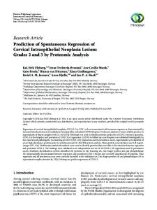
The presence of telomerase activity in normal and cervical precancer and cancer tissues was determined by standard TRAP assay (Fig 1). Twenty-two of 24 (91.7%) cervical cancer specimens and the single CIS tissues were found to contain telomerase activity when a standard 1.0-jig cell extract protein was used for the TRAP assays (Table 1). Both the CaSki (Fig 1, lanes P) and HeLa cells (data not shown) are very rich sources of telomerase activity, and the two cell lines yield similar results. The high prevalence of telomerase in cervical cancer was found even in lesions less than 4 cm in diameter (Table 1). Telomerase activity was undetectable in all nontumor cervical tissues obtained from the same 24 cervical cancer patients (data not shown). Telomerase activity was also negative in all 11 normal cervical tissues from the same number of control individuals. Under the same assay conditions for telomerase activity, one of four CIN-I (25%), two each of eight CIN-II (25%) and CINIII (25%), respectively, were found to contain telomerase
activity (Table 1). Telomerase activity found in the CIN tissues was weaker when compared with activity in cervical cancer and that of CaSki cells. But the CIN tissues clearly pre-
sented themselves with the characteristic 6-bp increment TRAP ladder (Fig 1). All telomerase signals or TRAP products, including those found in CIN tissues, were primer-dependent (data not shown) and RNase-sensitive (Fig 1, lanes R). The single band at the bottom of the gels in some of the samples is probably an artifact caused by primer-dimer formation. These data indicated that qualitative detection of telomerase was clearly correlated with cervical cancer progression, necessitating a quantitative, or at least semiquantitative, evaluation of these cervical tissue samples. The TRAP assays were then performed again with 0.1 jig, 1.0 jig, and 10 Lg extract protein per assay. Telomerase
Downloaded from ascopubs.org by 37.44.207.43 on January 17, 2017 from 037.044.207.043 Copyright © 2017 American Society of Clinical Oncology. All rights reserved.
1934
PAO ET AL
Fig 1. Detection of telomerase activity in cervical tissues by TRAP assay using 1.0 /ug extract protein. (A) Cervical cancer; (B) CIS, CIN-III, and CIN-II; (C) CIN-I and CIN-II; and (D) normal cervical cells. The lane numbers in A, B, and C correspond to patient no. in Table 1. Lanes R: 1.0 Xg CaSki cell extract protein treated with RNase before TRAP assay; lanes P: 1.0 pg CaSki cell extract protein that served as positive control. Numbers 51 to 61 in D were from 11 normal control individuals (not listed in Table 1).
activity remained positive in 10 of 24 cervical cancer tissue samples and in the single CIS tissue sample, even when only 0.1 ig extract protein was used for the TRAP assay (Table 1). On the other hand, the relative telomerase
activities in CIN samples varied considerably and became too low to be detected in two each of the CIN-II and CIN-III tissues when 0.1 jig instead of 1.0 jig extract protein was used for the assay (Table 1). There is clearly
Downloaded from ascopubs.org by 37.44.207.43 on January 17, 2017 from 037.044.207.043 Copyright © 2017 American Society of Clinical Oncology. All rights reserved.
1935
TELOMERASE AND CERVICAL CANCER
Table 1. Telomerase Activity in Cervical Precancer and Cancer Tissues Telomerase Detection PatientNo. Cervical cancer, stage lb and Ila 6 7 8 9 12 15 21 30 31 34 1 2 3 11
14 17 25 28 33 36 38 43 4 32 CIS 29 CIN-III 10 45 13 16 18 19 20 42 CIN-I1 27 40 5 22 23 35 41 44 CIN-I 26 24 37 39
0.1 pg
1.0 pg
10 pg
+
+
+
+
+
±
+
+
+
+
+
DNAt
Remarks
+
Node (+)
+
+
+
+
> 4 cm; node +) > 4 cm
+
+
+
+
+
+
+
+
+
+
+
+
+
+
+
+ +
+ +
+
+ +
+
+ -
±
-
+
+
+
-
+
+
+
-
+
+
+
-
+
+
+
-
+
+
+
-
+
+
+
-
+
+
-
+
+
+
> 4 cm; node (+)
+
-
+
+ +
-
+
+
+
-
+
-
--
_
_
+
+
+
+
-
+
+
-
+
+
+
-
+
-
_
_
_
Node (+)
+
-
-
Node (+) > 4 cm; node (+) Node (+) Node (+)
+
_-
-
q+
_
_
_
+
++
a progressive increase in telomerase activity based on the stage of the disease. The presence of HPV type 16 or 18 DNA sequences in these tissues is also shown in Table 1. Twenty-one of 23 (91.3%) telomerase-positive cervical cancer and CIS tissue samples and two of five (40%) telomerase-positive CIN tissue samples, respectively, were also positive for HPV (Table 1).
-
±+
+
+r
+~~~~
_
_
_
-
_
_
-
+
-
-
-
+
+
+
_
_
±-
+
*Shown for 0.1 pg, 1.0 g, and 10 pg of extract protein per assay. tPresence of either HPV type 16 or type 18 DNA sequences.
DISCUSSION Because earlier reports have suggested that telomerase may be important in cancer development, it would be very worthwhile to understand better the status of telomerase expression during cancer progression. The data here demonstrated that telomerase is undetectable in all normal cervical tissue but is clearly present in almost all cervical cancer and CIS tissue samples. Furthermore, relatively weak but distinct telomerase activity also can be found in significant numbers of CIN tissue samples. The study presented here is the first systemic examination for telomerase activity in cervical normal, precancer, and cancer tissues of the cervix. There were two cervical cancer tissue samples that were free of detectable telomerase activity even when 10 Ig extract protein was used for the assay. It has been reported that no telomerase activity was detected in significant proportion of human retinoblastomas2 0 and anaplastic astrocytomas and glioblastomas.2 1 It is possible that the development of malignant phenotype of certain populations of cancers is independent of telomerase and that telomerase activation may not be a strict requirement of carcinogenesis for all cervical cancers. A novel yet unidentified mechanism may be responsible for the development of these tumors. However, it should be underscored that telomerase was positive in 18 of 20 cervical cancer lesions smaller than 4 cm in diameter (Table 1). These results suggest that, when cervical cancers are detected at the clinical level, almost all are already telomerase-positive. The development of cervical cancer, like many other cancers, is thought to be a multistep process with multiple genetic changes, and with the involvement of multiple risk factors, including HPV infection. Keratinocytes that express both HPV E6 and E7 transforming genes undergo crisis periods and give rise to immortalized lines that show an increase in telomere length. 22 It has been reported that expression of the HPV E6 protein activates telomerase in early-passage human keratinocytes.2 2 23 Arrest of telomere shortening may be important in HPV-associated immortalization, and re-storation of telomere length may be advantageous to cells with regard to their ability to proliferate.2 2 Therefore, it is possible that
Downloaded from ascopubs.org by 37.44.207.43 on January 17, 2017 from 037.044.207.043 Copyright © 2017 American Society of Clinical Oncology. All rights reserved.
PAO ET AL
1936 telomerase activation is involved with HPV infection and cervical carcinogenesis. To assess the relative levels of telomerase expression in normal, precancer, and cancer tissues of the uterine cervix, it was necessary to determine the telomerase activity in at least a semiquantitative manner. It is clear from the data that when the telomerase activities of different types of cervical tissues are evaluated in relation to each other and to the activity of CaSki cells, both the intensity of telomerase activity and its frequency in telomerase-positive cases appear to correlate with progression of cervical carcinogenesis. Although telomerase activity in most of the CIN tissues is weak when compared with that of the cervical cancer and cell lines that were derived from cervical cancer, it is not an artifact. The TRAP assay products derived from the CIN tissues are both primer-dependent and RNase-sensitive and also clearly give the characteristic 6-bp ladder, indistinguishable from that observed in cervical cancer. There are a few reports of the presence of telomerase in nonmalignant tissues such as hematopoietic cells24'25 and nonmalignant skin cells.2 6 It has also been reported recently that although telomerase is present in almost all hepatocellular carcinoma cells, very weak telomerase activity can also be detected rather frequently in apparently cancer-free hepatitis liver cells." Both the intensity of telomerase activity and its frequency in telomerasepositive cases in these nonmalignant cells are also invariably much less than those detected in corresponding tumor tissues, as has been observed in the CIN tissues here. At the present time, it is not clear whether the telomerase activity in these tissues is shared by many cells with weak telomerase activity or by a small number of cells with strong activity. Although it is very unlikely, the possibility cannot be excluded that there are very small numbers of cervical cancer cells that were present in the CIN tissues but evaded histopathologic examination. It is unclear whether telomerase-positive CINs have a greater chance to develop into invasive cancer than those that do not exhibit telomerase activity. Prospective observation is ongoing for the occurrence of cervical cancer to see if telomerase activity in CIN patients correlates
with cervical cancer development. At present, it has also not been determined whether the increase in telomerase activity in CIN and cervical cancer may not be a consequence of increased cell proliferation. Elucidation of the role of telomerase in the development of cervical cancer is important in view of the suggested use of telomerase as a diagnostic marker for cancer and the therapeutic use of telomerase inhibitors.3 7 However, there are many questions that remain unanswered and that need to be examined. For example, because telomerase assay is not easily quantitated, a more definitive assessment of the role of telomerase in cervical cancer is still important.2 7 Additional studies are also necessary to develop a better understanding of the mechanism by which telomerase activity increases during cervical cancer progression. Investigation is currently underway to examine whether the arrest of telomere shortening has already taken place in CIN lesions; this seems to be important in HPV-associated immortalization and may be advantageous to cells with regard to their ability to proliferate. 2 2 In summary, human telomerase activity has previously been detected in more than 90% of immortal cell lines and approximately 85% of primary tumors.8 The study presented here is the first systemic examination of telomerase activity in cervical cells. Results suggest that telomerase is absent in all normal cervical cells but is differentially expressed in cervical precancer and cancer lesions. With increasing levels of telomerase expression (both the intensity of telomerase activity and frequency in telomerasepositive cases), it appears that telomerase is important in the development of cervical malignancies. Although the biologic significance of telomerase activity in cervical precancer tissues is unclear at present, it is possible that such activity may be an early indication of cancer development. It is also possible from these findings to postulate that most cervical cancer cells are already immortalized by the time they are detected clinically and that CIN cells are in a preparatory or early stage of acquiring immortalization. The fact that telomerase is already evident in cervical cancer tissues and in some cervical precancer lesions suggests that telomerase may play an important role in the associated generation of cervical cancer.
REFERENCES 1. Shay JW, Werbin H, Wright WE: Telomere shortening may contribute to aging and cancer: A perspective. Mol Cell Differ 2:121, 1994 2. Moyzis RK, Buckingham JM, Cram S, et al: A highly conserved repetitive DNA sequence (TTAGGG)n, present at the telomere of human chromosome. Proc Natl Acad Sci USA 85:66226626, 1988 3. Harley CB, Futcher AB, Greider CW: Telomeres shorten during aging of human fibroblasts. Nature 345:458-460, 1990
4. Hastie ND, Dempster M, Dunlop MG, et al: Telomere reduction in human colorectal carcinoma and with aging. Nature 346:866871, 1990 5. Greider CW, Blackburn EH: Identification of a specific telomere terminal transferase activity in Tetrahymena extracts. Cell 43:405-413, 1985 6. Morin GB: The human telomere terminal transferase enzyme is a ribonucleoprotein that synthesizes TTAGGG repeats. Cell 59:521-529, 1989
Downloaded from ascopubs.org by 37.44.207.43 on January 17, 2017 from 037.044.207.043 Copyright © 2017 American Society of Clinical Oncology. All rights reserved.
1937
TELOMERASE AND CERVICAL CANCER 7. Counter CM, Avilion AA, LeFeuvre CE, et al: Telomere shortening associated with chromosome instability is arrested in immortal cells which express telomerase activity. EMBO J 11:1921-1929, 1992 8. Kim NW, Piatyszek MA, Prowse KR, et al: Specific association of human telomerase activity with immortal cells and cancer. Science 266:2011-2015, 1994 9. Park TW, Fujiwara H, Wright TC: Molecular biology of cervical cancer and its precursors. Cancer 76:1902-1913, 1995 10. Schiffman MH: Recent progress in defining the epidemiology of human papillomavirus infection and cervical neoplasia. J Natl Cancer Inst 84:394-398, 1992 11. Hawley-Nelson P, Vousden KH, Hubbert NL, et al: HPV16 E6 and E7 proteins cooperate to immortalize human foreskin keratinocytes. EMBO J 8:3905-3910, 1989 12. Munger K, Phelps WC, Bubb V, et al: The E6 and E7 genes of the human papillomavirus type 16 together are necessary and sufficient for transformation of primary human keratinocytes. J Virol 63:4417-4421, 1989 13. Hudson JB, Bedell MA, McCance DJ, et al: Immortalization and altered differentiation of human keratinocytes in vitro by the E6 and E7 open reading frames of human papillomavirus type 18. J Virol 64:519-526, 1990 14. Maniatis T, Fritsch EF, Sambrook J: Molecular Cloning: A Laboratory Manual. Cold Spring Harbor, NY, Cold Spring Harbor Laboratory, 1982 15. Pao CC, Hor JJ, Wu CJ, et al: Human papillomavirus type 18 DNA in gestational trophoblastic tissues and choriocarcinomas. Int J Cancer 63:505-509, 1995 16. Pao CC, Lin SS, Lin CY, et al: Identification of human papillomavirus DNA sequences in peripheral blood mononuclear cells. Am J Clin Pathol 95:540-546, 1991 17. Pao CC, Lin CY, Chang YL, et al: Human papillomaviruses
and small cell carcinoma of the uterine cervix. Gynecol Oncol 43:206-211, 1991 18. Pao CC, Hor JJ, Tsai PL, et al: Inhibition of in vitro enzymatic DNA amplification by ultraviolet light irradiation. Mol Cell Probes 7:217-219, 1993 19. Tahara H, Nakanishi T, Kitamoto M, et al: Telomerase activity in human liver tissue: Comparison between chronic liver disease and hepatocellular carcinomas. Cancer Res 55:2734-2736, 1995 20. Gupta J, Han LP, Wang P, et al: Development of retinoblastoma in the absence of telomerase activity. J Natl Cancer Inst 88:1152-1157, 1996 21. Langford LA, Piatyszek MA, Xu R, et al: Telomerase activity in human brain tumours. Lancet 346:1267-1268, 1995 22. Klingelhutz AJ, Barber SA, Smith PP, et al: Restoration of telomeres in human papillomavirus-immortalized human anogenital epithelial cells. Mol Cell Biol 14:961-969, 1994 23. Klingelhutz AJ, Foster SA, McDougall JK: Telomerase activation by the E6 gene product of human papillomavirus type 16. Nature 380:79-81, 1996 24. Broccoli D, Young JW, de Lange T: Telomerase activity in normal and malignant hematopoietic cells. Proc Natl Acad Sci USA 92:9082-9086, 1995 25. Hiyama K, Hirai Y, Kyoizumi S, et al: Activation of telomerase in human lymphocytes and hematopoietic progenitor cells. J Immunol 155:3711-3715, 1995 26. Taylor RS, Ramirez RD, Ogoshi M, et al: Detection of telomerase activity in malignant and nonmalignant skin conditions. J Invest Dermatol 106:759-765, 1996 27. Wright WE, Shay JW, Piatyszek MA: Modifications of a telomeric repeat amplification protocol (TRAP) result in increased reliability, linearity and sensitivity. Nucleic Acid Res 23:3794-3795, 1995
Downloaded from ascopubs.org by 37.44.207.43 on January 17, 2017 from 037.044.207.043 Copyright © 2017 American Society of Clinical Oncology. All rights reserved.