Brain (2000), 123, 1092–1101
Differential adhesion molecule requirements for immune surveillance and inflammatory recruitment Michael D. Carrithers,1,3 Irene Visintin,1 Suk J. Kang1 and Charles A. Janeway, Jr1,2 1Section
of Immunobiology, 2Howard Hughes Medical Institute and 3Department of Neurology, Yale University School of Medicine, New Haven, Connecticut, USA
Correspondence to: Dr Michael D. Carrithers, Section of Immunobiology, Yale University School of Medicine, PO Box 208011, 310 Cedar Street, New Haven, CT 06520-8011, USA
Summary Activated CD4 Th1 lymphocytes can enter the brain in the absence of an inflammatory focus. However, the molecular mediators that regulate this early migration of lymphocytes into the brain have remained unclear. We hypothesized that the entry of these ‘pioneer’ lymphocytes into the brain is regulated by a set of molecular events that are distinct from those used once inflammation has been established. Using cells fluorescently labelled with the lipophilic dye DiI, myelin basic protein (MBP)-specific CD4 lymphocytes that expressed low or high levels of very late antigen-4 (VLA-4) and non-antigen-specific activated splenocytes homed to mouse brain in similar quantities 2 h after adoptive transfer. However, antigen specificity and VLA-4 expression were required for more robust
recruitment by 24 h. Immunocytochemistry revealed endothelial and microenvironmental upregulation of vascular cell adhesion molecule (VCAM), intercellular cell adhesion molecule 1 (ICAM-1), MHC class II and interferon-γ 48 h after transfer of MBP-specific cells. In contrast, expression of meningeal and choroid plexusassociated P selectin was upregulated 2 h after adoptive transfer, but not at 48 h. Monoclonal antibody to P selectin, but not to VLA-4, inhibited early migration of high VLA-4-expressing MBP-specific lymphocytes. These results suggest that early migration occurs independent of the lymphocyte integrin VLA-4 and endothelial VCAM, but does require increased surface expression of endothelial P selectin.
Keywords: brain; T lymphocytes; EAE/multiple sclerosis; homing; adhesion molecules Abbreviations: EAE ⫽ experimental autoimmune encephalomyelitis; EHAA ⫽ Eagle’s Hanks’ amino acid; ICAM ⫽ intercellular adhesion molecule; IFN-γ ⫽ interferon-γ; LFA-1 ⫽ lymphocyte function-associated antigen-1; MBP ⫽ myelin basic protein; MHC ⫽ major histocompatibility complex; MMP ⫽ matrix metalloproteinase; PSGL-1 ⫽ P selectin glycoprotein ligand-1; VCAM ⫽ vascular cell adhesion molecule; VLA-4 ⫽ very late antigen 4
Introduction The blood–brain barrier consists of endothelial tight junctions that permit stringent regulation of molecular transport and cell migration (Brightman and Reese, 1969). In addition, brain endothelial cells exist in a unique microenvironment that tightly regulates the expression of proteins relevant to cell migration and other transport processes. The perivascular space of the CNS contains a heterogeneous cell population, including bone marrow-derived perivascular microglia cells, which constitutively express MHC (major histocompatibility complex) class II (Hickey and Kimura, 1988); astrocyte foot processes, which form the glia limitans; and pericytes. The extracellular matrix further limits the ability of blood-borne cells to enter the brain parenchyma (Graeber et al., 1992). This complex microenvironment appears not only to regulate the expression of the tight junctions that form the physical blood–brain barrier but also to inhibit the surface expression © Oxford University Press 2000
of adhesion proteins such as vascular cell adhesion molecule (VCAM) and intercellular adhesion molecule 1 (ICAM-1). Breakdown of the blood–brain barrier occurs in CNS infections and inflammatory disorders, and appears to precede the development of an inflammatory focus as well as clinical symptoms in human multiple sclerosis (Kermode et al., 1990). Although the classic concepts of the blood–brain barrier have been useful in understanding the unique immune status of the brain, they have also hindered investigations of the normal immune surveillance of the CNS. It was thought for many years that immune cells do not enter this microenvironment in the absence of an inflammatory focus. However, the work of Hickey and colleagues clearly demonstrated that activated, but not naı¨ve, lymphocytes can enter the CNS to perform immune surveillance under normal conditions (Hickey et al., 1991), as had been
P selectin mediates early T-cell migration hypothesized previously (Wekerle, 1986). The steps necessary for entry have remained unclear, in part because the beststudied endothelial adhesion molecules, such as VCAM and ICAM-1, do not appear to be involved in this immune surveillance pathway (Hickey et al., 1997). As this migratory pathway is important not only in the immune surveillance of non-lymphoid organs but also in the initiation of tissuespecific inflammatory disease, the identity of the molecular steps responsible has relevance to the treatment of multiple sclerosis and the defence of the brain against noninflammatory infections. Lymphocyte migration is a multistep molecular process that requires at least four steps: an initial low-affinity contact associated with the rolling of lymphocytes on the endothelial cell surface; activation through chemokine G protein-linked receptors; activation-dependent arrest mediated by higheraffinity adhesion; and diapedesis into the extracellular matrix (Springer, 1994; Butcher and Picker, 1996). Interference with any of these steps will terminate transmigration, and the cell will return to the circulation. In general, binding of a lymphocyte carbohydrate ligand such as P selectin glycoprotein ligand-1 (PSGL-1) to selectins such as P and E selectin on the endothelial cell mediates the initial lowaffinity contact; the higher-affinity interactions necessary for arrest and diapedesis require the binding of the α4β1 integrin very late antigen 4 (VLA-4) to VCAM or of the αLβ2 integrin lymphocyte function-associated antigen (LFA-1) to intercellular adhesion molecules (ICAM-1, -2 and -3). We hypothesized that the entry of these ‘pioneer’ lymphocytes into the brain is regulated by a set of molecular events that are distinct from those used once inflammation has been established. For example, previous work in this laboratory demonstrated that VLA-4 was required for the recruitment of autoreactive CD4 T lymphocytes into the brain in the mouse model of human multiple sclerosis, experimental autoimmune encephalomyelitis (EAE) (Baron et al., 1993). However, since the endothelial counterligand VCAM is not expressed on the resting brain endothelium, a VLA-4–VCAM interaction is probably not relevant to early migratory steps. In contrast, P selectin does not appear to be necessary for the recruitment of inflammatory cells in EAE (Engelhardt et al., 1997), but can be expressed on the resting brain endothelium or rapidly induced on the endothelial surface in response to an acute immune stimulus. Here, using fluorescence-labelled T lymphocytes activated in vitro, our goal was to demonstrate that VLA-4, a molecule important for subsequent recruitment into the CNS, does not mediate pioneer lymphocyte migration into the brain, but that P selectin, an endothelial ligand not necessary for subsequent cellular recruitment in EAE, does facilitate entry into the CNS within the first few hours after adoptive transfer.
Material and methods Mice Female (SJL⫻PL/J)F1 and PL/J mice (6–8 weeks of age) were purchased from The Jackson Laboratory. PL/J.MBP
1093
mice were bred in our facility at the Howard Hughes Medical Institute, Section of Immunobiology, Yale University School of Medicine. Animal experiments were performed in accordance with the guidelines of the Yale Animal Care and Use Center and national authorities. Metofane inhalant anaesthesia was used for all procedures described.
Monoclonal antibodies The following biotinylated monoclonal antibodies (rat antimouse) for immunocytochemistry were obtained from PharMingen, San Diego, Calif., USA: 429 (anti-VCAM); 3E2 (anti-ICAM-1); XMG1.2 [anti-interferon-γ (IFN-γ)]; 10E9.6 (anti-CD4); and RB40.34 (anti-P selectin). Antibodies purified in our own hybridoma facility included R1–2 (antiVLA-4), C363.29B (anti-CD3) and biotinylated Y3JP (anti-I-Au).
T-cell clones and lines The Th1 clone C19lo, which expresses a T-cell receptor specific for Ac1–11 of myelin basic protein (MBP), was derived after immunization of PL/J mice as described previously (Baron et al., 1993). These clones express high levels of CD4, LFA-1 and Vβ8.2 and have a Th1 cytokine profile, but have low levels of VLA-4 and do not cause EAE when adoptively transferred to susceptible mice. Clones were stimulated every 14 days with 3⫻106 irradiated syngeneic splenocytes per well of a six-well plate, 5 µg/ml Ac1–11 peptide of MBP, and 5 U/ml rIL-2. Cells were maintained in Click’s Eagle’s Hanks’ amino acid (EHAA) medium with 5% foetal calf serum. T-cell lines expressing the same T-cell receptor were generated from PL/J.MBP transgenic mice (Hardardottir et al., 1995). Spleens were removed and stimulated with 5 µg/ml Ac1–11 peptide and 5 U/ml recombinant IL-2. After 7 days in culture, the cells were restimulated and used 4 days later for adoptive transfer. These cells were almost entirely CD4⫹ cells that expressed the clonotypic T-cell receptor specific for MBP and expressed high surface levels of VLA-4 and cause EAE when adoptively transferred (Dittel et al., 1999).
Fluorescence microscopy Cells were washed in Click’s EHAA medium and resuspended at a concentration of 1⫻107 cells/ml. The fluorescent lipophilic dye DiI (Molecular Probes, Eugene, Oreg., USA) was added from a ⫻1000 stock solution in 100% ethanol to a final concentration of 7.5 µg/ml. Cells were incubated at 37°C in the dark for 30 min and were then washed extensively in Click’s EHAA with 5% serum followed by phosphatebuffered saline. Labelled cells were injected intravenously (1⫻107 cells in 200 µl phosphate-buffered saline per animal). At the times indicated, mice were perfused and the tissue was fixed (paraformaldehyde lysine periodate) and prepared
1094
M. D. Carrithers et al.
for frozen sectioning. For brain tissue, eight sections (7 µm thickness) from three brain regions were counted manually, giving a total of 24 sections per animal. The total brain surface area examined for each mouse was ~800 mm2 and was similar in all treatment groups. For pancreatic tissue, a total of eight sections were counted per animal. The total pancreatic surface area examined for each mouse was ~200 mm2 and was similar in all treatment groups. Counted sections were separated by at least 20 µm to avoid counting the same cell twice. Approximately 2160 brain sections were analysed by fluorescence microscopy. Serial sections were used for immunocytochemistry. Cell density was expressed as the number of cells per unit calculated tissue volume. To detect differences between treatment groups and controls, interassay variance due to differences in labelling efficiency, perfusion and sectioning techniques was corrected by assigning matched controls, both untreated and isotype antibody-treated, to treatment animals for a given experiment and then expressing the data as a percentage of control migration. The density of cells in untreated mice was defined as 100%, and statistical differences were compared between isotype-treated animals and the treatment group. Differences between groups were compared using Student’s t-test. P ⬍ 0.01 was considered significant.
Immunocytochemistry Brain sections were stained according to standard protocols using bovine serum albumin (BSA) as preincubation blocker and Triton X-100 as solubilizer. Biotinylated antibodies were diluted in BSA and Triton X-100 solution and added to the sections for 2 h. Sections were washed and incubated with streptavidin–alkaline phosphatase (Zymed, South, San Francisco, Calif., USA). Sections were then washed, developed with alkaline phosphatase substrate Fast Red and counterstained with haematoxylin.
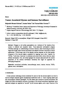
Results Migration of activated, DiI-labelled antigenspecific CD4 lymphocytes and antigen-nonspecific splenocytes into the brain and pancreas Within 2 h after adoptive transfer, MBP-specific, DiI-labelled C19lo cells, which express low levels of VLA-4, could be visualized within perivascular brain regions, tissue parenchyma and in the meningeal space (Fig. 1A, C and D). These fluorescent cells could also be detected by anti-CD4 immunocytochemistry (Fig. 1B). By 24 h, multiple labelled lymphoid perivascular cells could be visualized as well as occasional clusters of cells within the meninges and choroid plexus (Fig. 1D and E). However, large clusters of parenchymally located lymphocytes cells in the brain were seen only after transfer of cells that expressed high levels of VLA-4 and were specific for MBP (Fig. 2A). C19lo cells did not form these ‘hot spots’ in the brain, nor did cells with
high levels of VLA-4 accumulate in the pancreas 24 h after transfer (Fig. 2B). Antigen-non-specific anti-CD3-activated splenocytes were also visualized in the brain within 2 h but did not form small or large clusters at 24 h. When the number of cells in the sections was quantitated, as shown in Fig. 3A, C19lo cells and activated splenocytes were found to have entered the brain with similar efficiency within the first 2 h. As expected, there was a higher concentration of antigen-specific C19lo cells in the brain at 24 h than at 2 h, whereas most of the antigen-non-specific cells had disappeared; this result suggested antigen-specific recruitment or proliferation. Similarly, the numbers of C19lo cells entering the pancreas by 2 h was approximately the same as in the brain, but by 24 h there appeared to be greater numbers of cells in the brain than in the pancreas (Fig. 3B). The same trend was observed for the MBP-specific cell line (Fig. 4A). In addition, the ability of the low VLA-4-expressing C19lo clone to enter the brain within the first 2 h after adoptive transfer was similar to that of the high VLA-4-expressing MBP-specific T-cell line (Fig. 4B). However, recruitment or proliferation at 24 h was impaired in C19lo cells because much larger numbers of cells of the MBP-specific T-cell line were present at that later time. These observations suggested that VLA-4 is not necessary for the early migration but that subsequent recruitment or proliferation requires VLA-4 expression. Because the fluorescence intensity of the labelled cells declines with the number of cell divisions, a large percentage of the high VLA-4-expressing MBP-specific T cells probably represents recruitment, as judged by the fluorescence intensity in cell clusters (Fig. 2B).
Adhesion molecule, MHC class II and IFN-γ expression after adoptive transfer of C19lo cells or activated splenocytes In order to demonstrate that the C19lo cells can have a physiological effect and mediate the activation of the brain endothelium and immune microenvironment, we performed immunohistochemistry 4, 24, 48, 72, 96 and 168 h after adoptive transfer. As expected, endothelial VCAM and ICAM expression began to increase at 24 h, peaked at 48 h and began to decline at 96 h after adoptive transfer (Fig. 5A–D). Furthermore, at 48 and 72 h, MHC class II expression was elevated on both the endothelial surface and on parenchymal cells (Fig. 5E and F); meningeal IFN-γ staining was present at the same time points (Fig. 5G and H). Similar, but less intense, changes in VCAM and ICAM were observed after transfer of activated splenocytes. In contrast, P selectin was detectable 2 h after adoptive transfer in the choroid plexus and meninges but not at 48 h (Fig. 5I and J). Perivascular staining for P selectin in the brain parenchyma was not detectable.
Anti-P selectin monoclonal antibody, but not anti-VLA-4, inhibits early migration Our next goal was to attempt to block early migration with inhibitors of known adhesion events. To detect differences
P selectin mediates early T-cell migration
1095
Fig. 1 Migration of DiI-labelled C19lo cells. After transfer of 1 ⫻ 107 CD4⫹ MBP-specific T lymphocytes (C19lo cells), migration of single fluorescent cells into brain parenchyma could be visualized within 2 h (A, ⫻125, fluorescence microscopy with Nomarski optics). Using immunocytochemistry, these cells could also be identified by anti-CD4 staining (B, ⫻125, alkaline phosphatase counterstained with haematoxylin). Solitary perivascular (C, ⫻125, fluorescence microscopy with Nomarski optics) and occasional parenchymal doublets within the same microscopic field (D, ⫻62.5, fluorescence microscopy with Nomarski optics) were also seen within 2 h of transfer. Twenty-four hours after transfer, multiple perivascular (E, ⫻125, fluorescence microscopy with Nomarski optics) and meningeal (F, ⫻62.5, fluorescence microscopy with Nomarski optics) clusters of cells were observed. Parenchymal and perivascular cells were located predominantly in subcortical and periventricular brain regions. Meningeal cells were located in the lateral ventricles as shown (F), as well as in the cerebral convexities and the remainder of the ventricular system (not shown).
of a small number of cells between treatment groups and controls, we reasoned that interassay variance due to differences in labelling efficiency, perfusion and sectioning techniques could be corrected by assigning matched controls,
both untreated and isotype antibody-treated, to treatment animals for a given experiment and then expressing the data as a percentage of control migration. The density of cells in untreated mice was defined as 100%, and statistical differ-
1096
M. D. Carrithers et al.
Fig. 2 Migration of a DiI-labelled MBP-specific T-cell line. (A) Twenty-four hours after transfer, large numbers of high VLA-4expressing, MBP-specific T cells were observed within perivascular and parenchymal regions in the brain. Here a large cluster of periventricular cells is shown (⫻125, fluorescence microscopy with Nomarski optics). (B) At the same time point, only very rare cells were seen in the pancreas (⫻62.5, fluorescence microscopy with Nomarski optics).
Fig. 3 Quantitation of C19lo and splenocyte migration. Fluorescent cells were counted manually in tissue sections as described. (A) After transfer of 107 labelled cells, peak migration of C19lo cells (filled bars) (~20 cells/mm3) occurred at 24 h. In contrast, although antigennon-specific splenocytes (open bars) activated by anti-CD3 also entered the brain rapidly, peak migration occurred at 2 h, with significantly lower levels at 24 h. (B) Migration of labelled C19lo cells into the pancreas (open bars) and brain (filled bars) was similar at 2 h, but significantly higher levels were seen in the brain by 24 h. Each data point represents the mean from either 24 brain sections or eight pancreatic sections from a representative animal. A similar trend was noted in each of the three repetitions of each of the experiments.
ences were compared between isotype-treated animals and the treatment group. In these experiments, using the high VLA-4-expressing, MBP-specific T-cell lines, we examined the roles of P selectin and α4 integrin in pioneer T-lymphocyte migration 2 h after adoptive transfer. We hypothesized that P selectin, which appeared to be induced very rapidly on the endothelial surface
after adoptive transfer, would impair early migration, while blocking α4 integrin would not. The monoclonal antibody to VLA-4 (rat monoclonal antibody R1–2) has been shown previously to inhibit recruitment of lymphocytes to the CNS in EAE (Baron et al., 1993). The monoclonal antibodies used are summarized in Table 1, and the data are shown in Table 2. Control migration was 33 ⫾ 6 cells/mm3 (mean ⫾
P selectin mediates early T-cell migration
1097
Fig. 4 Quantitation of DiI-labelled MBP-specific T-cell migration. (A) Two hours after transfer of a VLA-4 expressing MBP-specific T-cell line, similar numbers of fluorescent cells were observed in the pancreas (open bars) and brain (filled bars) at 2 h, but significantly greater numbers were present in the brain by 24 h. (B) Compared with the VLA-low C19lo cells (open bars), recruitment at 24 h but not 2 h was significantly increased for the high VLA-4-expressing T-cell line (filled bars). Each data point represents the mean from either 24 brain sections or eight pancreatic sections from a representative animal. A similar trend was noted in each of the three repetitions of each of the experiments.
standard error of the mean). As shown, anti-VLA-4 at a dose of 250 µg per animal (~10 µg/g mouse weight) had little effect on migration at this time point, whereas anti-P selectin (rat monoclonal antibody RB40.34) at 125 µg per recipient (~5 µg/g mouse weight) inhibited migration to 49% of the control value. The differences between the anti-P selectin group and the isotype control were statistically significant (P ⬍ 0.01).
Discussion We have demonstrated in an in vivo model that endothelial P selectin but not lymphocyte VLA-4 facilitates migration of pioneer Th1 CD4 lymphocytes into the brain. Although previous studies have demonstrated that ICAM-1 and VCAM do not appear to be involved in CNS homing in the absence of an inflammatory focus (Irani and Griffin, 1996; Hickey et al., 1997), to our knowledge this study represents the first demonstration of an important molecular event in pioneer T-lymphocyte migration into the brain. Our quantitative results for the initial step of pioneer T-lymphocyte migration into the brain and the pancreas at 2 h were independent of cell type or specificity, whereas subsequent antigen-dependent recruitment by 24 h was very similar to the results of Hickey and colleagues (Hickey et al., 1991) if their data are converted to cells/mm3. For example, for all cell types tested in our experiments, ~10–40 cells were observed per mm3; based on section thickness, the range in the study of Hickey and colleagues was ~20–50 cells/mm3. Furthermore, although Hickey and colleagues were able to demonstrate trends in the number of
cells entering the tissue at various time points, the standard errors were relatively large and they were unable to detect statistical differences between time points. Despite the fact that that counting fluorescent cells proved easier than staining and counting CD4⫹ cells in the CNS, refinement of the technique was required to correct these problems. For this reason, we used isotype antibody-matched controls to permit statistical comparison between treatment groups that contained a relatively small number of observations. Similar endothelial and immune activation has been shown previously to correlate with leucocyte entry into the CNS (Cannella et al., 1990, 1991). Another study also demonstrated that MHC class II upregulation on brain astrocytes is hyperinducible by IFN-γ in rodent strains susceptible to EAE (Massa et al., 1987). Whether early P selectin expression or later VCAM or ICAM-1 endothelial expression is also hyperinducible is unclear. Further work would be clinically relevant because of genetic studies in EAE which suggest that endothelial factors may be involved in the pathogenesis of EAE and human multiple sclerosis (Teuscher et al., 1999). The present results demonstrating the surface expression of P selectin during early migration but not at 48 h after adoptive transfer are also consistent with previous studies. In most endothelial cells, P selectin is stored in Weibel–Palade bodies and can be transported rapidly to the cell membrane surface after early inflammatory signals such as thrombin and histamine (Wagner et al., 1982; Hattori et al., 1989). On CNS endothelium, constitutive expression of P selectin is very low or absent (Engelhardt et al., 1997), but expression can be induced by cytokine stimulation (Barkalow et al., 1996). Although
1098
M. D. Carrithers et al.
Fig. 5 Immunohistochemistry. Forty-eight hours after transfer of MBP-specific C19lo cells, increased expression of VCAM (B) and ICAM-1 (D) was observed on the endothelial surface compared with 2 h after transfer (A and C) (⫻62.5, alkaline phosphatase counterstained with haematoxylin). Significant but less robust changes were seen in VCAM and ICAM-1 expression 48 h after transfer of activated splenocytes (not shown). These changes in endothelial staining were most prominent in subcortical and periventricular brain regions. In addition, increased parenchymal expression of MHC class II (F) and meningeal IFN-γ (H) was noted 48 h after transfer of C19lo cells compared with 2 h (E and G) and compared with mice that received activated splenocytes (⫻62.5, alkaline phosphatase counterstained with haematoxylin). In contrast, staining for P selectin was observed in the choroid plexus of the lateral ventricle 2 h (I) after adoptive transfer but not at 48 h (J) (⫻62.5, alkaline phosphatase counterstained with haematoxylin).
Table 1 Summary of blocking monoclonal antibodies used Monoclonal antibody
Clone name
Isotype
Species source
Dose (µg/g mouse weight)
Isotype control Anti-α4 integrin (anti-VLA-4) Anti-P selectin (anti-CD62P)
R3–34 R1–2 RB40–34
IgG1, κ IgG2b, κ IgG1, κ
Rat anti-mouse Rat anti-mouse Rat anti-mouse
5 10 5
P selectin mediates early T-cell migration Table 2 Anti-P selectin monoclonal antibody, but not antiVLA-4, inhibits early migration Condition
Cells/mm3 (% of control ⫾ SEM)
n
Control Rat IgG1 Anti-VLA-4 Anti-P selectin
100 99 ⫾ 3* 92 ⫾ 7 49 ⫾ 2*
7 3 4 4
*P ⬍ 0.01.
brain endothelium appears to contain Weibel–Palade bodies, these bodies do not appear to contain P selectin. However, cytokines can mediate the upregulation of the surface expression of P selectin on brain endothelium through transcriptional upregulation (Barkalow et al., 1996). Based on our working model of pioneer T-lymphocyte migration into the brain, we hypothesize that P selectin is expressed constitutively, but at very low levels, on brain endothelium and can be upregulated in a cytokine-dependent fashion after adoptive transfer of activated Th1 lymphocytes. This speculation is supported by a recent study that demonstrated that membrane-bound or membrane-associated cytokines, particularly TNF-α (tumour necrosis factor α) from activated lymphocytes can mediate rapid endothelial activation within a few hours (Lou et al., 1996). In addition, a previous study showed that P selectin did not appear to be necessary for the recruitment of encephalitogenic cells in EAE and may be downregulated on brain endothelium during active disease (Engelhardt et al., 1997). We cannot discount the possibility that anti-P selectin treatment predominantly inhibited brain-epithelial migration of lymphocytes into the cerebrospinal fluid through the choroid plexus rather than migration across the brain endothelium (Steffen et al., 1996). A previous study demonstrated that, in cytokine-induced meningitis in mice, P selectin facilitates early migration of neutrophils and mononuclear cells into the cerebrospinal fluid (Tang et al., 1996). However, on the basis of our qualitative results there does not appear to be a clear reduction in the number of cells in the choroid–meningeal regions as opposed to the parenchymal– perivascular regions. Further studies are required to determine the differences in migration into these two unique compartments. On the basis of on these preliminary results, we have formulated the following working model. Initial attachment and rolling of activated pioneer T lymphocytes to the brain endothelium occurs via P selectin, which is either constitutively expressed at low levels or is rapidly induced throughout the endothelium by activated T cells. After this attachment, a chemokine receptor-dependent step occurs that can activate lymphocyte integrins. Firm adhesion does not appear to be mediated by VLA-4 but may be mediated by the binding of LFA-1 to a member of the ICAM family. Subsequent migration and antigen presentation within the perivascular space
1099
causes endothelial and microenvironmental activation, which leads to higher-affinity VLA-4-dependent recruitment within 24 h. Further elucidation of these steps is necessary to fully understand homing to the brain by lymphocyte subsets during immune surveillance and in disease states. One may expect, for example, that disease-causing Th1 CD4 cells use a different molecular pathway than Th2 lymphocytes. After activation, Th1 cells, but not Th2, cells express the enzyme α(1,3)fucosyl transferase VII, which is required for the fucosylation of PSGL-1 and for subsequent binding to P and E selectin (Maly et al., 1996; Austrup et al., 1997; Borges et al., 1997). This functional difference permits Th1 cells to home specifically to inflamed skin (Austrup et al., 1997; Borges et al., 1997). This model also suggests that therapies for multiple sclerosis designed to inhibit T-lymphocyte migration into the brain should target multiple molecules at different time points during lesion development if they are to achieve optimal efficacy. For example, in addition to their numerous other biological effects, β-interferons may be effective in the treatment of individuals with relapsing–remitting and secondary progressive disease because of their inhibitory effect on gelatinase B, a matrix metalloproteinase (MMP-9) (Leppert et al., 1996; Stuve et al., 1996). Gelatinase B is expressed by activated T lymphocytes and is required for the migration of cells across the perivascular basement membrane and into tissue parenchyma. MMPs and other proteases may be responsible for the breakdown of the blood–brain barrier in acute multiple sclerosis lesions, which can be demonstrated by gadolinium enhancement of MRI images. It is not surprising, therefore, that inhibition of MMP activity by β-IFNs results in a marked decrease in the number of new enhancing lesions in multiple sclerosis patients (Stone et al., 1995). Similarly, a recent short-term study of an antiVLA-4 monoclonal antibody (Antegren, natalizumab) also demonstrated a reduction in new active and enhancing lesions during the first 12 weeks of follow-up (Tubridy et al., 1999). As VLA-4 ligation by VCAM can initiate MMP activation in T lymphocytes to facilitate migration (Romanic and Madri, 1994), both MMP inhibitors and blockers of VLA-4 are probably acting during the recruitment phase of a developing multiple sclerosis lesion. In other words, these agents seem to be most efficacious at a time point in pathogenesis after pioneer T lymphocyte entry but before breakdown of the blood–brain barrier severe enough to be visualized by gadolinium enhancement on MRI. Although inhibition of early lymphocyte entry would not be useful for the treatment of acute clinical multiple sclerosis exacerbations, it may be of benefit in combination therapy with an IFN-β or natalizumab to prevent clinical relapses. Clearly, additional laboratory studies are required to identify mediators of early migration that can be blocked without interfering with immune surveillance of other tissues. The greatest challenge will be to design rational combination treatments that can be tested with sufficient statistical power in clinical trials.
1100
M. D. Carrithers et al.
Acknowledgements We wish to thank B. Dittel, D. Graesser, J. Madri, P. Preston-Hurlburt and S. Wong for helpful discussions, G. Losyev for the preparation of monoclonal antibodies, C. Annicelli for animal care and L. Carrithers for assistance with the graphics. This work was supported by the Howard Hughes Medical Institute and National Institutes of Health grants AI/AR 36529 (to C.A.J.) and KO8 NS 02124–01 (to M.D.C.). M.D.C. was previously an Advanced Postdoctoral Fellow of the National Multiple Sclerosis Society (FA1237-A-1).
References Austrop F, Vestweber D, Borges E, Lohning M, Brauer R, Herz U, et al. P- and E-selectin mediate recruitment of T-helper-1 but not T-helper-2 cells into inflamed tissues. Nature 1997; 385: 81–3. Barkalow FJ, Goodman MJ, Gerritsen ME, Mayadas TN. Brain endotheliums lack one of two pathways of P-selectin-mediated neutrophil adhesion. Blood 1996; 88: 4585–93. Baron JL, Madri JA, Ruddle NH, Hashim G, Janeway CA Jr. Surface expression of α4 integrin by CD4 T cells is required for their entry into brain parenchyma. J Exp Med 1993; 177: 57–68. Borges E, Tietz W, Steegmaier M, Moll T, Hallmann R, Hamann A, et al. P-selectin glycoprotein ligand-1 (PSGL-1) on T helper 1 but not on T helper 2 cells binds to P-selectin and supports migration into inflamed skin. J Exp Med 1997; 185: 573–8. Brightman MW, Reese TS. Junctions between intimately apposed cell membranes in the vertebrate brain. J Cell Biol 1969; 40: 648–77. Butcher EC, Picker LJ. Lymphocyte homing and homeostasis. [Review]. Science 1996; 272: 60–6. Cannella B, Cross AH, Raine CS. Upregulation and coexpression of adhesion molecules correlate with relapsing autoimmune demyelination in the central nervous system. J Exp Med 1990; 172: 1521–4. Cannella B, Cross AH, Raine CS. Adhesion-related molecules in the central nervous system; upregulation correlates with inflammatory cell influx during relapsing experimental autoimmune encephalomyelitis. Lab Invest 1991; 65: 23–31. Dittel BN, Merchant RM, Janeway CA Jr. Evidence for Fasdependent and Fas-independent mechanisms in the pathogenesis of experimental autoimmune encephalomyelitis. J Immunol 1999; 162: 6392–400. Engelhardt B, Vestweber D, Hallmann R, Schulz M. E- and Pselectin are not involved in the recruitment of inflammatory cells across the blood–brain barrier in experimental autoimmune encephalomyelitis. Blood 1997; 90: 4459–72. Graeber MB, Streit WJ, Buringer D, Sparks DL, Kreutzberg GW. Ultrastructural location of major histocompatibility complex (MHC) class II positive perivascular cells in histologically normal human brain. J Neuropathol Exp Neurol 1992; 51: 303–11.
Hardardottir F, Baron JL, Janeway CA Jr. T cells with two functional antigen-specific receptors. Proc Natl Acad Sci USA 1995; 92: 354–8. Hattori R, Hamilton KK, Fugate RD, McEver RP, Sims PJ. Stimulated secretion of endothelial von Willebrand factor is accompanied by rapid redistribution to the cell surface of the intracellular granule membrane protein GMP-140. J Biol Chem 1989; 264: 7768–71. Hickey WF, Kimura H. Perivascular microglia cells of the CNS are bone marrow-derived and present antigen in vivo. Science 1988; 239: 290–2. Hickey WF, Hsu BL, Kimura H. T-lymphocyte entry into the central nervous system. J Neurosci Res 1991; 28: 254–60. Hickey WF, Lassmann H, Cross AH. Lymphocyte entry and the initiation of inflammation in the central nervous system. In: Keane RW, Hickey WF, editors. Immunology of the nervous system. New York: Oxford University Press; 1997. p. 200–25. Irani DN, Griffin DE. Regulation of lymphocyte homing into the brain during viral encephalitis at various stages of infection. J Immunol 1996; 156: 3850–7. Kermode AG, Thompson AJ, Tofts P, McManus DG, Kendall BE, Kingsley DP, et al. Breakdown of the blood brain barrier precedes symptoms and other MRI signs of new lesions in multiple sclerosis. Brain 1990; 113: 1477–89. Leppert D, Waubant E, Burk MR, Oksenberg JR, Hauser SL. Interferon beta-1b inhibits gelatinase secretion and in vitro migration of human T cells: a possible mechanism for treatment efficacy in multiple sclerosis. Ann Neurol 1996; 40: 846–52. Lou J, Dayer JM, Grau DE, Burger D. Direct cell/cell contact with stimulated T lymphocytes induces the expression of cell adhesion molecules and cytokines by human brain microvascular endothelial cells. Eur J Immunol 1996; 26: 3107–13. Maly P, Thall AD, Petryniak B, Rogers CE, Smith PL, Marks RM, et al. The alpha(1,3)fucosyltransferase Fuc-TVII controls leukocyte trafficking through an essential role in L-, E-, and P-selectin ligand biosynthesis. Cell 1996; 86: 643–53. Massa PT, ter Meulen V, Fontana A. Hyperinducibility of Ia antigen on astrocytes correlates with strain-specific susceptibility to experimental autoimmune encephalomyelitis. Proc Natl Acad Sci USA 1987; 84: 4219–23. Reese TS, Karnovsky MJ. Fine structural localization of a blood– brain barrier to exogenous peroxidase. J Cell Biol 1967; 34: 207–17. Romanic AM, Madri JA. The induction of 72-kD gelatinase in T cells upon adhesion to endothelial cells is VCAM-1 dependent. J Cell Biol 1994; 125: 1165–78. Springer TA. Traffic signals for lymphocyte recirculation and leukocyte emigration: the multistep paradigm. [Review]. Cell 1994: 76; 301–14. Steffen BJ, Breier G, Butcher EC, Schulz M, Engelhardt B. ICAM1, VCAM-1, and MAdCAM-1 are expressed on choroid plexus epithelium but not endothelium and mediate binding of lymphocytes in vitro. Am J Pathol 1996; 148: 1819–38. Stone LA, Frank JA, Albert PS, Bash C, Smith ME, Maloni H, et al. The effect of interferon beta on blood–brain barrier disruptions
P selectin mediates early T-cell migration
1101
demonstrated by contrast-enhanced magnetic resonance imaging in relapsing–remitting multiple sclerosis. Ann Neurol 1995; 37: 611–9.
susceptibility to monophasic remitting/nonrelapsing experimental allergic encephalomyelitis. J Immunol 1999; 163: 2262–6.
Stuve O, Dooley NP, Uhm JH, Antel JP, Francis GS, Williams G, et al. Interferon beta-1b decreases the migration of T lymphocytes in vitro: effects on matrix metalloproteinase-9. Ann Neurol 1996; 40: 853–63.
Tubridy N, Behan PO, Capildo R, Chaudhuri A, Forbes R, Hawkins CP, et al. The effect of anti-α4 integrin antibody on brain lesion activity in MS. Neurology 1999; 53: 466–72.
Tang T, Frenette PS, Hynes RO, Wagner DD, Mayadas TN. Cytokine-induced meningitis is dramatically attenuated in mice deficient in endothelial selectins. J Clin Invest 1996; 97: 2485–90. Teuscher C, Butterfield RJ, Ma RZ, Zachary JF, Doerge RW, Blankenhorn EP. Sequence polymorphisms in the chemokines Scya1 (TCA-3), Scya2 (monocyte chemoattractant protein (MCP)-1), and Scya12 (MCP-5) are candidates for eae7, a locus controlling
Wagner DD, Olmsted JB, Marder VJ. Immunolocalization of von Willebrand protein in Weibel–Palade bodies of human endothelial cells. J Cell Biol 1982; 95: 355–60. Wekerle H, Linington G, Lassmann H, Meyermann R. Cellular immune reactivity within the CNS. Trends Neurosci 1986; 9: 271–77. Received September 28, 1999. Revised January 3, 2000. Accepted January 24, 2000