Research Article For reprint orders, please contact:
[email protected]
d -
Development of mannose-anchored thiolated amphotericin B nanocarriers for treatment of visceral leishmaniasis
Aim: Our goal was to improve treatment outcomes for visceral leishmaniasis by designing nanocarriers that improve drug biodistribution and half-life. Thus, longacting mannose-anchored thiolated chitosan amphotericin B nanocarrier complexes (MTC AmB) were developed and characterized. Materials & methods: A mannoseanchored thiolated chitosan nanocarrier was manufactured and characterized. MTC AmB was examined for cytotoxicity, biocompatibility, uptake and antimicrobial activities. Results: MTC AmB was rod shaped with a size of 362 nm. MTC AmB elicited 90% macrophage viability and 71-fold enhancement in drug uptake compared with native drug. The antileishmanial IC50 for MTC AmB was 0.02 μg/ml compared with 0.26 μg/ml for native drug. Conclusion: These studies show that MTC can serve as a platform for clearance of Leishmania in macrophages. First draft submitted: 2 September 2016; Accepted for publication: 26 October 2016; Published online: 23 November 2016 Keywords: amphotericin B • macrophage nanoparticle targeting • mannose receptors • thiolated chitosan • visceral leishmaniasis
Visceral leishmaniasis (VL; kala-azar) remains a common tropical infectious disease. Leishmania amastigotes and promastigotes target mononuclear phagocytes (MP; monocytes and tissue macrophages), and parasite growth is sustained by the cell’s microenvironment. Infection remains high in prevalence, morbidity and mortality in the developing world [1] . The obligate intracellular Leishmania donovani amastigotes replicate within membrane-bound MP subcellular organelles. Current medical management is not effective for eliciting microbial clearance due to drug resistance, toxicity, bioavailability and cost [2] . Chemotherapy for VL is based on the use of antimony. However, the emergence of resistance has transformed medical management to the use of amphotericin B (AmB) for VL treatment [2,3] . Notably, targeted intracellular delivery of AmB has now emerged as a first-line medical strategy to facilitate pathogen clearance. The develop-
10.2217/nnm-2016-0325 © Benson Edagwa
ment of improved drug delivery formulations that include nanotechnology-based targeted therapeutics that target infected cells and tissues could improve treatment outcomes [3,4] . It is well accepted that mannose-based carriers can be harnessed to improve antileishmanial drug delivery. This idea is based on the target macrophage surface receptor distribution [2–4] . Proof of concept for such an approach includes, but is not limited to, drugs such as muramyl dipeptide, efavirenz and rifampin [5–7] . Each has yielded encouraging results. Mannose receptors recognize corresponding sugars and facilitate cellular uptake of drug-encased particles [8,9] . Consequent internalization of the therapeutic carrier facilitates drug accumulation at sites of active parasitic infection [8] . Such receptormediated MP drug nanoparticle targeting is linked to the effectiveness of the ligandanchored therapeutic carrier. This serves to improve drug cell entry, retention and cargo
Nanomedicine (Lond.) (2017) 12(2), 99–115
Gul Shahnaz1,2, Benson J Edagwa*,2, JoEllyn McMillan2, Sohail Akhtar3, Abida Raza4, Naveeda A Qureshi5, Masoom Yasinzai6 & Howard E Gendelman2 1 Department of Pharmacy, Quaid-i-Azam University Islamabad, 45320, Pakistan 2 Department of Pharmacology & Experimental Neuroscience, University of Nebraska Medical Center, Omaha, NE 68198, USA 3 Department of Entomology, University College of Agriculture & Environmental Sciences, The Islamia University, Bahawalpur, Pakistan 4 Nuclear Medicine, Oncology & Radiotherapy Institute, Islamabad, Pakistan 5 Department of Animal Sciences, Quaid-i-Azam University, Islamabad, 45320, Pakistan 6 Centre for Interdisciplinary Research in Basic Sciences, International Islamic University, Islamabad, Pakistan *Author for correspondence: benson.edagwa@ unmc.edu
part of
ISSN 1743-5889
99
Research Article Shahnaz, Edagwa, McMillan et al. release in an infected or bystander cell [9] . However, the question of finding the optimal vehicle for drug-based macrophage carriages has not yet been addressed. One answer is chitosan-based nanocarriers. These carriers have gained considerable interest due to their tunable functional groups, biocompatibility and biodegradability [10] . Thiolated chitosan (TC) such as chitosan–thioglycolic acid conjugates can be generated by immobilization of thiol groups on the chitosan polymer backbone [11] . Formation of inter- and intramolecular disulfide bonds within the thiolated polymer matrix results in improved properties, such as in situ gelling/cohesion that facilitates the drug delivery system stability [12,13] . Specifically, mannosylation of TC polymer (MTC) is believed to be an appealing strategy for targeted intracellular MP delivery of AmB as it would not only aid in parasite clearance but also help reduce the dosing volume and frequency. To date, there is only limited research that has been conducted to investigate the potential of mannose-anchored thiolated nanocarriers for VL and linked parasitic disease therapy [2,3,14] . In this study, improved mannoseanchored TC (MTC) nanocarriers were developed to facilitate stability, biocompatibility, controlled release and cell uptake by macrophages for antileishmanial therapy. Materials & methods Materials
Low MW chitosan, cysteine, hydrogen peroxide, Ellman’s reagent (5,5-dithiobis[2-nitrobenzoic acid]), AmB, acetonitrile, methanol, mannose and DMSO were purchased from Sigma-Aldrich (MO, USA). Pooled human serum was obtained from Innovative Biologics (VA, USA). Solvents used were HPLC-grade materials. TC polymer synthesis
The synthesis of the thiolated polymer was made through covalent linkage of thioglycolic acid (TGA) to chitosan by amide bond formation between the polymer amino groups and carboxylate groups of the sulfhydryl moiety [12] . Briefly, chitosan 1% (w/v) was hydrated with 1% (v/v) acetic acid solution. This was by followed the addition of 500 mg of TGA after which 1-ethyl-3-(3-dimethylaminopropyl) carbodiimide hydrochloride-coupling reagent at a final concentration of 100 mM was added to activate the carboxylic acid moieties of TGA. The pH of the reaction mixture was adjusted to 5.5 with 10 M NaOH, and mixtures were incubated for 3.5 h under continuous stirring. The solution was dialyzed five-times in a membrane tubing (MW cut-off 12–14 kDa) for 3 days under protection from light at 10°C in order to separate unbound sulf-
100
Nanomedicine (Lond.) (2017) 12(2)
hydryl moieties from the thiolated polymer. Briefly, the crude thiolated polymer was dialyzed once against 5 mM HCl (5 l), then twice against the same media of 1% (w/v) NaCl to break ionic interactions between the positively charged polymer and the negatively charged sulfhydryl moieties. Finally, the samples were dialyzed thoroughly twice against 1 mM HCl to adjust the pH of the TC solution. The dialyzed product was lyophilized and stored at 4°C for subsequent use. MTC synthesis & characterization
Mannose was linked onto TC as described with modifications [15] . Mannose immobilization was carried out by reductive amination of TC. Two percent TC was dissolved in 1% v/v aqueous acetic acid by stirring at room temperature. Sodium 0.12 M cyanoborohydride and 0.33 M D-mannose were then added to the resulting viscous polymer solution with vigorous stirring. The resulting reaction mixture was continuously stirred until a white foamy gel was formed. The gel was smashed up then rinsed four-times with 150 ml methanol and once with diethyl ether (150 ml). The light pink solid product was then dried for several hours. Quantitation of primary amine functional groups at each level of modification was determined by a colorimetric 2,4,6-trinitrobenzenesulfonic acid (TNBS) assay. In this assay, the polymer (0.5 mg) was dissolved in 500 μl NaCl (0.5%, w/v) solution. After incubation at 25°C for 30 min, 500 μl of TNBS (0.1%, w/v) comprising NaHCO3 (4%, w/v) was incorporated into each hydrated aliquot. After 3 h of incubation at 37°C and centrifugation (33,527 × g; 5 min; 4°C), absorbance was recorded at 410 nm on a microtiter plate reader (Molecular Devices, CA, USA). L-cysteine HCl standards were used for the calculations. The degree of conjugation as assessed by the amount of thiol groups anchored to TC and MTC was determined by Ellman’s reagent. First, conjugates (0.5 mg) were hydrated in 500 μl of phosphate buffer (0.5 M; pH 8.0). Then 500 μl of Ellman’s reagent (3 mg in 10 ml of 0.5 M phosphate buffer) was added into each aliquot. The mixtures were incubated for 3 h at room temperature. The thiol content on TC and MTC was then determined by measuring the absorbance of 300 μl of the mixture at 410 nm on a microtiter plate reader (Molecular Devices). L-cysteine HCl was used as a standard to quantify the sulfhydryl on the MTC. The extent of disulfide bond formation due to air oxidation during polymer modifications was measured as previously reported [12] . TC and MTC (0.5 mg) were swelled in Falcon tubes (15 ml) containing 1 ml of 0.05 M Tris buffer, pH 6.8. After 30 min of incubation at room temperature, 1 ml of a 4% (w/v) sodium-borohydride solution was added to the reaction mixtures. The
future science group
Mannose-anchored thiolated amphotericin B nanocarriers for treatment of visceral leishmaniasis
Research Article
Table 1. Composition (polymer, surfactant, drug and cross-linking agent ratio) and physicochemical characterization including polydispersity index, particle size, payload and ζ-potential of the ionically cross-linked nanocarriers used in study (designated as 1, 2 and 3). Formulations
Polymer % (m/v)
Tween % (v/v)
TPP (μM)
Drug % (m/v)
Size (nm)
PDI
Payload %
ζ-potential (mv)
Unmodified chitosan UC -1
0.3
16.6
0.35
0.1
1048 ± 54
0.83 ± 0.72
21.0
33 ± 2
UC -2
0.3
16.6
0.40
0.1
827 ± 12
0.65 ± 0.31
35.0
30 ± 7
UC -3
0.3
16.6
0.54
0.1
449 ± 38
0.31 ± 0.01
47.0
28 ± 3
Thiolated chitosan TC -1
0.3
16.6
0.4
0.1
654 ± 41
0.71 ± 0.06
24.5
29 ± 2
TC -2
0.3
16.6
0.6
0.1
495 ± 13
0.29 ± 0.03
64.8
23 ± 0.9
TC -3
0.3
16.6
0.7
0.1
1032 ± 14
0.61 ± 0.02
35.0
21 ± 4
Mannose-anchored thiolated chitosan MTC -1
0.3
16.6
0.4
0.1
1012 ± 18
0.42 ± 0.01
53.8
29 ± 4
MTC -2
0.3
16.6
0.6
0.1
478 ± 9
0.25 ± 0.01
72.9
20 ± 1
MTC -3
0.3
16.6
0.7
0.1
951 ± 27
0.91 ± 0.08
26.0
19 ± 0.8
Indicated values are mean ± SD of at least three experiments. MTC: Mannose-anchored thiolated chitosan; PDI: Polydispersity index; SD: Standard deviation; TC: Thiolated chitosan; UC: Unmodified chitosan; TPP: Sodium tripolyphosphate.
samples were then oscillated for 3 h in a shaking water bath at 37°C. The mixture was quenched by the addition of 200 μl of 5 M HCl. Ellman’s reagent (500 μl of 0.4% [w/v]) was added to determine the remaining thiol content as described above. Fourier transform IR & proton NMR spectroscopy
Immobilization of mannose on TC was elucidated further by Fourier transform IR (FTIR; Schimadzu, Model 8400, Japan) and proton NMR (1H NMR; 500 MHz; Varian Medical Systems, Inc., CA, USA) spectroscopy. FTIR spectroscopy was acquired by the KBr disk method over the range of 4000–400 cm-1. 1 H NMR spectroscopy was conducted in deuterated DMSO. Preparation of the nanocarriers
Nanocarriers of unmodified chitosan (UC), TC and MTC were synthesized using in situ gellation. Specifically, AmB was dissolved in 2.5% (v/v) aqueous acetic acid solution and Tween 80 to a final concentration shown in Table 1. The polymer (0.33%, w/v) was then added to the mixture and the reaction allowed to proceed for 2 h. Afterward the product was mechanically mixed using high-pressure homogenization. The purified nanocarriers were then stabilized by adding an aqueous solution of sodium tripolyphosphate crosslinker at the final concentrations shown in Table 1. The synthesized particles were categorized as either clear, opalescent or agglomerates based on qualitative visual
future science group
evaluation. The nanocarriers were then partially oxidized with 0.5% (v/v) H2O2 solution after incubation for 1 h under continuous stirring at room temperature. The resultant opalescent product of desired physicochemical parameters was selected for further characterization and investigations as shown in Table 2. To prevent particle aggregation during differential centrifugation (33,527 × g; 10 min; 4°C), 5% (w/v) trehalose was added to the nanoformulation before this step. After centrifugation, the supernatant was removed and the pellet washed twice with water before resuspending in 0.1% (v/v) aqueous acetic acid solution (2 ml) and stored at 4°C for subsequent use. Particle size, surface charge and polydispersity index (PDI) of the nanocarriers were determined by dynamic light scattering using a Malvern Zetasizer Nano Series Nano-ZS (Malvern Instruments, Inc., MA, USA). Encapsulation efficiency of AmB in UC, TC and MTC was determined after centrifugation (14,000 × g, 60 min, 4°C) of nanoparticle suspensions to remove unentrapped insoluble drug. The pelleted nanocarriers were dissolved in methanol and ultrasonicated for 10 min to facilitate particle rupture. The drug released into the supernatant after centrifugation (14,000 × g, 5 min, 4°C) was quantified by HPLC [16] . Drug encapsulation efficiency was calculated using the formula: EE = Total amount of drug in formulation - Unentrapped drug # 100 Total amount of drug
www.futuremedicine.com
101
Research Article Shahnaz, Edagwa, McMillan et al.
Table 2. Characterization in terms of thiol groups, disulfide bonds, polydispersity index, particle size, payload and ζ-potential of covalently cross-linked (oxidized) nanocarriers. Formulations
-SH [μMol/g]
-S-S- [μMol/g]
Size (nm)
PDI
Payload %
ζ-potential (mv)
UC
-
-
449 ± 38 *
0.31 ± 0.01
47.0 **
28 ± 4
TC
1283 ± 96
82 ± 27
495 ± 13
0.29 ± 0.03
64.8
23 ± 0.9
Thiolated chitosan (Ox)
857 ± 45
389 ± 52
390 ± 26 §*
0.21 ± 0.02
69.5 §*
21 ± 0.6
MTC
1269 ± 101
62 ± 18
478 ± 9
0.25 ± 0.01
72.9
20 ± 1
Mannose-anchored Ox
781 ± 95
454 ± 113
362 ± 27‡*
0.22 ± 0.03
78.3‡***
19 ± 3
†
†
Values are mean ± SD of at least three experiments. Statistically significant differences were determined between UC and ox TC†; UC and Ox MTC‡; Ox TC and Ox MTC§. *p < 0.05; **p < 0.01; ***p < 0.001. MTC: Mannose-anchored thiolated chitosan; Ox: Oxidized; PDI: Polydispersity index; SD: Standard deviation; TC: Thiolated chitosan; UC: Unmodified chitosan.
Stability of the designed nanocarriers was evaluated under various storage conditions. For stability testing, 100 μl aliquots of the nanocarriers were stored at -20, 4 and 37°C over 4 weeks then analyzed for surface charge, size and PDI. Scanning electron microscopy of the nanocarriers was performed using a Hitachi S4700 Field-Emission Scanning Electron Microscope (Hitachi High Technologies America, Inc., IL, USA). Nanoparticle drug release
The release profile of AmB from UC, TC and MTC nanocarriers was evaluated at macrophage endosomal and physiological pH, 5.5 and 7.4, respectively. Either sodium acetate buffer or phosphate-buffered saline (PBS), each containing 1% (v/v) Tween 80, was used as the release medium. The release rate of native AmB powder was evaluated as a control. An appropriate volume of each nanocarrier suspension was reconstituted in 10 ml of release media to achieve a final concentration of 10 mg/ml and transferred into dialysis bags (MW cut-off 12–14 kDa). The dialysis bags were then immersed in 100 ml of release media and stirred at 37°C. At predetermined time intervals, an aliquot of the medium was drawn and diluted in methanol. The concentration of drug released was determined by HPLC [16] . Parasites, cells & nanoparticle treatments
The WHO reference strain of L. donovani promastigotes (MHOM/IN/80/DD8) was obtained from American Type Culture Collection (ATCC® 50212) and is sensitive to AmB. The resistance in the acquired strain was provoked by incremental increases in AmB concentration enabling the pathogen to grow at 500 μM drug concentration. Resistant and sensitive L. donovani clinical isolate parasites (1–2 × 10⁶ cells/ml) were grown at 25°C in Medium 199 with Hank’s Balanced Salts (Thermo Fisher Scientific, MA, USA), plus 10% (v/v) heat-inactivated fetal bovine serum; American Type Culture Collection cat. no. 30–2020) and 50 mg/l gen-
102
Nanomedicine (Lond.) (2017) 12(2)
tamicin, in a 75 cm2 tissue culture flask (14 ml medium) for >4 days to stationery phase (50–100 × 106 cells/ ml). Macrophage J774 cells were grown in RPMI-1640 medium (HyClone; GE Healthcare Life Sciences, UT, USA) supplemented with 15% (v/v) heat-inactivated fetal bovine serum and 50 mg/l gentamicin at 37°C. The effects of the nanoformulations on cell viability of J774 cells and primary human monocyte-derived macrophages were evaluated using the 3-(4,5-dimethyl2-thiazolyl)-2,5-diphenyl-2H-t etrazoliumbromide (MTT) assay with minor modifications [2] . Briefly, noninfected J774 cells were seeded at a density of 1 × 10⁶ cells/ml in 96-well plates at a volume of 200 μl culture medium/well and incubated for 24 h at 37°C. Depending on surface chemistry nanoformulations had variant encapsulation/associations of AmB with the polymeric nanocarriers; the drug loading capacities were 80%, 70%, and 47% for MTC, TC, and Chito, respectively. To maintain equivalent concentrations, nanoformulations of MTC, TC and UC were dissolved in 100% DMSO and diluted to reach concentrations of 25, 50 and 75 μg/ml AmB in the culture medium. The maximum concentration of DMSO after dilution was 0.1%. Cells treated with 25, 50 or 75 μg/ml of native AmB or no drug constituted control groups. The cells were incubated for 24, 48 or 72 h at 37°C in a 5% CO2 incubator after nanocarrier treatment. Following incubation, the supernatant was replaced with culture medium containing MTT (500 μg/ml) and further incubated for 4 h at 37°C. To dissolve formazan crystals, 100 μl of DMSO was added and absorbance was measured at 450 nm using a microplate reader. J774 cell uptake and retention of nanocarriers, AmBisome® and native drug were evaluated as previously described [17] . Briefly, cells were incubated with nanocarriers at 100 μM drug concentrations, and cellular uptake measured over a 12 h period. Adherent cells were washed three-times with PBS and scraped. Cell pellets were collected by centrifugation at 950 × g for 10 min then resuspended in 200 μl of methanol,
future science group
Mannose-anchored thiolated amphotericin B nanocarriers for treatment of visceral leishmaniasis
sonicated and centrifuged. Triplicate 20-μl samples of methanol extracts were assessed by HPLC [16] . For determination of efflux pump inhibition, J774 cells infected with L. donovani were treated with 5 μM verapamil, an adenosine triphosphate-binding cassette (ABC) transporter inhibitor. Verapamil was added to the cells with or without parasites and incubated for 2 h prior to treatment with nanoformulations or AmB. After treatment with verapamil the cells were washed with PBS (pH 7.2) and treated with nanoformulations, AmBisome or AmB then processed for microbial uptake [17] . For cell drug retention studies, J774 cells were incubated with nanoformulations for 24 h and washed three-times with PBS. Fresh medium without nanoformulations was added and the cells were cultured for a further 10 days with half medium changes every other day. On days 2, 4, 8 and 10 following nanoformulation treatment, J774 cells were scraped then processed for drug quantitation by HPLC as described for cell uptake. Antileishmanial activities
An amastigote model in a macrophage cell line was used to evaluate antileishmanial activity of the developed formulations and AmBisome, as reported previously [18] . Briefly, the J774 cells were resuspended (2.5 × 10⁵ cells/ml) in RPMI-1640 culture medium without serum. The cells were plated onto 8-well Lab-Tek CCR 2 tissue culture slides (NUNC; Thermo Fisher Scientific) at a density of 200 × 103 cells/well and incubated at 37°C for 24 h in a humidified incubator. The cells were then washed twice with serum-free medium and infected with 100 μl metacyclic stage of L. donovani at an infection ratio of 10:1 (parasites/macrophages) in 200 μl of whole medium (RPMI 1640 + 10% heatinactivated fetal calf serum + 50 mg/l gentamicin) and incubated for 12 h. Nonphagocytosed parasites were removed by washing three-times with PBS and the wells were resupplemented with RPMI-1640 complete medium. Stock solutions of native AmB and UC, TC, MTC nanocarriers were prepared in 100% DMSO at 1 mg/ml AmB. AmBisome was reconstituted consistent with the manufacturer’s protocol to achieve a 5 mg/ml stock of AmB. Working concentrations were prepared in whole medium (RPMI 1640 + 10% heatinactivated fetal calf serum + 50 mg/l gentamicin). The cells were treated with AmB formulations (Chito, CT, MCT and AmBisome) at six different drug concentrations (1–0.004 μg/ml AmB) prepared by serial dilution. Untreated infected macrophages were used as positive controls. The drug activities of free UC, TC and MTC nanocarriers were also investigated. Each formulation concentration was tested in quadruplicate. The maximum DMSO concentration of 0.1%
future science group
Research Article
was found to have no influence on macrophage/amastigote clearance. After 72 h of incubation (5% CO2 at 37°C), slides were fixed with 100% methanol for 1 min and stained for 10 min with 10% Giemsa’s solution. Giemsa-stained intramacrophage amastigotes slides were visualized under a light microscope (Zeiss, AXIO, NY, USA). Percent inhibition from test formulations and AmB were calculated as cells/100 nucleated nontreated control cells. Data were fitted using the nonlinear dose–response sigmoidal curve, and the IC50 values were estimated by using Microsoft xl/fit. In vivo therapeutic efficacy evaluations of the developed nanoformulations were performed according to a protocol approved by the Institutional Animals Ethical Committee of Quaid-i-Azam University, Islamabad, Pakistan. Thirty-six BALB/c mice (20–25 g) were infected by intracardiac injection of 1 × 10⁸ promastigotes of L. donovani. After 4 weeks, infection was confirmed in three randomly selected mice by Giemsa staining of splenic tissue. Native-free drug AmB (group I) and nanoformulated UC (group II), TC (group III), MTC (group IV) and drug-free nanocarrier (group V) were administered orally at 1 mg/kg body weight per day for 7 days; whereas the control group (group VI) received an equal volume of saline. In order to validate the data, a single intravenous dose of AmB was administered at 1 mg/kg body weight/day for 7 consecutive days. Treated groups were sacrificed on day 7 posttreatment and compared with untreated infected control animals. The spleen weight was calculated after autopsy and tissue smears were prepared for microscopic examination by Giemsa staining. The percent suppression of splenic parasite load and parasite burden and the percent inhibition of parasite replication were determined using the following formula:
PI =
PP - PT # 100 PP
where PI is the percent inhibition, PP is the amount of amastigotes/100 macrophage nuclei pretreatment while PT is the amount of amastigotes/100 macrophage nuclei post-treatment in spleen tissue smears. Statistical analyses
Data sets were investigated for normal distribution by using the Kolmogorov–Smirnov test. Normally distributed data were analyzed using one-way analysis of variance followed by the Tukey’s posthoc test. Kruskal–Wallis tests were followed by Dunn’s post hoc evaluations for non-normally distributed data. All results were expressed as mean ± standard deviation (SD), with n representing the number of repeats. Twotailed statistical analyses were considered significant at p < 0.05 or highly significant at p < 0.01.
www.futuremedicine.com
103
Research Article Shahnaz, Edagwa, McMillan et al.
CH2OH
CH2OH CH2OH O
H
OH
H
OH
O
H
H OH
H
H
H H
H
OH
O
H
OH
NaCNBH3
O
H
O
H
H
NH2
NH2
OHO
HO
NH
H
HO
n
H HS
Thiolated polymer
Mannose
O
O
H OH
H
H
NH
OH
O
H
OH
O
H
O
H
H
OH
O
H H
H H
OH
CH2OH
CH2OH CH2OH
H
NH2 n
N H CH H
HS
H
OH
HO O
H
H
Mannose-modified thiolated polymer
OH
HO HO
H
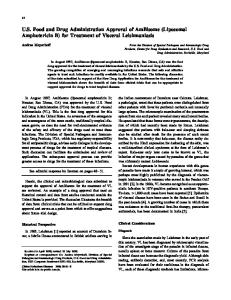
Figure 1. Synthetic scheme for the establishment of mannose-modified chitosan–thioglycolic acid conjugate. The Schiff’s base (R-CH = N-R) mediated amide bonds formation between the aldehyde group of mannose and the amino group of the thiolated polymer.
Results TC synthesis & characterization
Evaluation of the thiol groups immobilized to the thiolated polymer by the Ellman’s test demonstrated that on average 1301 ± 194 μmol of thiol groups were attached/gram of polymer (mean ± SD; n = 3). In addition, the amounts of primary amino groups and disulfide bonds within the TC were estimated by TNBS and disulfide bond assays, respectively. According to the data, there were 429 ± 62 μmol primary amino groups per gram of polymer (mean ± SD; n = 3) and 107 ± 12 μmol disulfide bonds/gram of polymer (mean ± SD; n = 3). The reduced disulfide bond formation implied reduced oxidation that had occurred during the conjugation reaction. This demonstrated the high efficiency of the thiolation process. The efficiency of the purification step for the TC polymer was con-
firmed by comparison to control polymers synthesized similarly but without 1-ethyl-3-(3-dimethylaminopropyl) carbodiimide hydrochloride during the conjugation step. The control polymers exhibited negligible quantities of thiol groups per gram of polymer. The purified polymers were frozen and lyophilized to give white fibrous structures that were readily soluble in aqueous solutions. MTC nanocarriers
We next sought to determine the utility of thiolated polymer for MP targeting as a therapeutic strategy as a therapy for VL. Mannose modification of the TC polymer was initiated by ring opening of mannose followed by reductive amination reaction of the resultant aldehyde with free amino groups on the TC polymer. The synthetic scheme for this mannose-modified chi-
Figure 2. Characterization of the polymers by nuclear magnetic resonance spectroscopy (see facing page). 1H-NMR spectra comparing unmodified polymer (A) and thiolated chitosan (B) polymer controls to mannose-anchored thiolated chitosan (C). The presence of methylene peaks (3.5-3.9 ppm and 4.5-5.1 ppm) in the spectrum of the mannose-anchored thiolated chitosan confirmed the attachment of the sugar to the polymer.
104
Nanomedicine (Lond.) (2017) 12(2)
future science group
5
future science group
4
3
2
www.futuremedicine.com
0.10
(rel)
2 0.42
0.69
(rel)
-1.117
-1.243
-2.093
-2.644 2.512 2.509 2.505 -2.369
2
-1.240
0.60
0.50
27.82
0.56
3.340
3
0.09
3
-1.915 -1.854
-2.370
-2.509
-2.643
86.08
-3.661
4
0.42
3.70
4
-3.072
-3.214
-3.359
-3.511
1.00
5
-3.882
-3.978
-4.465
-4.632
-4.827
-5.087
5
22.13
1.64
1.59
1.14
0.04
0.02
0.83
0.02
0.03
1.00
0.03
1.5 (rel)
-1.242
-1.680
-1.808
-1.934
-2.644 2.512 2.508 2.505 -2.370
-4.223
Mannose-anchored thiolated amphotericin B nanocarriers for treatment of visceral leishmaniasis
Research Article
3.0
2.5
2.0
1.0
0.5
-0.0
(ppm)
0.8
0.6
0.4
0.2
-0.0
(ppm)
2.0
1.5
1.0
0.5
(ppm)
-0.0
105
Research Article Shahnaz, Edagwa, McMillan et al.
0.10
0.05
Absorbance
0.00
-0.05
-0.10
-0.25 3500
3000
2500
2000
1500
1400.04
-0.20
1431.36
1652.12
-0.15
1000
Wave numbers (cm-1) Figure 3. Characterization of the polymers by fourier transform infrared spectroscopy. Fourier transform IR spectra of thiolated chitosan (A) and mannose-modified thiolated chitosan (B). The presence of amide carbonyl and hydroxyl bands in FTIR spectrum of the mannose-anchored thiolated polymer further confirms the association of the mannose sugar and the polymer backbone.
tion was induced by the addition of an aqueous solution of H2O2. Consequently, a decrease in the density of thiol groups within the nanocarriers was confirmed using the Ellman’s reagent. Stability of the developed nanoformulations in terms of size, charge and PDI was evaluated under various storage temperatures (-20, 4 and 37°C) in a time-dependent manner. No notable changes in size, ζ-potential and PDI, were observed when the nanoformulations were kept for a period of 4 weeks at 4 and 37°C. However, statistically significant increases in average size and PDI (∼2- and 1.5fold, respectively) were observed for nanoformulations after 4 weeks at -20°C. Drug release profiles
In this study, AmB release profiles of native drug powder, UC, TC and MTC nanocarriers were evaluated for 10 days at endosomal pH of 5.5 and physiological pH of 7.4. When assessing in vitro drug release of a hydrophobic drug, sink conditions were maintained. These conditions were maintained by inclusion of 1% (v/v) Tween in the phosphate buffer [17,19] . This allowed the total quantity of drug can be eluted from the nanocarriers and to reduce the adhesion of AmB
106
Nanomedicine (Lond.) (2017) 12(2)
onto the dialyzing membrane wall. The percentage of AmB released from the nanocarriers was evaluated in a time-dependent manner (Figure 5) . The release patterns were biphasic (i.e., an initial burst release of 40% within 2 days followed by an extended sustained release for 10 days). Moreover, as observed from native drug powderm 100% drug was released in both settings within 1 h (data not shown). Therefore, release media provides a sufficient hydrophobic environment for complete dissolution of drug. The initial burst release of the drug might be the result of diffusion of loosely bound drug near the particle surface. During the second phase, both TC and MTC nanocarriers exhibited a slow sustained release of AmB for up to 10 days, compared with >80% of drug released from UC nanocarriers within 4 days, possibly due to poor stability of the UC polymeric network. It is also worth noting that both TC and MTC nanocarriers exhibited the lowest initial drug release as compared with UC. Biocompatibility screening studies
The biocompatibility of the developed nanoformulation was evaluated in J774 cells using the MTT assay.
future science group
Mannose-anchored thiolated amphotericin B nanocarriers for treatment of visceral leishmaniasis
tosan–TGA conjugate is illustrated in Figure 1. Successful synthesis of the conjugate was confirmed by 1 H NMR and FTIR spectroscopy. The 1H NMR spectrum of MCT is shown in Figure 2. Peaks in the region of 3.5–3.9 ppm and 4.5–5.1 ppm correspond to methylene protons (CH 2 group) of the mannose sugar. These were present in the final product spectrum but not in the starting material ( Figure 2A & B, respectively). The FTIR spectra of the TC and mannose-modified TC are illustrated in Figure 3. A characteristic band at 3363 cm-1 corresponds to stretching vibrations of –NH2 and –OH functional groups. The band at 2375 cm-1 corresponding to thiol groups was observed in the TC spectrum (Figure 3) . For the mannose-modified TC, new peaks at 1652, 1431 cm-1 and 1400 cm-1 corresponds to amide bond formation and –OH stretching vibrations, an indication that the mannose sugar was incorporated on the polymer backbone.
Research Article
AmB loaded UC, TC and MTC nanocarriers were manufactured by a blend of embedding and diffusion. The payload of AmB-loaded nanocarriers was from 21 to 72% (Table 1) . Based on our prior experience, 0.54 μM TPP is required for cross-linking Chito and 0.60 μM for TC and MTC nanocarriers to stabilize the particles. The morphologies of the synthesized nanocarriers were rod-shaped (Figure 4) . Nanoparticle surface chemistry and parameters influence nanoformulation stability and attachment onto biological surfaces. The average ζ-potentials of MTC (19 ± 3) and TC (21 ± 0.6) were found to be lower than that of UC (28 ± 4) (p < 0.05) nanocarriers. The decrease in the ζ-potential of MTC was the result of mannose immobilization onto the polymer backbone. Moreover, all the nanoparticles were found to have a moderate PDI and narrow size distribution as illustrated in Table 2. To further stabilize TC and MTC nanocarriers, inter- and intramolecular disulfide bond forma-
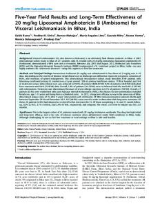
Figure 4. Nanoparticle morphologies. SEM images of unmodified chitosan (A), thiolated chitosan (B) and mannose-anchored thiolated chitosan (C) nanocarriers before centrifugation and SEM image of mannoseanchored thiolated chitosan (D) after centrifugation. SEM: Scanning electron microscopy.
future science group
www.futuremedicine.com
107
Research Article Shahnaz, Edagwa, McMillan et al.
** 100
Release (%)
80 *
60 40
MTC TC UC
20 0
0
2
4 6 Days (n)
8
10
Figure 5. Release profile of developed nanoformulations. Dissolution studies were performed in phosphate buffer pH 7.2 at 37°C. Values are mean ± SD of three experiments. Statistically significant differences in the release profile of MTC were determined compared with TC and UC at significance levels of *p < 0.05; **p < 0.01. MTC: Mannose-anchored thiolated chitosan; SD: Standard deviation; TC: Thiolated chitosan; UC: Unmodified chitosan.
J774 cells were incubated with various concentrations of AmB nanoformulations (0, 25, 50 and 75 μg/ml AmB). Differences were observed in the cytotoxicity profiles between TC and MTC, UC (p ≤ 0.05) and native drug (p ≤ 0.05). With MTC >90 ± 4.3% cell survival over 72 h was observed at the highest concentration tested (75 μg/ml AmB), while native drug solution reduced cell viability to 34.5 ± 5.6% (n = 3 ± SD) as shown in Figure 6. The values for the negative control Triton X-100 (2% v/v) and the positive-control medium without fetal calf serum/phenol red showed 0.40 ± 0.03% and 100.8 ± 4% viability, respectively. Internalization of MTC AmB nanocarriers by macrophages
Cellular internalization of targeted (TC and MTC), nontargeted (UC), AmBisome and native drug after 12 h incubation was compared in sensitive and resistant L. donovani infected or uninfected control cells following drug nanoformulation treatments (100 μM). UC nanocarriers, AmBisome and native drug showed reduced uptake into infected cells with the resistant VL strain after 12-h incubation (p ≤ 0.05) (Table 3) . To determine whether MDR1 is involved in the efflux of AmB, pretreatment with verapamil, an inhibitor of MDR1 was tested. Here retention of AmB from UC, AmBisome and native drug solution in infected and
108
Nanomedicine (Lond.) (2017) 12(2)
uninfected macrophages was evaluated (Table 3) . Pretreatment with verapamil demonstrated partial reversal of the efflux process, as the uptake of AmB was 2-, 2.5-, 1.2-fold higher from UC, AmBisome and native drug solution, respectively, for the resistant strain-infected macrophages. However, the uptake of the uninfected macrophages and sensitive strain-infected macrophages was not altered significantly by pretreatment with verapamil. These observations suggest that the lower intracellular AmB concentration in the resistant straininfected macrophages compared with noninfected macrophages and sensitive strain-infected macrophages may be the result of upregulated drug efflux machinery. TC and MTC nanocarriers (100 μM) were rapidly internalized into the noninfected macrophages and drug concentrations of up to 12.9 ± 2.3 μg of AmB/106 cells and 28.6 ± 1.4 μg AmB/106 cells over 12 h, respectively. We observed the uptake of TC and MTC to be 4.2- and 9.6-fold higher, respectively, in resistant strain-infected macrophages compared with unmodified nonthiolated nanocarriers (Table 3) . The results reported in this work therefore suggest that the thiolated surface-modified nanocarriers contribute to inhibition of AmB efflux in resistant strain-infected macrophages. MTC nanoparticles showed 71.5-, 26.0-, 4.4- and 2.2-fold increases in uptake by noninfected macrophages at 12 h compared with equimolar concentrations of TC (p ≤ 0.05), UC (p ≤ 0.05), AmBisome (p ≤ 0.05) and native drug (p ≤ 0.001), respectively, Table 3. The amount of drug lost during the washing steps of cellular internalization was determined by HPLC analysis of the supernatants. It was observed that loss of drug in the washed volume was decreased with longer incubation time (data not shown). Interactions of the nanocarriers with the cell surface seem to be time dependent. Moreover, UC, AmBisome and native drug displayed higher drug loss in contrast to TC and MTC nanocarriers. We also investigated the drug retention profile of the developed nanocarriers over 10 days (Figure 7) . There were significant differences in the drug retention profile (p ≤ 0.05) between AmBisome and native drug when cells were treated with equimolar drug concentrations. Specifically, 0.34 μg of AmB/106 cells was detected 2 days after treatment with AmBisome, compared with none for the native drug. UC nanocarriers were found to keep the drug inside the cells for 8 days. The TC and MTC nanocarriers retained and slowly released AmB over the 10-day period. Antileishmanial activities
In this study, various AmB nanoformulations were synthesized and investigated against L. donovani amastigotes in a concentration-dependent manner (Figure 8) .
future science group
Mannose-anchored thiolated amphotericin B nanocarriers for treatment of visceral leishmaniasis
future science group
120
100
80
60
40
20
MTC
TC
UC
AmB
Positive control
0 Negative control
Discussion For a spectrum of infectious diseases that include leishmaniasis, tuberculosis and HIV, macrophages serve as critical reservoirs and site of microbial replication for the intracellular microorganisms. Limited access of therapeutic agents to intracellular sites remains a major obstacle to effective anti-infectious disease therapy. In addition to reducing nonspecific drug toxicity, selective targeting of intramacrophage parasites with ligandanchored nanocarrier delivery systems would facilitate pathogen clearance by ensuring that drug concentrations at these sites remain within the desired therapeutic range. In order to achieve this goal, mannose receptor-mediated endocytosis of drug nanocarriers by macrophages has become increasingly attractive. In the present study, MTC nanocarriers were synthesized, characterized and evaluated against intramacrophage amastigote parasites. Chitosan-based nanocarriers are widely used for drug delivery applications owing to their efficient
intracellular release of drug payloads and biocompatibility. Thiolated polymers such as TC [20] promote nanoparticle internalization owing to their unique properties. Mannose modification of the TC polymer was used to improve uptake of the drug carrying particles by macrophages where Leishmania amastigotes reside. Nanocarriers for macrophage delivery may offer numerous advantages over traditional delivery, including enhanced treatment rates, reduced off-target adverse effects, amplified drug stability and successful intracellular targeting. The MTC nanocarriers were manufactured and characterized by various analytical techniques, including FTIR, 1H NMR and scanning electron microscopy. The results of mannose anchoring were consistent with the earlier described mannosylation of nanocarriers with amino groups of polymer [2] . UC, TC and MTC nanocarriers were formulated by homogenization followed by ionic gelation with TPP and generation of intra- and intermolecular disulfide bonds. Rod-shaped morphologies of the nanocarriers could be due to a par-
Cell viability (%)
Free AmB and AmBisome were used as controls. Drugfree nanocarriers (UC, TC and MTC) were used as additional controls. As shown in Figure 8A, AmB, AmBisome UC, TC and MTC nanocarriers at doses equivalent to 1.0 μg/ml AmB showed 48% ± 6%, 60% ± 11%, 76% ± 9%, 84% ± 7% and 96% ± 2% inhibition of parasites, respectively. The measured IC50 of AmB, AmBisome, UC, TC and MTC nanocarriers was found to be 0.256 ± 0.013 μg/ml, 0.208 ± 0.01 μg/ml, 0.164 ± 0.03 μg/ml, 0.096 ± 0.004 μg/ml and 0.019 ± 0.007 μg/ml, respectively. Macrophage targeting through mannose-anchored thiolated nanocarriers significantly improved the antileishmanial activity of AmB against intracellular parasites. Further evaluation of the potential application and future development of the synthesized nanoformulations as slow acting antileshmania therapies were conducted over a 10-day period. The native drug exhibited limited antimicrobial responses (data not shown). In contrast, MTC showed extended activity against L. donovani, suppressing parasite replication by 3.4-fold on the 10th day. The in vivo therapeutic efficacy of MTC nanocarriers at a single dose of 1 mg AmB/kg was evaluated in L. donovani-infected mice and compared with native AmB. The observed findings were then validated with AmBisome. The results showed that MTC nanocarriers were considerably more effective (89% ± 7% inhibition) than AmB (17% ± 4% inhibition) (p < 0.001). Whereas TC, UC, AmBisome and nanocarriers alone had 63% ± 5%, 36% ± 2%, 19% ± 2.4% and 11% ± 1% reductions in parasites, respectively. Among all tested formulations, MTC demonstrated the greatest antileishmanial efficacy.
Research Article
Figure 6. Biocompatibility of nanoformulations in J744A.1 cells after 72 h incubation. To retain equivalent concentrations, nanoformulations of MTC (0.93 μg/ml), TC (1 μg/ml) and UC (1.6 μg/ml) were dissolved to achieve concentrations of 75 μg/ml AmB in the culture medium. Results are expressed as mean ± SD of three replicate wells at significance levels of **p < 0.01; ***p < 0.001. MTC: Mannose-anchored thiolated chitosan; SD: Standard deviation; TC: Thiolated chitosan; UC: Unmodified chitosan.
www.futuremedicine.com
109
Research Article Shahnaz, Edagwa, McMillan et al. ticular characteristic of homogenized hydrophobic drug particles. For in vivo applications, it is highly desirable to design a carrier that is stable and provides sustained release of therapeutic agents during systemic circulation. In the systemic circulation, the stability of the drug carrier is key to minimizing rapid elimination of the drug from the body. Hence, drug release was carried out at endosomal pH of 5.5 and physiological pH of 7.4 containing 1% (v/v) Tween-80. An initial burst release of drug (∼ 40% within 2 days) was observed with TC and MTC nanocarriers followed by a slowrelease phase, which continued for 10 days. In contrast, UC nanocarriers released 80% of drug within 4 days. A possible explanation for the observed sustained drug
release from TC and MTC nanocarriers may be due to covalent cross-linking of disulfide bonds formed within the modified polymer matrix during the swelling process [21] . Moreover, all developed nanocarriers released >55% of drug at endosomal pH 5.5 (data not shown). This faster release at endosomal pH may be the result of increased solubility of AmB and the polymer under acidic conditions. The extent of oxidation of immobilized thiol groups via thiol (-SH)/disulfide (-S-S-) exchange reactions was found to be greater at physiological pH due to thiolate anion (RS -) formation at high pH. Therefore, these findings support the use of modified chitosan nanoparticles for sustained and controlled release of drug payloads at intracellular infection sites.
Table 3. Comparison of uptake of amphotericin B from targeted (thiolated chitosan and mannoseanchored thiolated chitosan) nanocarriers, unmodified chitosan nanocarriers, AmBisome® and native drug in uninfected macrophages. Formulations
Uptake studies (μg AmB/10 6 cells)
p-value
Uninfected macrophages
Sensitive strains-infected macrophages
Resistant strainsinfected macrophages
AmB
0.4 ± 0.01
0.38 ± 0.02
0.04 ± 0.006
0.058x 0.0002y 0.0003z
AmBisome
1.1 ± 0.07
1.2 ± 0.08
0.3 ± 0.05
0.061x 0.007y 0.0073z
UC
6.5 ± 0.9
6.3 ± 0.7
2.9 ± 0.4
0.055x 0.0004y 0.0003z
TC
12.9 ± 2.3
11.5 ± 1.1
12.3 ± 2.2
0.056x 0.071y 0.069z
MTC
28.6 ± 1.4†***,‡**,§*,¶*
27.9 ± 1.6†***,‡**,§**,¶*
28.1 ± 1.7†***,‡**,§**,¶*
0.059x 0.063y 0.064z
Pretreatment with verapamil AmB
0.39 ± 0.02
0.36 ± 0.03
0.084 ± 0.01
0.061x 0.003y 0.004z
AmBisome
1.2 ± 0.06
1.3 ± 0.09
0.76 ± 0.05
0.079x 0.009 y 0.008z
UC
6.6 ± 0.8
6.2 ± 0.5
3.5 ± 0.9
0.062x 0.004y 0.003z
Both resistant and sensitive Leishmania donovani were used to infect macrophages. Data are expressed as three replicates in all tests, and the data are expressed as mean ± SD of three independent experiments. Statistically significant differences of MTC were determined in relation to AmB solution (†), AmBisome (‡), UC (§) and TC (¶) at significance levels of *p < 0.05; **p < 0.01; ***p < 0.001. x = probability value between uninfected macrophage group and sensitive strain-infected macrophage group; y = probability value between uninfected macrophage group and resistant strain-infected macrophage group; z = probability value between sensitive strain-infected macrophage group and resistant strain-infected macrophage group. AmB: Amphotericin B; MTC: Mannose-anchored thiolated chitosan; SD: Standard deviation; TC: Thiolated chitosan; UC: Unmodified chitosan.
110
Nanomedicine (Lond.) (2017) 12(2)
future science group
Mannose-anchored thiolated amphotericin B nanocarriers for treatment of visceral leishmaniasis
future science group
35
MTC TC UC
30
Concentration (µg/ml)
The biocompatibility induced by nanoformulations and native drug was assessed at various concentration (0, 25, 50 and 75 μg/ml AmB) in J774 cells by using the MTT cell proliferation assay. The rank order of biocompatibility at highest concentration was MTC>TC>UC> AmBisome > AmB. Higher biocompatibility of thiolated nanoformulations (TC and MTC) could be due to the surface modification, where less ionizable-free amino groups are available in TC and MTC as compared with UC. Moreover, surfacemodified nanocarriers resulted in an enormously significant decline (p ≤ 0.05) in the cytotoxicity of AmB. Our findings are similar to those previously reported where surface-modified nanocarriers were used to deliver AmB [2] . The toxicity of AmB has been associated with the formation of drug aggregates [2] . It has been demonstrated that the nonaggregated form of AmB is biocompatible toward mammalian cells but causes porosity of parasitic cells. The aggregated form of AmB forms pores in both mammalian and parasitic cell membranes. The decline in cytotoxicity of AmB encapsulated into surface-modified nanocarriers could be linked, in measure, to the slow release of nonaggregated drug. Since macrophages serve as reservoirs for Leishmania amastigotes, receptor-mediated endocytosis of drug nanocarriers is essential in ensuring the delivery of high intracellular drug concentrations required to clear the parasite. We therefore sought to design and study MTC nanocarriers for cellular internalization of antileishmanial agents. The cellular internalization of nanoformulations and native drug was compared in sensitive and resistant L. donovani-infected or -uninfected control cells. Overexpression of ABC transporters especially MDR1 is well known to be an important mechanism of drug efflux for L. donovaniresistant strains [18] . Hence, lower cellular internalization of UC nanocarriers, AmBisome and native drug into cells infected with the resistant VL strain could be due to upregulated drug efflux machinery. In contrast, no significant difference in cellular internalization profile of TC and MTC was seen in sensitive and resistant L. donovani-infected or -uninfected control cells. Efflux pump inhibitory capability of thiolated polymers has been shown in mammalian cells [11] . Dünnhaupt et al. demonstrated that thiolated polymers inhibit the efflux pump by blocking the efflux pump drug-binding sites [22] . Accordingly, TC and MTC may be also responsible for the inhibition of drug efflux machinery in the resistant VL strain. Thiol-reactive groups have also been shown to promote binding and cellular internalization of nanomaterials [13,23,24] . A recent study on thiolated surfacestabilized superparamagnetic iron oxide nanoparticles
Research Article
25 20 15 10 5 0 0
2
4
6
8
10
Retention of drug (days) Figure 7. Intracellular retention of MTC, TC, UC nanocarriers, AmBisome® and native drug in macrophages determined over 10 days. Retention of drug was observed along with sustained release. Data are shown as mean ± SD of three replicate wells. ***p < 0.001 (comparison of MTC versus AmB and AmBisome); **p < 0.01 (comparison of MTC versus TC and UC nanocarriers). AmB: Amphotericin B; MTC: Mannose-anchored thiolated chitosan; SD: Standard deviation; TC: Thiolated chitosan; UC: Unmodified chitosan.
demonstrated efficient internalization of thiol surfacemodified nanoparticles by stem cells. It was found that decorating the magnetic particles with thiol moieties resulted in a 17-fold increase in uptake by progenitor cells when compared with nonthiolated superparamagnetic iron oxide nanoparticles [20] . These findings provide additional support to the concept that the presence of thiol groups on the surface of the particles could enhance cellular internalization. Accordingly, a 1.98-fold increase in uptake of TC by noninfected macrophages was observed over equivalent concentrations of UC nanocarriers. Further improvement in AmB cellular uptake was seen when MTC was used as the drug carrier. The increase in uptake for the mannose-anchored MTC over the TC may be due to the mannose receptor-mediated endocytosis of the drug particles. The macrophage-targeted MTC drug delivery system therefore represents a promising approach to Leishmania eradication that would allow for delivery of effective drug concentrations at sites of amastigote replication. Efficient internalization of MTC into macrophages may proceed by either specific or nonspecific mechanisms that include: access and initial interaction
www.futuremedicine.com
111
Research Article Shahnaz, Edagwa, McMillan et al.
AmB UC MTC
AmBisome TC 0.30
100
Concentration (µg/ml)
60
40
20
0.20
0.15
0.10
0.05
0
Concentration (µg/ml)
MTC
TC
UC
AmBisome
0.0041
0.0123
0.037
0.111
0.333
1
0.00 AmB
Inhibition of amastigotes (%)
0.25 80
Figure 8. Pharmacological evaluation of antileishmanial activities. Percent inhibition of Leishmania donovani amastigotes by escalating concentration (ug/ml) of AmB, AmBisome, UC, TC and MTC (A). For (B) the concentration (ug/ml) required to achieve an in vitro (IC50 ) activity against L. donovani amastigotes. All experiments were performed with human MDM infected with the parasite for a 12 h incubation (B). Concentration of the polymer used in nanoformulation was biocompatible to the target macrophage. Data expressed as mean ± SD for a representative of one or three performed experiments. AmB: Amphotericin B; MDM: Monocyte-derived macrophages; MTC: Mannose-anchored thiolated chitosan; SD: Standard deviation; UC: Unmodified chitosan.
of nanocarriers with the surface membrane of macrophages; interaction of reactive thiol moieties on the surface of MTC with the macrophage membrane; recognition of MTC by mannose phagocytic machinery expressed on the macrophage surface; and configurational changes in the cell membrane structure (projection or invagination), vacuolization and internalization of the drug particles. Therefore, increased intracellular drug concentrations and prolonged retention exhibited by MTC are all important aspects in the development of nanoformulations that would target and eradicate intracellular infections. Superior cellular retention profiles of the UC, TC and MTC nanocarriers as compared with AmBisome and native drug could be due to cationic chitosan polymeric backbone. Chitosan-based nanocarriers are known to escape from the endosomes thereby able to escape lysosomal degradation and deliver the drug payload to the target site [25] . This phenomenon that is common to cationic polymers is referred to a proton sponge effect [26] . In addition, the extended sustained and slow release of drug for TC and MTC nanocarriers might be the result of interactions between thiol moi-
112
Nanomedicine (Lond.) (2017) 12(2)
eties of the nanocarriers and the disulfide bonds of the lysosomal enzyme via thiol/disulfide exchange reaction (SH → -S-S-; -S-S- → -SH). This prolonged retention profile would allow for a reduced dosing frequency. Leishmania parasites have two main life-cycle phases: the extracellular motile promastigotes phase, found in the phlebotomine sandflies vector and the intracellular immotile amastigotes phase that thrives in the mammalian host. Amastigotes proliferate in the macrophage parasitophorous vacuoles, phagolysosome-like compartments with hydrolytic and acidic surroundings. Parasitophorous amastigotes lead to destruction of the host cell but manage to escape into the systemic circulation where they are adept at infecting new phagocytic cells and responsible for the pathogenesis of leishmaniasis. Targeted drug delivery to parasite reservoirs such as macrophages is anticipated to increase the therapeutic efficacy and diminish off-target drug toxicity. For antileishmanial activity against parasitophorous L. donovani amastigotes-infected macrophages, MTC again provided maximum inhibition of the parasite. Low drug concentrations of the MTC formulation were able to inhibit intracellular
future science group
Mannose-anchored thiolated amphotericin B nanocarriers for treatment of visceral leishmaniasis
Conclusion Long acting macrophage-targeted MTC nanocarriers loaded with AmB were successfully synthesized and evaluated for cellular uptake and antileishmanial responses. The mannosylated nanocarriers displayed enhanced uptake of the encapsulated AmB by macrophages when compared with uptake of native drug or nontargeted particles. Interestingly, the mannosylated system was found to retain the drug inside the macrophages for up to 10 days. The MTC nanocarriers were also found to be highly effective at inhibiting the growth of intracellular amastigotes in infected macrophages when compared with native drug. These encouraging in vitro data provide proof-of-concept that macrophagedirected TC nanocarriers hold promise as potential therapies for parasitic and other intracellular infections.
future science group
*** *
100 Growth inhibition of amastigotes in mice (%)
80
60
**
40
20
MTC
TC
UC
AmBisome
AmB
Blank MTC
0 Control
parasite replication when compared with the clinically used AmBisome formulation (p < 0.05). The IC50 of MTC was 13.0-, 10.6-, 8.4- and 4.9-fold lower than that of AmB (p < 0.05), AmBisome (p