―Original―
Ultrasound Evaluation of the Transverse Movement of the Flexor Pollicis Longus Tendon on the Distal Radius during Wrist and Finger Motion in Healthy Volunteers Mitsuhiko Nanno1, Takuya Sawaizumi2, Norie Kodera2, Yuji Tomori2 and Shinro Takai2 1
Department of Orthopaedic Surgery, Nippon Medical School, Musashi Kosugi Hospital 2
Department of Orthopaedic Surgery, Nippon Medical School
Purpose: This study aimed to evaluate the kinematics of the flexor pollicis longus tendon (FPL) at the wrist by examining the movement of the FPL on the distal radius during various wrist and finger motions using transverse ultrasound in healthy volunteers. Methods: Forty-eight wrists of 24 asymptomatic volunteers were examined by transverse ultrasound to observe the location of the FPL on the distal radius at 5 wrist positions (neutral, 60° dorsal flexion, 60° palmar flexion, 40° ulnar deviation, and 10° radial deviation) with all 5 fingers in full extension and full flexion, and isolated thumb in full flexion, respectively. Results: We found that the FPL was situated statistically significantly more ulnodorsally at the wrist dorsal and ulnar deviation positions, more ulnopalmarly at the wrist palmar flexion position, and more radiopalmarly at the wrist radial deviation-position than at the wrist neutral position with all 5 fingers at full extension. Especially, it moved statistically significantly most ulnodorsally at the wrist dorsal flexion position during finger motion. The FPL moved most statistically significantly ulnopalmarly at the wrist palmar flexion position with all 5 fingers in full extension among all wrist positions during finger motion. During finger motion, the wrist dorsal flexion position induced significant displacement of the FPL to the distal radius and compressed it between the flexor tendons and the distal radius. The average distance between the FPL and the volar surface of the distal radius in the palmar-dorsal direction at wrist dorsal flexion position in all fingers at full flexion was 1.9 mm, the smallest among all wrist positions during finger motion. Conclusions: There is a significant relationship between the transverse movement of the FPL at the distal radius and wrist and finger motions. Our findings indicated that the irritation of the FPL caused by the movement of both the FPL itself and of the flexor digitorum superficialis and profundus is most induced with the wrist in dorsal flexion with all 5 fingers at full flexion compared to other wrist positions during finger motion. This wrist position might be the optimum one at which to evaluate the irritation of the FPL from volar locking plates in patients with distal radius fracture. We believe that our transverse ultrasound results can play a role in the gaining of a better understanding of the kinematics of the FPL. Moreover, they have potential to lead to improved diagnosis of and treatment for fractures of the distal radius and help to minimize the risk of FPL rupture related to volar locking plates. (J Nippon Med Sch 2015; 82: 220―228) Key words: flexor pollicis longus, ultrasound, wrist position, finger motion, distal radius
Introduction
tures of the upper extremity, are usually treated with vo-
Distal radius fractures, which are the most common frac-
lar locking plate fixation1―5. After this operation, a major
Correspondence to Mitsuhiko Nanno, Department of Orthopaedic Surgery, Nippon Medical School Musashi Kosugi Hospital, 1― 396 Kosugi-cho, Nakahara-ku, Kawasaki, Kanagawa 211―8533, Japan E-mail:
[email protected] Journal Website (http:! ! www.nms.ac.jp! jnms! ) 220
J Nippon Med Sch 2015; 82 (5)
Movement of Flexor Pollicis Longus at Wrist
complication1―11, which occurs in 1.8% to 12% of cases6,10,12, is rupture of the flexor pollicis longus tendon (FPL). These ruptures are reportedly caused by fracture malunion or plate prominence due to incomplete reduction or poor plate positioning1,3―5,11,12. However, even if efforts are made to prevent tendon irritation through the design, careful
positioning,
and
application
of
low-profile
anatomic volar locking plates, FPL tendon ruptures still occur1,3,5,11. To prevent the FPL tendon from rupturing, its movement on the distal radius has recently been evaluated. Several studies using longitudinal ultrasonography have found a relationship between the position of the volar plate and the movement of the FPL at the distal radius during thumb motion in patients who had undergone volar locking plate fixation of distal radius fractures13,14. Moreover, these studies have found that the FPL runs longitudinally and also shifts dorsally toward the volar
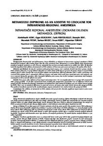
Fig. 1 Transverse ultrasound examination setup
plate in active isolated thumb flexion13. Ultrasonography is a useful for examining and elucidating the various tendon movements noninvasively and quantitatively at the
Materials and Methods
wrist in vivo. Thus, ultrasonographic evaluation of FPL
The subjects of this study were 24 healthy male volun-
movement during wrist and finger motions is considered
teers (mean age, 35.8 years; age range, 24―56 years). Sub-
useful for providing detailed information about FPL
jects were excluded if they had a history of traumatic in-
kinematic changes in healthy subjects and in patients
juries, such as fractures or tendon damage to the upper
who have undergone volar locking plate fixation for dis-
extremities, or had neurological deficit, hand pain, or
tal radius fractures. However, with longitudinal ultra-
numbness. Ultrasound examinations were performed for
sonography, holding the transducer on the wrist, except
both wrists after signed consent forms were obtained
at the wrist neutral or slightly dorsal flexion position, is
from the subjects.
difficult, and a transverse examination at varied wrist po-
Subjects sat with the forearm in supination, the elbow
sitions is necessary to further detail the kinematics of the
at about 45° flexion, and the shoulder in neutral position.
FPL.
The subject’s forearm was fixed to a custom-made table.
Few studies have evaluated the transverse ultra-
An ultrasound scanner (Mylab Five, Hitachi Medical
sonographic displacement of the FPL in relation to varied
Corporation, Tokyo, Japan) equipped with an LA332 3.5!
wrist and finger motions. If the location of the FPL could
12-MHz hi-definition linear array transducer (LA332)
be evaluated as a coordinate on the transverse plane this
was used. The study protocol was approved by our insti-
would offer a more detailed understanding of the FPL
tutional review board.
orientation at the distal radius. Therefore, in the present
All ultrasonographic examinations were performed by
study we performed a quantitative analysis with trans-
a single orthopedic surgeon (MN). The transducer was
verse ultrasonography of the displacement and location
placed on the distal radius of the wrist (Fig. 1). A
of the FPL as coordinates on the distal radius at varied
custom-made device fastened to the transducer was at-
wrist positions during finger motion in healthy volun-
tached at the subjects’s wrist to hold the transducer sta-
teers.
ble during all examinations, and the transducer was held
We hope that this study could help further the knowl-
at a 90° angle to the skin of the wrist proximal to the
edge of FPL kinematics, and moreover could help to pro-
wrist crease to identify the ulnar edge of the most distal
vide precise diagnosis of and suggest the optimum treat-
portion of the distal radius without applying extra pres-
ments for distal radius fractures using volar locking plate
sure to the tissue above the distal radius. This bony land-
fixation, while helping to prevent rupture of the FPL.
mark was easy to confirm by palpation and transverse ultrasonography in all wrists. The tendons of the FPL,
J Nippon Med Sch 2015; 82 (5)
221
M. Nanno, et al
Fig. 2 Measuring the location of the flexor pollicis longus tendon on the distal radius as a coordinate FPL: the flexor pollicis longus tendon, FCR: the flexor carpi radialis tendon, FDS: the flexor digitorumsuperficialis tendon, FDP: the flexor digiti profundus tendon, MN: the median nerve, RA: the radial artery ‘P’: the reference point defined as the vertex of the palmar bony prominence of the radiolunate fossa of the distal radius on the watershed line, Distance ‘x’: The distance between the ‘P’ and the centroid of FPL on the X-axis, Distance ‘y’: The distance between the ‘P’ and the centroid of FPL on the Y-axis
the flexor digitorum superficialis (FDS), and the flexor
pronator fossa distally2. The P can be easily identified
digitorum profundus (FDP) and the median nerve were
with transverse ultrasonography or magnetic resonance
identified with transverse ultrasonography during full
imaging (Fig. 3a and b). The FPL has been previously re-
flexion and full extension of all 5 fingers and full flexion
ported to always be located on the radial side of point P1.
of the isolated thumb. Transverse ultrasonographic im-
We measured the distances between P and the centroid
ages were obtained respectively at each of 5 wrist posi-
of the FPL on the X-axis (x) and on the Y-axis (y). The
tions (neutral, 60° dorsal flexion, 60° palmar flexion, 40°
position of the FPL was shown as a coordinate point (x
ulnar deviation, and 10° radial deviation) during finger
mm, Y mm). As the distance x or y increased, the loca-
motion. Each wrist angle was measured with a goniome-
tion of the FPL was more deviated to the radial side or
ter (SD1-01, Suzukiiryo, Inc., Tokyo, Japan) positioned on
the palmar side. Thus, the FPL displacement in ulnar-
the wrist. Subjects were asked to move from full exten-
radial and palmar-dorsal directions could be calculated
sion to full flexion of all 5 fingers or from full extension
with a comparison of the starting and ending coordinates
of all 5 fingers to flexion of the thumb individually at
of the FPL on the distal radius.
each wrist position. The images of 2 cycles of the flexion-
The results were calculated as the averages of 3 cycles.
extension finger motion at each wrist position were ex-
All data were analyzed with the software program IBM
amined.
SPSS Statistics 21.0J (IBM Japan Ltd., Tokyo, Japan), and
All recorded images of the initial and final frames of
the FPL displacement from the neutral wrist position to
each finger and wrist motion were also evaluated. Fur-
the other 4 wrist positions in the extension of all 5 fin-
thermore, the coordinates of the FPL were determined as
gers and from the extension to the flexion of all 5 fingers
follows (Fig. 2). The reference point (P) was defined as
and the isolated thumb at each of the 5 wrist positions
the vertex of the palmar bony prominence of the radiolu-
were examined statistically with the paired t-test. A P-
nate fossa of the distal radius on the watershed line pre-
value of