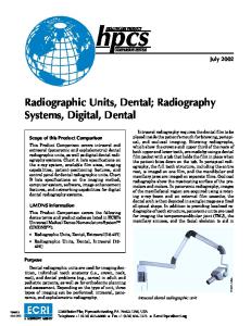
Dental Radiography Series Quality Assurance in dental fim radiography.
A complete quality assurance program is easy to establish and maintain. Quality assurance will pay for itself, not only in dollars and cents, but by reducing exposure to patients and personnel, and by helping the dental community provide better patient care. Quality assurance is a plan of action to ensure that a diagnostic x-ray facility will produce consistent, highquality images with a minimum of exposure to patients and personnel. Quality assurance means regular testing to detect equipment malfunctions, planned monitoring and scheduled maintenance for consistent film processing and regular assessment of other variables that affect image quality.
This document provides some tips on setting up a dental radiography quality assurance program for your practice.
The benefits derived from a quality assurance program far outweigh implementation efforts and costs. In addition to aiding in diagnosis, a well-executed quality assurance program can result in minimized radiation dosage to patients because radiographs are produced under the most favorable conditions. Fewer repeat radiographs mean time and cost savings for both patients and operators. Regular testing also helps ensure compliance with state and federal regulations.
Important Factors in Quality Control
4 Annual testing
5 Film processing Manual processing
6 Dark images Light images Replenish chemicals
Automatic Processing 7 Mix chemicals Use the full cycle Replenish chemicals
Tips for Quality Control in Film Processing
9 1. Create a reference radiograph 2. Create monitor strips 3. Use a dental radiographic normalizing and monitoring device
Daily Routine 11 First thing in the morning At the end of the day Maintain a daily log Store and handle films Improper film handling
Tips for Extraoral Screens and Cassettes 11 Test screen-film contact
Maintain the Viewbox Suggested Guidelines for Quality Assurance 14 Processor maintenance Film storage and handling Darkroom Radiographic screens Viewbox maintenance
Important Factors in Quality Control One of the most important facets of quality assurance is quality control. Quality control refers to the tests used for routine assessment of x-ray systems. These include tests of the basic performance of x-ray units, of manual and automatic processing, of image receptors and viewing conditions.
Annual testing In addition to regular calibration, the American Academy of Oral and Maxillofacial Radiology recommends that the following eight tests of x-ray generating equipment be conducted annually. • �X-ray output
Accurate calibration of x-ray machines promotes consistent equipment performance and reduces the number of film retakes due to equipment malfunctions. All machines, both new and old, must be recalibrated at regular intervals by a qualified technician.
• �Kilovoltage (kVp) calibration • �Half value layer • �Timer • �Milliamperage (mA) • �Collimation • �Beam alignment • �Tube head stability
All of these tests can be performed by dental practitioners, and require only film and test logs to provide written records of quality control checks and some basic testing materials. The equipment manufacturer’s service representative can also complete the tests.
4
Film processing One of the most critical areas for quality control, and most frequently overlooked, is film processing. Even the best exposure conditions can be compromised with poor processing technique.
Manual processing For manual processing, it is crucial to adhere to the times and temperatures recommended by the film manufacturer. The chart below shows those for Carestream Dental intraoral and extraoral dental films with GBX chemicals. A common error in manual processing is failing to check and adjust the temperature of the solution before development. Unless temperature control units are used, solution temperature may vary significantly during the day because it is influenced by incoming water and by the surrounding environment. Even a slight increase in temperature can cause films to darken.
Extraoral Radiographs Carestream Dental Extraoral Films EVG T-MAT G/RA T-MAT L/RA T-MAT H/RA X-OMAT DBF Intraoral Radiographs Carestream Dental Intraoral Films INSIGHT Ultra-speed
Develop
Rinse
Fix
Wash
Fresh Running Water
GBX Fixer and Replenisher
Clean Running Water (Approx. 8 Volume Changes per Hour)
60-85 °F (15.5-29.5 °C)
60-85 °F (15.5-29.5 °C)
60-85 °F (15.5-29.5 °C)
30 sec agitate continuously
2-4 min or twice the clearing time, intermittent agitation
5 min
Develop
Rinse
Fix
Wash
GBX Developer and Replenisher
Fresh Running Water
GBX Fixer and Replenisher
Clean Running Water (Approx. 8 Volume Changers per Hour)
60-85 °F (15.5-29.5 °C)
60-85 °F (15.5-29.5 °C)
60-85 °F (15.5-29.5 °C)
30 sec agitate continuously
2-4 min or twice the clearing time, intermittent agitation
10 min
GBX Developer and Replenisher 68 °F (20 °C) 70 °F (21 °C) 72 °F (22 °C) 76 °F (24.5 °C) 80 °F (26.5 °C)
68 °F (20 °C) 70 °F (21 °C) 72 °F (22 °C) 76 °F (24.5 °C) 80 °F (26.5 °C)
5 min 4½ min 4 min 3 min 2½ min
5 min 4½ min 4 min 3 min 2½ min
5
Use the following process for intraoral films when rapid processing is desirable – for example, during endodontic procedures.
Light images Light images can result from: • �Development time too short
Special rapid process recommendations for intraoral film Solution/Step
• �Underactive or exhausted solutions
Temperature
Time
Agitation
Rapid Access Developer
75 °F (22 °C)
15 sec
Continuous
Rapid Access Fixer
60-85 °F (15.5-29.5 °C)
15 sec or until clear
Continuous
Wash
60-85 °F (15.5-29.5 °C)
Quick rinse
Running water
Note: Discard developer and fixer after processing the equivalent of 60 intraoral films (No. 2 size packets) in 5 ounces (148 mL) of solution or after 3 days, whichever comes first. Keep developer covered when not in use to reduce the rate of oxidation and evaporation and to prevent contamination. If you plan to keep the radiographs, refix for 2 to 4 minutes in GBX fixer or Rapid Access Fixer, and rewash in running water for 10 minutes. Dry film in a dust-free area at room temperature or in a suitable drying cabinet (temperature not to exceed 120 °F [49 °C]).
Dark images As shown in the accompanying image, dark images may be caused by:
• �Developer solution too cold • �Developer incorrectly mixed (diluted) • �Under exposure
Replenish chemicals Use fresh or replenished chemicals designed for manual processing in manual systems. GBX Developer and Replenisher and GBX Fixer and Replenisher are available in a combination twin pack. The concentration and purity of the chemical solutions in your manual processing tanks are important to the image quality of the resulting radiograph. Always keep separate, labeled, capped bottles of developer replenisher and fixer replenisher readily available. We suggest adding 8 ounces of the appropriate replenisher (236 mL) per one gallon (3.8 L) of developer and fixer working solutions every morning, regardless of the solution levels visible in the tanks. Do not replenish or “TOP OFF” with water. This will help ensure that the correct processing solution strength is present for the radiographs to be processed that day. It may be necessary to remove some of the existing solution before adding the replenisher, so that the processing tanks do not overflow.
• �Development time too long • �Over exposure • �Developer solution too hot
To guard against possible cross-contamination, always use separate paddles to mix each type of processing solution. Label the paddles to indicate the solution they are used for, and always wash and dry the paddles after each use. NEVER “TOP OFF”or replenish processing solutions with water. Using water will dilute the chemicals, making them less effective. This can result in images with poor density and contrast and a yellow appearing image. This could also result in images turning brown over time due to poor fixing.
6
Automatic Processing During automatic processing, the film is transported through the processing sequence mechanically. It moves through the developing, fixing and washing stages at a controlled speed. With proper care, most mechanical processors can be efficient, reliable and consistent. The chemicals used in automatic processing are specially designed to be used with mechanical transport systems. They operate at higher temperatures and do not require film rinsing between development and fixing. In some roller transport systems, the temperature of the chemicals is thermostatically controlled and the processing time is regulated by controlling the speed of the rollers in the transport mechanism. The amount of time required to complete the
Check to see if a starter solution is recommended by the chemical manufacturer. A starter solution helps to avoid large swings in density from fresh chemistry to seasoned chemistry. READYMATIC and READYPRO chemicals are ready to use and do not need a starter solution. They are designed to provide a consistent image throughout the life of the solution.
Use the full cycle Some film processors employ an endo cycle. Typically a 2½-minute processing cycle, this provides a quick-look for non-archival quality periapical films. This process is used for dental surgical procedures where rapid processing is desired. For best image quality and archival results, use the full processing cycle or process manually using the guidelines stated in the manual processing section.
Replenish chemicals
processing cycle and the temperature setting for the developer varies from processor to processor and is set by the manufacturer. In today’s equipment, the time required to complete the processing cycle for Carestream Dental intraoral films, for example, ranges from 4 minutes at 85 °F to 6½ minutes at room temperature.
Mix chemicals Mix chemical solutions according to directions. Chemicals are available mainly in two forms: concentrate and ready-to-use. The concentrate requires mixing with water. Ready-to-use chemicals, such as READYMATIC Developer and Replenisher and READYMATIC Fixer and Replenisher, do not require any mixing. Temperature and time in developer are inversely related: the higher the temperature, the shorter the time in the developer. READYMATIC chemicals can be used in slower systems at room temperature as well as in faster systems at higher temperatures.
While some automatic processors provide replenishment automatically, other equipment requires operator intervention to maintain the correct chemistry levels and strengths. For those intraoral film processors without automatic replenishment, it is recommended that 8 ounces (236 mL) of replenisher be added to each working solution daily, based on an average daily run of 20 to 30 intraoral films. Even if no films are processed for a period of time, replenishment is necessary. If you process more than 30 intraoral films per day, you should increase the amount of daily replenisher solution at the rate of 0.25 fluid ounce (7 mL) per additional film processed. For example, 50 intraoral films per day would require that 5 additional ounces (148 mL) be added to the daily 8 ounces, for a total of 13 ounces (384 mL). All replenished solutions should be changed every three to four weeks under normal conditions. (Normal use is defined as 30 intraoral films per day.) Manufacturers of automatic non-roller type processors recommend that all non-replenished solutions be changed every two weeks. 7
For those processors incorporating daylight film-loading features, it is important that the processing unit be placed in subdued lighting (no direct, bright light). This will help to prevent film fogging through the filter of the loading units.
Note: Soaking rollers in water for a period of time is not an effective means of cleaning or rinsing.
NEVER “TOP OFF” or replenish processing solutions with water. Using water will dilute the chemicals, making them less effective. This can result in images with poor density and contrast and a yellow appearing image. This could also result in images turning brown over time due to poor fixing.
4. Always fill the fixer tank first. Rinse out the empty developer tank in case any fixer splashed into it while filling the fixer tank. Then fill the developer tank. This prevents contamination of the developer.
Tips for Quality Control in Film Processing The majority of problems in radiography can be traced to improper film processing procedures. Quality control of processing is the most effective way to avoid these problems, and the most effective way to achieve highquality radiographic images consistently. Effective quality control for automatic processing begins at each solution change: 1. Wearing rubber gloves, a rubber apron and protective goggles/glasses, clean the tanks and rollers thoroughly to remove chemical deposits and contaminants.
For normal weekly or monthly cleaning, we recommend using warm water and a non-abrasive brush to prevent scratching of the rollers. Periodically (2–4 times a year), use the systems cleaner recommended by your processor manufacturer to remove heavier buildup.
2. After cleaning with a systems cleaner, rinse for twice as long as the manufacturer recommends to remove any trace of cleaner. If not completely rinsed off, residual systems cleaner will contaminate your fresh chemicals resulting in poor image quality. 8
3. Inspect the rollers, gears and turning mechanism for signs of wear, and reassemble.
5. After filling the processor, turn it on. If the processor has a thermostat to adjust temperature, let it reach the pre-determined operating temperature. Periodically double-check the temperature of the unit by inserting a thermometer into each tank. 6. Process one or more Carestream Dental Roller Transport Cleanup Films to remove any cleaning residue – NEVER reuse cleanup film, reuse can redeposit dirt and contaminate process. Simple quality control procedures assure high-quality patient radiographs: When large numbers of radiographs are being processed, optimum quality control can be achieved using a sensitometer, which measures light, and a densitometer, which measures film density. A dental office, in general, does not process enough film to justify purchase of these tools. In the dental office, there are three simple and inexpensive procedures that can be used for quality control of processing. 1. Create an intraoral or stepwedge reference radiograph and tape it to the viewbox. 2. Use an aluminum stepwedge and periapical or larger film to create test monitor strips. 3. Use a dental radiographic normalizing and monitoring device.
1. Create a reference radiograph The reference radiograph can easily be created in the dental office. The image that appears on the reference radiograph is a personal choice. It could be of an intraoral image that you feel reflects the quality you seek or it could be of a stepwedge that records the range of densities from white to black. Whatever choice you make, it is very important to produce the reference radiograph under optimum conditions: • F� resh, unexpired film that was properly stored •C � orrect exposure under the proper conditions • F� reshly mixed chemicals •C � hemical temperatures at recommended settings An intraoral or a stepwedge radiograph processed under these conditions will yield a quality image that can be used as a comparative/reference radiograph.
To help you isolate the problem, use the appropriate publication referred to on the back cover of this booklet.
2. Create monitor strips To prepare monitor strips, expose about 20 films with a dental aluminum stepwedge using the same exposure factors and focal film distance. The exposure factors for a molar projection (INSIGHT: 65 kVp/7 mA/.13-.14 seconds, Ultra-speed: 65 kVp/7 mA/.29-.32 seconds) should produce a satisfactory image of the wedge. Save 19 films by placing them in a cool, dry place protected from radiation. Process one film in your recently cleaned processor containing fresh chemicals. Use this film as the standard. View the film and select a density approximately in the middle of the step for monitoring purposes. The following day, after replenishing the solutions and allowing the chemicals to reach their proper operating temperature, take out one monitoring strip and allow it to reach room temperature. Then process the pre-exposed monitoring film strip. Compare the processed film with the standard processed the day before.
The radiograph should be viewed only when surrounded by an opaque frame to prevent incidental light from diminishing the overall intensity of the black, gray and white tones.
Compare density ranges Each day, as you process x-ray film, select one of the radiographs and compare the new image to the range of densities (the black, gray and white tones) that appear on the reference radiograph. Minor or major changes in densities indicate problems with exposure, processing or solutions, and you should take corrective action.
The films should be very close in appearance and density, since they were exposed under identical conditions. If the films look different, their difference can be attributed to film fog or faulty processing conditions. Repeat this procedure daily. If the daily film strips differ from the standard by more than two steps on the stepwedge, corrective action should be taken.
9
3. Use a dental radiographic normalizing and monitoring device A third procedure is to expose a D- or F-speed intraoral film, using your usual settings, while it is inserted under a copper filter provided with the dental radiographic normalizing and monitoring device. This simple monitoring method helps maintain consistency in the imaging process. After processing in fresh solutions, the film is compared with a seven-step film strip, also provided with the device. If the processed film matches the mid-range of the film strip, skin exposure per film is within an acceptable range. If the exposure is below or above the mid-range, suggestions for correcting the exposure or processing are provided with the device.
The illustration represents films used to monitor a processor for five days. The matching film densities indicate that processing is affecting all the films in a consistent way. However, between days four and five there was a drastic reduction in film density. After careful inspection, it was found that someone had changed the temperature setting. The temperature was adjusted and a second strip was processed; a matching density was achieved. Ordering information for the normalizing and monitoring device recommended by the American Academy of Oral and Maxillofacial Radiology can be found on the inside back cover of this booklet.
Corrective actions Should any of the quality control checks indicate a problem, here are some of the corrective actions to take: • �Check • �Add
operating temperature.
replenisher daily.
• �Change • �Check
the processing solutions completely.
darkroom for light leaks.
The advantage of performing daily processing checks is that the changes are often detected before they affect patient radiographs.
10
Daily Routine To maintain the stability and consistency of the processing chemicals, replenish the developer and fixer daily. Regardless of whether or not films are processed, chemicals should be replenished daily to compensate for oxidation. If the processing volume is heavy, more replenishment may be needed. Consult the literature provided by the chemical manufacturers for recommendations. Also, see pamphlet Exposure and Processing for Dental Radiographs, (publication number 8170).
First thing in the morning Processing solutions should be allowed to reach working temperature. To remove any residual gelatin or dirt on the rollers, process 1-2 full sheets of Carestream Dental roller transport cleanup film through each side of the processor making sure to cover the full width of the processor. Note: these are single use. Reuse can cause chemical contamination and can redeposit dirt on the rollers.
At the end of the day Remove the main cover and the developer and fixer covers, unless otherwise stated by the manufacturer, and allow chemical fumes to escape. This prevents condensation droplets from forming under the covers of automatic processors. These droplets soil the rollers, corrode metal parts and create film artifacts. In manual systems, cover the chemicals to reduce the oxidation process.
Maintain a daily log Record the number of films processed and record replenishment, if manual, list the date that the processing solutions were changed, problems that were encountered and any corrective action taken. Consistent care and preventive action promotes a reliable, predictable, high-quality work environment.
Store and handle films It is extremely important to store and handle film properly. Unexposed and unprocessed film should be kept in a cool, dry place. Film should be stored at temperatures ranging between 50°F (10 °C) and 75 °F (24 °C), and between 30% and 50% relative humidity. DO NOT freeze or refrigerate dental film. Films kept at a very low temperature and brought out into room temperature can cause condensation on the film. Another possible impact of refrigerating film is that it can dry out the film and cause static artifacts. High temperatures decrease contrast and increase gross fog. Film should be used before its date of expiration. Use the oldest film first. Observe this rule of thumb for using film: first in, first out. Storing film in an operatory or near an x-ray unit can fog film.
11
Improper film handling
Test screen-film contact
Improper handling can result in artifacts such as streaks, lines and marks that may interfere with the diagnostic value of the radiograph. Films should be handled with care. Avoid bending the packet of film or touching it with wet hands. Handle film by the edges, and always make sure it is protected from potential sources of fogging.
To test for screen-film contact, you will need a 1/8-inch brass or copper piece of wire mesh cut to fit on top of the cassette as the subject. Since the wire mesh will be handled frequently and you will want to protect it from damage, mount it between two sheets of cardboard or composition board. Follow these steps:
Tips for Extraoral Imaging Screens and Cassettes Proper care of screens is also important. They should be cleaned with X-OMAT Screen Cleaner and inspected on a regular basis to assess film-screen contact and to check for light leaks and artifacts. Because some cleaning agents produce residues which seriously affect the emission of the screen, use only denatured ethyl alcohol if X-OMAT Screen Cleaner is not available. To clean a cassette: 1. Open the cassette. 2. Look for worn or stained areas. 3. Dampen a clean gauze square or lint-free cloth with X-OMAT Screen Cleaner. 4. Wipe one screen at a time. Wipe in one direction only. 5. After cleaning, wipe each screen with a dry gauze square or lint-free cloth. 6. Leave the cassette open until the screens are thoroughly dry. Note: placing a film onto a damp screen can cause permanent screen stains and damage.
12
Good contact
1. Place the wire mesh on top of the cassette on a flat surface; check for evenness. 2. Position the tube at a 40 in. focal film distance, making the central ray perpendicular to the wire mesh and cassette. 3. Make an exposure using about 10 mA, 0.25 seconds at 70 kVp. 4. Process the film. 5. View the processed radiograph in a dimly lit room on an x-ray viewbox at a distance of about 6 feet. 6. Cassettes that exhibit areas of poor contact in the central position should be repaired or replaced.
Poor contact
These illustrations show a cassette exhibiting good film-screen contact with sharp, uniform density, and a cassette exhibiting poor film-screen contact with blurred, fuzzy areas. Areas of poor contact will appear darker than areas of good contact.
Maintain the Viewbox The condition of the viewbox or illuminator can have an effect on the perceived density and contrast of images. Variations or distortions can result from: • �Dirt
on the plexiglas
• �Discoloration • �Type
of the plexiglas front
or age of the bulb
As a bulb approaches the end of its useful life, it will begin to blacken. If this happens, the lamp should be replaced before it burns out totally. The entire bank of lights should be replaced simultaneously and the illuminator fronts cleaned regularly.
13
Suggested Guidelines for Quality Assurance A well-planned and well-executed quality assurance program can increase the likelihood of producing the best possible radiographs with the lowest possible radiation exposure to patients, the lowest cost and the least number of retakes. Dental offices can very easily establish and maintain quality control procedures as part of their overall management system. Remember that our service and sales representatives are always available for further consultation about quality control measures. You should perform annual quality control tests on dental x-ray generating equipment that your practice owns and operates. The best quality control procedures are invalid, however, without careful monitoring of film processing conditions. The highest quality images are usually produced under optimum processing conditions. To complement the quality control tests recommended for dental x-ray generating equipment, follow these suggested routines:
Processor maintenance •C � lean
processors and work areas at regular intervals. • Change solutions at scheduled time. •R � eplenish fixer as well as developer daily. •F � or roller transport processors, run Carestream Dental roller transport sheets daily. •M � onitor time and temperature daily. •C � heck the operating time and temperature of processors and the temperature of solutions daily. •P � erform quality control every morning (after replenishment and chemicals reach proper temperature). •U � se one of the three quality control methods described on pages 9 and 10 daily.
Film storage and handling •S � tore
and handle film properly. film stock regularly.
•R � otate
Darkroom GBX-2 or ruby red safelight filters •P � osition safelight at no less than 4 feet (1.2 m) from the film. Use a 15-watt frosted or 7.5 watt clear bulb. •C � heck safelighting conditions every six months. •C � heck for light leaks each month. •M � ake sure ventilation is adequate. •
Radiographic screens •C � lean
with X-OMAT Screen Cleaner monthly. screens monthly.
• I�nspect
Viewbox maintenance •C � lean
the glass weekly. • I�nspect the fluorescent bulbs and replace them when necessary.
14
To obtain the dental radiographic normalizing and monitoring device recommended by the American Academy of Oral and Maxillofacial Radiology, order from: Dental Radiographic Devices P.O. Box 9294 Silver Spring, Maryland 20916
15
Other Publications in the Dental Radiography Series • �Exposure and Processing in Dental Film Radiography • �Guidelines for Prescribing Dental Radiographs • �Radiation Safety in Dental Radiography • �Successful Intraoral Radiography • �Successful Panoramic Radiography
Would you like to know more? For more information call 1-800-933-8031 or visit carestreamdental.com.
© Carestream Health, Inc., 2014. Carestream, INSIGHT, Ultra-speed, READYMATIC, READYPRO, T-MAT and X-OMAT are trademarks of Carestream Health. 8661 DE Intraoral Film BR 1216 CHSP-8544; Rev: 4