Perspectives in Biology and Medicine
Spring 1998 v41 n3 p359(32)
Page 1
Current perspectives on the function of sleep. by Allan Rechtschaffen A number of sleep theories have been put forth and fluctuations in biologic patterns have been measured during sleep, but the function of sleep is not yet understood. Sleep can be understood as fulfilling many different functions but intuition suggests there is one essential function. The discovery of this function will open an important door to the understanding of biologic processes. © COPYRIGHT 1998 University of Chicago Press Introduction Sleep researchers are often confronted with such reasonable questions as the following: How much sleep do we need? What is the most efficient sleep schedule? Does anything substitute for sleep? What is the most important sleep stage? Do we need less sleep when we get older? Genuine answers to these questions require some understanding of the function of sleep. How can one say which sleep is most efficient or how much sleep we need unless we know what sleep is supposed to accomplish? Sleep researchers and clinicians can retain some posture of expertise with answers like, "Get enough sleep to keep from feeling sleepy during the day." But this temporizing answer addresses the issue of relieving discomfort, not the issue of need fulfillment. Would we feel comfortable advising patients to "Eat enough to keep from feeling hungry"? More genuine answers are difficult to come by because our understanding of sleep function is quite uncertain..this paper aims to review for biologists the ground that sleep researchers-have covered in their quest for the function of sleep and to convey our own contention that the issue is not yet resolved. This overview will not permit a detailed examination of all the theories of sleep function or the full merits of any of the theories, but we will note certain features of theoretical or empirical weakness to show why these theories have not enjoyed overwhelming acceptance. Greater attention will be given to the more recently advanced theories which have not been subjected to critical review. There are several indications that sleep is indeed functionally important, i.e., that it ultimately enhances survival. 1. Sleep is ubiquitous among mammals, birds, and reptiles. The occasional, rare reports of sleepless species or individuals probably result from insensitive or insufficiently prolonged observation. Sleep may also be ubiquitous among lower forms, e.g., amphibians, fish, and invertebrates, but this is uncertain because they do not show the characteristic electroencephalographic sleep patterns of the higher orders[1]. Their quiescent states might be simple reductions of activity characteristic of rest rather than sleep, which is much more organizationally complex. 2. Sleep has persisted in evolution even though it is apparently maladaptive with respect to other functions. While we sleep we do not procreate, protect or nurture the young, gather food, earn money, write papers, etc. It is against the logic of natural selection to sacrifice such important activities unless sleep serves equally or more important functions. 3. Accommodations are made to permit sleep in different environments and life styles. For example, the maintenance of critical muscle tone permits perching birds to sleep[2]. Some marine mammals sleep with one half of the brain at a time, which enables the wake half to continue regulation of periodic surfacing for air[3]. Many prey species select safe sleeping sites. 4. Sleep is homeostatically regulated. Sleep deprivation is usually followed by sleep compensation. The rebound sleep almost never compensates completely for lost sleep time, but it may be more "intense" than normal sleep. The urge to sleep following sleep deprivation may be so overpowering as to be life threatening, as is all too frequently seen in driving accidents. Selective deprivation of specific sleep stages is often followed by selective rebounds of the lost stage, suggesting that the different sleep stages serve at least partially different functions. 5. Serious physiological changes result from prolonged sleep deprivation of animals. Rats die after about two to three weeks of total sleep deprivation -- not much longer than the average of 16 days of total food deprivation[4]. Rats also die after about five weeks of selective deprivation of rapid eye movement (REM) sleep[5], which usually comprises only about 7 to 10 percent of the adult rat’s existence. - Reprinted with permission. Additional copying is prohibited. -
GALE GROUP Information Integrity
Perspectives in Biology and Medicine
Spring 1998 v41 n3 p359(32)
Page 2
Current perspectives on the function of sleep. Given the apparent importance of sleep and its accessibility to scientific study, one would think that its function would long since have been resolved, but this has not been the case. Many theories have been proposed as explanations for limited features of sleep, but to date no theory has explained the diverse data of sleep so parsimoniously as to inspire confidence that the essence of sleep function has been captured. This review will be organized mostly in terms of the data revealed by the classical scientific strategies of description, stimulation (experimentation), correlation, and deprivation. As applied to the study of function, each strategy has its own strengths and weaknesses. Descriptive features may identify, functional targets, but these features may also be epiphenomenal to less obvious but more critical outcomes. (A prone position is typical of human sleep, but lying prone is not a major functional target of sleep. Lying prone is easily achieved without sleep, and persons who are bedridden still need to sleep.) Correlates of sleep may offer clues about function but they could reflect causes of sleep, effects of sleep, or effects of third variables which are not related to function. (A correlation between amount of sleep and a personality trait could mean that people with that personality need much sleep, or that sleep time predisposes to that personality, or that both are independent responses to certain constitutional features.) Hopefully, the stimuli which affect sleep would suggest the kind of internal changes to which sleep processes respond; however, the stimuli may excite or inhibit sleep mechanisms independent of need state. (Sleeping pills might stimulate sleep mechanisms, but not the sleep need.) Ideally, sleep deprivation would reveal what goes wrong without sleep, but it might also reflect independent responses to the sleep-preventing stimuli. (If animals are kept awake by continuous forced locomotion, ensuing pathology could result from motor fatigue rather than sleep loss.) Almost 20 years ago, we suggested that, since the sins of one method are not necessarily visited upon the data from other methods, a confluence of findings from the different methods would constitute strong support of an indicated function[6]. We can now update the major findings of the different methods to see whether any such confluence has developed over the past two decades. Although the presentation will be largely organized according to the yields of the different research strategies, these boundaries will be violated where data from more than one strategy are relevant to a particular theoretical position. For the most part, we will consider the functions of non-rapid eye movement (NREM) and rapid eye movement (REM) sleep separately, because their functions may be different. It is now widely known that NREM sleep is characterized by a distinctive electroencephalographic (EEG) pattern of "sleep spindles" (12 to 14 Hz in humans) and/or high amplitude, slow (circa 0.5 to 1.0 Hz in humans) "delta" waves; that REM sleep is characterized by the occurrence of rapid eye movements, a low voltage-mixed frequency EEG pattern, and muscle atonia; that NREM sleep constitutes the major portion of the sleep period (about 75 to 80 percent in humans); and that sleep usually starts with NREM sleep which is interr-upted periodically by episodes of REM sleep (at about 90 min. intervals in humans). NREM sleep may be further divided into four substages according to EEG criteria[7]. Reports of dreaming can be elicited on awakenings from all stages but are the most vivid and most frequent on awakenings from REM sleep. The Evidence from Descriptive Studies Major descriptive features of NREM sleep are abbreviated in Table 1. The systemic features of NREM sleep (including motor, metabolic, vascular, and sympathetic aspects) all show the reductions of activity which have produced the widely held view that the purpose of sleep is to rest. However, there are several reasons why these descriptive features are not convincing evidence that sleep is for rest. Table 1 Descriptive Features of NREM Sleep Systemic Features Reductions in motor activity, postural tonus, behavioral responsiveness, metabolic rate, heart rate, respiration rate, ventilatory response to [CO.sub.2], vasomotor tone, arterial blood pressure, brain and body temperatures, thermoregulatory setpoint, renal function, decreased intestinal motility General parasympathetic dominance Endocrine Features Reductions in release of cortisol and thyrotropin Increased secretion of growth hormone, aldosterone, testosterone, prolactin, insulin Increased glucose levels
- Reprinted with permission. Additional copying is prohibited. -
GALE GROUP Information Integrity
Perspectives in Biology and Medicine
Spring 1998 v41 n3 p359(32)
Page 3
Current perspectives on the function of sleep. Cerebral Features Drifting, unfocused thought; occasional dreams; occassional reports of no mental activity Decreased activation of forebrain by reticular system Hyperpolarization of thalamo-cortical neurons Decreased neuronal firing in some areas; increases in others Burst-pause firing pattern of neurons in several major brain areas "Sleep-active" neurons in anterior hypothalamus, basal forebrain, amygdala, and nucleus of the solitary tract Reduced cerebral metabolism during slow wave sleep Cerebral blood flow varies regionally Cerebral temperature decreases Note. -- For detailed information on features described above, see references[86-89]. The reductions in energy expenditure during sleep are relatively modest. The metabolic savings of sleep over quiet wakefulness has been estimated at approximately 10 to 15 percent; from the standpoint of energy savings, a night of sleep for a 200-pound person is worth a cup of milk[8]. With those savings, why should overweight people sleep at all? Of course, reductions of activity might be functionally targeted to specific organ systems, but specific organ that require sleep in order to rest have not been identified. As shown in Table 1, the endocrine system certainly does not shut down during sleep, when, in fact, the release of several hormones is increased. Contrary to some popular beliefs, the brain does not shut down during sleep. According to one recent, comprehensive study of human sleep, cerebral metabolism is substantially reduced from waking levels only during the high-voltage, slow-wave portion of NREM sleep, which normally constitutes only about 20 percent of total sleep[9]. There is no pronounced, widespread decrease in neuronal firing. Rather, there are complex changes in neuronal firing which vary with state and brain site[10]. Neurons in thalamic and cortical areas tend to show modest decreases in rate during NREM sleep and then increases to the waking level or above during REM sleep. In many of these neurons, the most striking change is the development of a burst-pause firing pattern, which has been interpreted by some as functionally important (see below). Limbic and hypothalamic neurons may show increases or decreases in firing rates in NREM or REM sleep, depending on the specific nuclei recorded. Brain stem neurons generally show decreased firing rates during NREM sleep, but in REM sleep they may show their highest firing rates in some areas, such as the mesencephalic or pontine reticular formation, or their lowest firing rates in other areas, such as the dorsal raphe or locus coeruleus nuclei. In the passage from waking to NREM sleep, there is increased firing in neurons of the anterior hypothalamus, nucleus of the solitary tract, and amygdala, which are believed to be involved in sleep generation or behavioral inhibition. Complex changes in patterns of neural activity can occur without changes in average firing rates: in the visual cortex, association cortex, and brain stem of the cat, neurons that fire rapidly during NREM sleep tend to fire even more rapidly during waking, while neurons which fire more slowly during NREM sleep tend to fire even more slowly during waking[11]. Clearly, there is no simple descriptive characteristic of brain activity that points to sleep function. The quiescence of sleep and the refreshed feeling that often follows it have inspired a popular corollary of the "sleep is for rest" theory, i.e., the idea that something is restored or rejuvenated during sleep. But theories of what may be restored during sleep are challenged by inconsistent evidence from correlative and sleep deprivation studies. Although a tissue restoration function for sleep is suggested by the well-documented increase of growth hormone release during the slow wave portion of NREM sleep, other studies have shown no effect of sleep on total protein synthesis[12,13]. Neither is there convincing evidence that energy stores (ATP) are replenished by sleep[14]. A recent theory has proposed that sleep functions to restore brain glycogen levels which are depleted during wakefulness[15]. An older study showed that brain glycogen levels in the rat are increased during the early portions of sleep periods and decreased during the early portions of wake periods, but there is little further change as sleep or wake periods are extended, i.e., there is very limited proportionality between brain glycogen and amounts of sleep and wakefulness[16]. Based on the available data, rats should require only several minutes of sleep to restore brain glycogen levels, leaving the functional utility of most of rat sleep unexplained. (Rats sleep about 14 hours a day.) Also, one must ask, if the function of NREM sleep is to restore brain glycogen, why is NREM sleep regularly interspersed with the relatively high cerebral metabolic rates of REM sleep[9]? Other theories consider the rest and energy-conserving features of sleep as incidental correlates of its major behavioral attributes -- the motor quiescence and decreased response to sensory stimulation, which are viewed as its major functional features. One theory views sleep as a behavioral adaptation which gets the animal out of harm’s way once it enters the phase of the diurnal cycle when it can no longer behave adaptively (e.g., the hours of darkness for most animals)[17]. A related theory views sleep as protection from predation[18]. Central to such theories is the idea that sleep simply fills time that is not otherwise useful. These theories are hard pressed to explain sleep rebounds following prior sleep deprivation. After staying awake on a Monday night, what protective behavioral adaptation would be served by - Reprinted with permission. Additional copying is prohibited. -
GALE GROUP Information Integrity
Perspectives in Biology and Medicine
Spring 1998 v41 n3 p359(32)
Page 4
Current perspectives on the function of sleep. sleeping longer on Tuesday night? Indeed, the added sleepiness during the d&y on Tuesday would appear to be behaviorally maladaptive. Descriptive features of Rem sleep are shown in Table 2. (The distinction between descriptive and correlative features is partly arbitrary and not very important for present purposes.) These descriptive features obviously fail to converge on a common function; they are so diverse as to challenge creative ingenuity. For example, the reader is invited to construct a functional theory of REM sleep that includes dreaming; intense bursts of neuronal activity in certain pontine nuclei; spike potentials in the pons, lateral geniculate body, and visual cortex (PGO spikes); neuronal silence in the raphe nuclei and locus coeruleus; increased brain temperature, but a profound reduction in thermoregulatory responses to thermal challenges; active inhibition of spinal motoneurons, but frequent twitches of the extremities and vigorous eye movements; penile erections. The obvious difficulty in formulating a parsimonious theory of REM sleep has not deterred theorizing about its function. The quest for parsimony has simply been largely ignored, and partial theories have been proposed for each of several isolated features of REM sleep, as shown in Table 3. The implications of such a plethora of partial theories will be considered later. Table 2 Descriptive Features of REM Sleep Motor and Vegetative Features Rapid eye movements Diminished baroreflexes Pupil constriction and phasic dilation Reduced behavioral responsiveness Irregular heart rate Irregular respiration Irregular blood pressure Penile erections Actively induced motor inhibition, interrupted by phasic twitches Decreased temperature regulation Further reductions of ventilatory response to [CO.sub.2], during phasic REM Vasodilation in tonic REM (except in red muscle); constriction in phasic REM Whole body metabolic rate increased in man, but not in cat Cerebral Features Abundant dreaming Suspension of reflective thought Increased cerebral metabolism Increased cerebral blood flow Increased intracranial pressure Increased brain temperature Hypersynchronous hippocampal theta EEG Increased neuronal activity in pyramidal tract; visual cortex; brainstem reticular formation; laterodorsal tegmental nucleus; pedunculopontine nucleus Decreased neuronal activity in dorsal raphe and locus coeruleus PGO spikes and associated phenomena Note. -- For detailed information on feature, described above, see[86,87]. Table 3 REM Characteristics and REM Theories Characteristic Emotional contents of dreams Follows NREM sleep Cerebral arousal
Motor inhibition Increased brain temperature Eye movements Locus coeruleus quiescence Behaviors of cats without REM sleep motor inhibition Hypersynchronous theta EEG in hippocampus Increased brain protein synthesis Bizarre dreams that are mostly forgotten
Theoretical Function Emotional adaptation[90] Compensates for NREM restorative process[29] Compensates for NREM quiescence Provides periodic endogenous stimulation[92] Prepares for awakening (sentinal function)[19] Promotes cerebral maturation[53] Protects infants when brain activity is high[53] Warms brain after NREM cooling[62] Exercises binocular coordination[24] Upregulates catecholamine receptors[27] Rehearses genetically programmed behaviors[93] Facilitates memory consolidation[76] Protects neural circuitry subserving memory[94] Weakens useless memory traces[21]
- Reprinted with permission. Additional copying is prohibited. -
GALE GROUP Information Integrity
Perspectives in Biology and Medicine
Spring 1998 v41 n3 p359(32)
Page 5
Current perspectives on the function of sleep. Even in their explanation of isolated REM phenomena, most of the theories suffer theoretical or empirical weaknesses. For example, the theory that REM sleep serves a sentinel function that prepares the sleeper for rapid arousal when confronted by predatory threat suffered from subsequent evidence that REM sleep was selectively reduced in rats which slept near cats[19,20]. Because we usually have several dreams a night that are forgotten, Crick and Mitchison proposed that vigorous bursts of random neural activity during REM sleep function to erase or unlearn useless memories to make room for new learning[21]. Acknowledging that a direct test of the theory by examining structural and chemical correlates of the unlearning is not now possible, the authors turned for support to comparative data. They suggested that the echidna, a primitive egg-laying mammal, may need its relatively large cerebrum to store new memories because it does not have REM sleep to erase old memories. However, this formulation did not fit with an earlier report of little relationship across mammalian species between amount of REM sleep and brain weight (corrected for a measure of metabolic rate based on body weight)[22]. Neither is it consistent with the more recent report of little relationship between REM sleep and encephalization, or with another recent report of REM sleep-like neuronal activity in the brainstem of the echidna[8,23]. Also, the theory defies the common sense experience that, before we enjoy our nightly REM sleep, we are quite capable, especially if we are old, of forgetting most of the thousands of noncritical sense impressions that have bombarded us during the day. Furthermore, we don’t have the impression that major areas of forgotten mental contents are typically represented in dreams. Most of us in academia forget thousands of lectures and papers, yet when awakened in the laboratory for freshly recalled dreams, students and faculty subjects rarely report dreams with the contents of such lectures or papers. Positive correlations between amounts of REM sleep and conjugate eye mobility in phylogenesis inspired the theory that REM sleep facilitates the development and maintenance of binocularly coordinated eye movements during wakefulness[24]. This theory was then supported by evidence that binocular depth perception was better on awakenings made at the ends of REM periods than on awakenings made at the beginnings of REM periods[25]. Subsequent reports of REM sleep, (without eye movements) in the owl, which has immobile eyes, and the mole, which is virtually blind, were explained as possible vestiges from ancestors with functional eye movements, or as indicative of multi-functional determination of REM sleep[26]. The hypothesis that REM sleep functions to upregulate central noradrenergic receptors certainly fits with the dramatic reduction of neuronal firing in the locus coeruleus nucleus during REM sleep, but it is challenged by the failure of chronic sleep deprivation to substantially affect these receptors[27,28]. Largely on the basis of the usual appearance of REM sleep episodes after NREM sleep and the positive relationship between REM sleep occurrence and prior NREM sleep accumulation in rats, Benington and Heller proposed the purpose of REM sleep was to reverse some unspecified consequence of the neural activity of NREM sleep[29]. To maintain the argument for the dependence of REM sleep on prior NREM sleep, the authors proposed that the occurrences of REM sleep shortly following wakefulness in narcoleptic and depressed patients were abnormal phenomena; that the predominant REM sleep of infants was morphologically and functionally different than the REM sleep of adults; and that correlations between the durations of adjacent NREM and REM episodes were not found in the human because "it is not the optimal species" for establishing such correlations. One previous study was specifically directed at the issue of whether REM was generated during wakefulness and NREM sleep or only during NREM sleep. During a day in bed, human experimental subjects were allowed unlimited NREM sleep and almost no REM sleep while control subjects were kept awake and had no REM sleep. The two groups had similar REM sleep that night[30]. Benington, Woudenberg, and Heller explained this finding on the basis that control subjects still accumulated 49 minutes of NREM sleep (and associated drowsiness) during the day, during which time REM need could have accumulated. Their argument obfuscates the issue, since the experimental subjects accumulated almost five hours of NREM sleep during the day, which should have produced a much greater propensity for subsequent REM sleep according to their hypothesis. In a similar vein, Endo, et al., more recently reported that REM sleep deprivation of rats produced REM rebounds, regardless of whether NREM sleep or wakefulness dominated the deprivation period[31]. Finally, the theory would have difficulty explaining the huge, extended rebounds of REM sleep following total sleep deprivation in the rat[32]. The Evidence from Stimulation (Experimental) Studies If sleep is a functional response to the demands of waking life, then one would expect that certain classes of waking stimulation should strongly increase the need for sleep and promote its occurrence. Table 4 summarizes several major findings on the effects of waking stimuli on subsequent sleep. The list is certainly not complete, since some relevant studies have undoubtedly escaped our attention or memory. However, it is unlikely that we have failed to capture major findings that would undo or reverse our major conclusions. - Reprinted with permission. Additional copying is prohibited. -
GALE GROUP Information Integrity
Perspectives in Biology and Medicine
Spring 1998 v41 n3 p359(32)
Page 6
Current perspectives on the function of sleep. Table 4 Effects of Stimulation on Sleep Stimulation That Has Affected Sleep Increases or decreases of ambient temperature from thermoneutrality decreased sleep in humans, cats, and rats; generally, REM sleep is more strongly affected than NREM sleep by deviations from thermal neutrality[61] Brief skin temperature changes towards thermoneutrality triggered REM sleep in rats[95] Cold markedly increased REM sleep in pontine cats with prepontine brainstem transections[96] Presleep heating by exercise or warm baths increased subsequent slow wave sleep (the fraction of NREM sleep that is dominated by high amplitude, long wave-length EEG waves)[43,97,98] High carbohydrate meals increased sleep in the rat; Food deprivation reduced rat sleep in one study, but not in another; human subjects given food at lunchtime had longer postprandial naps than unfed subjects, but difference could have, resulted from arousal in unfed subjects; high vs. low caloric intake did not affect total postprandial sleep; four days of food deprivation had minimal effect on total sleep in humans, but it increased slow wave sleep; however, slow wave steep was also increased during weight gain in refed anorectics[99-105] Short intervals of darkness triggered REM sleep in albino but not brown rats[106] Copulation and vaginal stimulation triggered REM sleep in rabbits; sexual activity increased NREM sleep in rats[107,108] The majority of relevant studies have shown increased REM sleep in laboratory animals following learning or exposure to enriched environments[33] Auditory clicks increase REM sleep in cats, rats, and humans[109] Sleep is affected by pathogens; a frequent pattern is an initial increase and subsequent decrease in slow ware sleep and a decrease in REM sleep[39,40] Stimulation That Has Not Significantly Affected Sleep In rats, total sleep has not been significantly affected by prior exercise, administration of thyroxine (to increase metabolic rate); or an array of intense sound, light, and olfactory, stimulation[110-113] In cats, exercise increased sleep, but the effect was modest, short-lived, and partly attributable to increased temperature; three hours of continuous electrical stimulation of the brain stem of cats (presumedly fatiguing the reticular activating system) did not affect sleep latency or total amount of sleep; there was a modest, short-lived increase in REM sleep which probably resulted from the specific location of the stimulating electrodes in brain areas controlling REM sleep generation[46,114-116] In humans, total sleep has not been substantially affected by exercise, prolonged bedrest, increased visual load, or stimulating daytime activities; profound perceptual deprivation produced initial increases in total sleep, but no substantial continuing effect; early night sleep characteristics were not significantly affected by intense presleep intellectual work[117-121] Note. -- By no means does the above list include all studies of stimulates effects on sleep. We know of no comprehensive review of this topic. For additional references, see Horne’s book on sleep function[14]. An overview of stimulation that successfully affects sleep reveals a pot-pourri of miscellaneous effects with no obvious central, organizing themes. Few of the effects are very powerful, some have been demonstrated only in one species, some have been ruled out in other species, and very few have been demonstrated to persist on a chronic basis if the stimulation is repeated daily over many days or weeks. One fairly consistent stimulus effect has been the increase in REM sleep following exposure to enriched environments or during "time windows" following learning trials[33]. These studies have been taken as evidence for a role of REM sleep in the consolidation of memories. Such a role would also predict memory impairments following REM deprivation, but the relevant studies have been methodologically complicated have produced mixed results[34-36]. In fact, in certain learning situations, acquisition is followed by decreases in REM sleep, and post-learning REM deprivation reliably increases retention (reviewed in[37]). A relatively recent and remarkable finding has suggested that sleep processes may be responding more to internal than to external stimulation. Pigarev reported that certain neurons in visual cortex stop responding to visual stimuli during NREM sleep, but then begin to respond to electrical stimulation in the area of the stomach and small intestine[38]. In fact, he suggested that the burst firing of cortical cells during NREM sleep might derive from the burst patterns observed in some afferent visceral fibers. Pigarev further suggested that the cortex might participate in homeostatic regulation by processing interoceptive signals during sleep, although he recognized that much more information was needed about the specific properties of these responses. The slowing of intestinal motility during sleep and the mixed data on the effects of food intake on sleep offer no clear indication of what information the digestive system might be sending to the visual cortex during sleep or of what the cortex might be doing with that information. Another stimulation effect, the initial increases in NREM sleep and the amount of slow wave activity within NREM sleep following the introduction of pathogens, is consistent with the popular idea that sleep is important for immune function[39]. - Reprinted with permission. Additional copying is prohibited. -
GALE GROUP Information Integrity
Perspectives in Biology and Medicine
Spring 1998 v41 n3 p359(32)
Page 7
Current perspectives on the function of sleep. However, these sleep changes do not inevitably occur with all pathogens or in all species, and it is not entirely clear whether they are mediated by host defense mechanisms, direct stimulation of hypnogenic centers, or, in some cases, by temperature increases[40]. Also, sleep occurs regularly in the absence of serious illness. Therefore, more critical evidence for a role of sleep in either combatting illness or maintaining normal immune function would be the impairment of immune function during sleep deprivation. However, sleep deprivation studies have produced increases, decreases, and no change in indicators of immuno-competence[40]. Based on data indicating that sleep deprivation-induced increases in bacterial cell wall products may have hypnogenic effects, a recent specific immunity theory has proposed that sleep evolved to enhance the blood-brain barrier against invasion of these products from the gut[41]. The theory presentation offered neither direct evidence of increased brain protection during sleep nor an explanation of how or why a state like sleep might provide such protection. The second half of Table 4, which shows stimulation that has not-significantly affected sleep, reveals an impressive resistance of sleep to an array of basic, generalized, waking stimulation parameters. Thus, amount of sleep has not been substantially affected by induced elevations of metabolic rate or the intensity and variety of sensory stimulation. A recent metaanalysis of 38 studies on exercise and human sleep showed a significant effect of exercise on total sleep, but the mean difference in total sleep between exercised and control subjects was only 10 minutes[42]. Commonplace experience also argues against the stimulation of sleep by exercise. If we have had an adequate night of sleep, two sets of tennis the next morning might leave us very tired, but not very sleepy. (The frequent failure to distinguish between feelings of physical fatigue and feelings of sleepiness contributes to the widespread idea that sleep is for rest.) Several studies have reported an increase in "slow wave sleep" after body heating prior to sleep[43]. However, the meta-analysis cited above found no effect of heat load during exercise on subsequent human slow wave sleep. One study found that several hours of elevated ambient temperature sufficient to raise brain temperature increased subsequent slow wave sleep in the rat, but another rat study failed to confirm the effect[44,45]. Stimulation studies do not support the idea that it is brain fatigue rather than body fatigue that promotes sleep; sustained elevation of brain activity by direct electrical stimulation failed to influence subsequent total sleep in the cat[46]. These results, along with the data on continuing neuronal activity during NREM sleep and increased activity in several brain areas during REM sleep, certainly fail to support Pavlov’s classical idea that sleep is a state of cortical inhibition which protects it from excessive fatigue[47]. On the whole, most of the stimulus effects reviewed are either nonexistent or of small to modest magnitude. None are so large, reliable, and sustained as to leave the impression that sleep exists primarily in response to such variations in daily life. The stimulus effects are most reasonably seen as influences upon sleep rather than the major regulators of sleep, which usually remains fairly stable from night to night in spite of large variations in daily experience. Although we are fond of social chatter about having had a night of long or short sleep, percentage-wise these variations rarely amount to very much. Most of us have much greater day-to-day variations in food intake, exercise, stress, and mood than we have in sleep amount. These results should raise skepticism about the common assumption that sleep is the price we pay for wakefulness. New doubts about this assumption have been raised by recent findings that upon awakening from prolonged hibernation, which is certainly not a state of wakefulness, ground squirrels go to sleep and do not return to hibernation until certain sleep quotas have been filled[48,49]. Following daily torpor, Djungarian hamsters show increased slow wave EEG activity similar to that seen during recovery from sleep deprivation[50]. One implication of these results is that we may need to sleep, not because we have been awake, but because sleep is a biological necessity in its own right. This possibility fits the generalization that the most consistently effective sleep-inducing experimental manipulations across a wide variety of situations and species are not those that vary the type and/or intensity of waking stimulation and motor output, but those that restrict prior sleep. The Evidence from Correlational Studies The strongest correlates of total sleep or amount of NREM sleep have been constitutional parameters as they vary across mammalian species. Table 5, which has been adapted and condensed from an analysis by Zepelin, shows correlations between constitutional and sleep parameters for 84 mammalian taxa (species or genera)[8]. Of the correlations between total sleep and other variables, the most important is the correlation of -0.53 between total sleep and body weight. Obviously, small animals tend to sleep the most. For example, rats sleep about 14 hours a day, whereas elephants sleep - Reprinted with permission. Additional copying is prohibited. -
GALE GROUP Information Integrity
Perspectives in Biology and Medicine
Spring 1998 v41 n3 p359(32)
Page 8
Current perspectives on the function of sleep. about four hours a day. When the relationship between body weight and total sleep is controlled by partial correlation techniques, the correlations between total sleep and the other constitutional variables retreat from significance, or, in the case of metabolic rate, actually reverse sign. Thus, when the contribution of body size to the correlation between sleep and metabolic rate is partialed out, that correlation is changed to -0.49, i.e., high metabolic rates are associated with less sleep. [TABULAR DATA 5 NOT REPRODUCIBLE IN ASCII] We observed earlier that the descriptive features of sleep lend some support to the popular idea that sleep was for rest, but other descriptive features are inconsistent with this idea. A similar state of affairs obtains with respect to the correlates of sleep. The strong correlation between body size and sleep fits with the theory that sleep functions to conserve energy. Larger animals have more stored energy and a relatively lower cost of locomotion. Because they have lower surface area to body mass ratios, and because they tend to have more insulating surface hair, larger animals suffer relatively lower heat loss. On the other hand, the energy conservation theory needs to explain the negative correlation between sleep and metabolic rate, as well as why the same function could not be satisfied simply by resting. Sleep is a complex state which requires active processes for its induction maintenance. Why would this unique, complex, active process evolve and persist in evolution just to permit inactivity and rest -- responses to fatigue which are available on a simpler basis? It has been argued that sleep enforces rest in situations which might otherwise elicit vigorous activity. But vigorous activity in the face of fatigue is likely to have important survival value, e.g., response to impending predation, escape from thermal challenge, search for critical food or water, response to the distress of infants. The enforcement of sleep under such conditions could be severely maladaptive. One would think that rest could most often be delayed to more opportune times. Enthusiasm for the rest hypothesis is also restrained by the magnitude of the size-sleep relationship, which is highly significant but explains only a small percentage of the sleep variance nevertheless. Finally, it may be noted that dolphins, which must swim constantly to maintain a stable posture in rough waters, show the typical brain waves of sleep while in constant motion. Apparently, sleep exists in these mammals even though it provides no motoric rest. Table 5 shows stronger relationships between constitutional variables and cycle length, i.e., the time between the start of a NREM sleep episode and the end of the next REM episode. (Cycle lengths can vary from as little as seven minutes in the bat and guinea pig to 90 minutes in man.) The critical variable here is brain weight, since all the other relationships are reduced to nonsignificance when the contribution of brain weight is partialed out. The brain weight -- cycle length relationship may relate more to mechanism than to function, since cerebral cortex samples of small-brained animals show relatively high levels and rates of production of cortical acetylcholine and increased cholinesterase. Acetylcholine is known to be involved in the initiation of REM sleep[51]. Cycle length might also possibly relate to neuronal density, which increases per unit volume of cortex as brain size decreases. The functional significance of the alternation of NREM and REM sleep in mammals remain’s one of the major mysteries of sleep. Most theories of either NREM sleep or REM sleep have not dealt with the regular alternation of the two states, and no theory has really capitalized on the remarkably strong relationship between cycle length and brain weight. Because total amount of REM sleep depends strongly on total sleep amount, relationships of constitutional variables with total REM sleep are predictably similar to those with total sleep (see Table 5). The more telling data on how REM sleep relates to constitutional variables are those that involve REM% (REM sleep as a percentage of total sleep), and those turn out to be inconsequential. On the other hand, strong correlations between amount of REM sleep and immaturity, both within and across species, have suggested an important role for REM sleep in development. In several species that are born immature, including cats, rats, and humans, it has long been known that both total REM sleep and REM sleep as a percentage of sleep time are very high in early infancy and then decrease precipitously during infant development. In the cat and rat, REM percentage of total sleep is near 90 percent in the first few days after birth and decreases to below 40 percent by 40 days of age[52]. REM% is about 50 percent in the human newborn and decreases to less than half of that in the young adult[53]. Zepelin has described an impressive array of data that indicate higher totals and percentages of REM sleep in species that are born immature than in species that are born relatively well developed, as indicated by such features as long gestation period, small litter size, open eyes, fur growth, capacity for locomotion, and percentage of adult brain weight[8]. The REM-immaturity relationship has been variously interpreted. Roffwarg and colleagues have proposed that the - Reprinted with permission. Additional copying is prohibited. -
GALE GROUP Information Integrity
Perspectives in Biology and Medicine
Spring 1998 v41 n3 p359(32)
Page 9
Current perspectives on the function of sleep. cerebral activation of REM sleep optimizes neural growth, while the motor inhibition that is characteristic of the state protects the infant from getting into trouble while the brain is activated[53]. This theory probably has more correlative support going for it than any other functional theory of sleep. However, there are other possible interpretations. Because REM sleep is generated by nuclei in the lower and middle brainstem, it may be the default state for organisms which have only poorly matured the more rostral brain centers for NREM sleep and organized wakefulness. Therefore, the developmental hypothesis of REM function requires corroboration from other strategies. Deprivation studies have supported an important role for REM sleep in development. It was recently reported that the effects of monocular patching on lateral geniculate cells (cells connected to the patched eye became smaller, while cells connected to the seeing eye became larger) were increased in REM-deprived kittens[54]. The magnitude of the REM deprivation effect was only modest, but other deprivation studies have contributed to support of the developmental hypothesis. Another recent study has reported similar effects after total sleep deprivation (which, of course, includes REM sleep deprivation)[55]. There are other strong indications of an important role for REM sleep in development. Vogel and associates showed that when neonatal rats were given the REM sleep-inhibiting drug cloimipramine, as adults they exhibited depressive-like symptoms both in their sleep patterns (short latency from waking to the start of a REM period, frequent sleep onset REM periods, and an abnormal course of post-deprivation REM sleep rebound) and in their behaviors (decreased sexual, aggressive, and intracranial self-stimulation activities and motor hyperactivity under stress)[56]. Similarly, Mirmiran and associates found that cloimipramine or clonidine (another REM sleep suppressant) administered to rat pups produced later REM sleep abnormalities, increased open field ambulation and decreased sexual behavior[57]. The ambulation effect was also produced by nonpharmacological suppression of REM sleep. Some clues to mediation were provided by both groups. Mirmiran, et al., reported that the REM-deprived rats showed regional reductions in brain weight, DNA, and proteins, while the Vogel group reported decreased doral raphe neuronal firing[58]. Taken together, the very strong correlations between REM sleep and immaturity, both across and within species, combined with the effects of early REM sleep deprivation on later behavior, brain structure, and brain electro-physiology support an important role for REM sleep in development. This combined evidence may well be the best made case for a sleep function yet developed. But even so, we need more meat on this skeletal proposition. We need to see more powerful effects of nonpharmacological REM deprivation, or -- given that very severe REM sleep deprivation is extremely difficult in neonates -- it would be reassuring to see at least dose-dependent effects of instrumental deprivation procedures. We need a more precise idea of what REM sleep does for the neonate; if stimulation per se is the critical mediator, then it should be possible to reverse the deprivation-induced effects in the neonate with electrical or other stimulation. And we need a harmonious explanation of why adults retain so much REM sleep and respond to its loss with subsequent rebounds. Relationship between sleep and temperature have inspired thermoregulatory theories for both NREM and REM sleep. One theory proposes that the characteristic declines of brain temperature during NREM sleep, the hypnotic effects of moderate warming, and the increased firing of heat-sensitive hypothalamic neurons at sleep onset are indicative of a brain and body-cooling function for NREM sleep[59]. This theory is notable for its attempt to explain both correlative and stimulation data and to specify homeostatic mediation in mechanistic terms, but several questions are raised. Why wouldn’t simple motor quiescence suffice to reduce brain and body temperatures without the elaborate mechanisms of sleep and the potentially dangerous loss of vigilance? Why should reptiles, which can tolerate wide variations in temperature, have NREM sleep[1]? If NREM sleep serves to reduce brain temperature below potentially dangerous levels, why is it regularly followed (in most mammalian species) by the raised brain temperatures of REM sleep? Why should chronic sleep deprivation in rats reduce the characteristic decline of temperature in the passage from wake to sleep[60]? Also, it has been pointed out by Glotzbach and Heller that if sleep is for cooling, small animals, which can dissipate heat quickly, should need the least sleep[61]. Instead, as we have seen above, small animals tend to sleep the most. Because REM sleep has been observed in almost all examined mammals and birds, but not convincingly in reptiles (from which birds are believed to have evolved), it has been suspected by many that REM sleep may have evolved to serve some autonomic thermoregulatory function. Increases in brain temperature during REM sleep in most recorded species and the heightened propensity for REM sleep under certain cool conditions have been taken as evidence for a brain-warming function for REM sleep[62]. In apparent contradiction to this hypothesis, in some species, brain temperatures increase during REM sleep when ambient temperatures are high and decrease during REM sleep when ambient temperatures are low. However, this has been considered debatable evidence against the theory because it was - Reprinted with permission. Additional copying is prohibited. -
GALE GROUP Information Integrity
Perspectives in Biology and Medicine
Spring 1998 v41 n3 p359(32)
Page 10
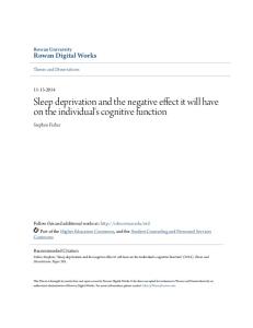
Current perspectives on the function of sleep. gathered under laboratory conditions which might obscure natural relationships[62]. Sleep Deprivation As reported in a comprehensive review, the physiological effects of sleep deprivation in humans have been quite negligible[63]. However, most human sleep deprivation studies have been carried out for only one to four days, which might not be sufficient to reveal significant physiological changes. Because the urge to sleep is so overpowering after only one night of sleep loss, we have been led to expect that even short periods of sleep loss might produce prominent effects. However, this strong sleep urge might reflect the operation of a powerful appetitive mechanism that prevents the development of a serious functional deficit. (By analogy, missing even one meal leaves us ravenously hungry, but without serious physiological consequences.) Longer sleep deprivation periods have been used in several animal studies over the past 100 years. These studies have produced mixed results, ranging from minor changes to severe pathologies and death (see Kleitman[64] for review). However, the sleep loss has been confounded with the strong stimulation -- most frequently enforced locomotion -- required to keep the animals awake, so it has been impossible to attribute the changes unambiguously to sleep deprivation. About 15 years ago, we devised a procedure that enforces sleep deprivation in rats by minimal, gentle stimulation that can be applied equally to control rats. An experimental rat and a control rat are simultaneously housed each on one side of a divided horizontal disk suspended over a shallow pool of water (Fig. 1). As soon as continuous electrophysiological monitoring reveals sleep onset in the experimental rat, the disk is slowly rotated, which forces both rats to walk opposite to disk rotation to avoid being carried into the water. Thus the experimental rat is almost totally deprived of sleep, whereas the control rat can sleep whenever the experimental rat is spontaneously awake and the disk is still. In several replications of these experiments (see[65] for review), rats never survived unrelenting total sleep deprivation; survival time averaged two to three weeks, which is similar to survival time for total food deprivation. FIGURE 1 ILLUSTRATION OMITTED] Perhaps even more remarkable is that selective deprivation of REM sleep, which normally occupied only about 7 percent of total time in our rats, also resulted inevitably in death, usually in about four to six weeks. These results, which have been replicated in several experiments, probably constitute the most consistent and imposing evidence of the biological importance of sleep, but they have shed only limited light on what the function of sleep might be. In spite of extensive physiological monitoring, histological studies, and biochemical analyses, a clear cause of death has not become evident. Everson has reported that most near-terminal rats deprived of sleep by these methods showed bacterial invasion of the bloodstream, suggesting that death might have resulted from septicemia[66]. However, in a subsequent study we showed that the elimination of bacterial invasion with antibiotics did not prevent the deaths of the sleep-deprived rats[67]. The Everson result is clear evidence of a breakdown in host defense in severely sleep-deprived rats. An earlier study using the same sleep deprivation procedure revealed no abnormality in mitogen responses or in vitro or in vivo antibody responses, suggesting that the failed host defense may have resulted from a breakdown of nonspecific defenses such as tissue barriers rather than a failure of specific immune system components, e.g., lymphocytes and antigen-processing cells[68]. These chronic sleep deprivation experiments in rats have revealed changes in thermoregulation. Totally sleep-deprived rats show an increased thermoregulatory setpoint and excessive heat loss; REM sleep-deprived rats show only the excessive heat loss[65]. These results suggest that one function of sleep in the rat is thermoregulation, but there is little evidence as yet on whether thermoregulation is similarly affected by sleep deprivation in larger animals which, by virtue of their relatively enhanced heat retention and generally lower metabolic rates, may have less difficulty maintaining stable temperatures. Certainly in humans, and to a certain extent in animals, the earliest appearing and most frequently observed effects of sleep deprivation have been to worsen performance. Because many of these performance deficits can be mitigated by increasing rewards, interest value, or self-pacing, it appears that they reflect homeostatically driven tendencies to fall asleep rather than deprivation-induced damage to learning and memory mechanisms[69]. One recent study points to deprivation-induced attention deficits which are independent of "microsleeps", but these lapses of attention might be in the service of facilitating sleep onset nevertheless[70]. A few recent studies (e.g.,[71]) have suggested that even small amounts of sleep loss produce impairments of creative and complex thinking that cannot be readily explained by sleepiness or attention lapses. These results suggest that sleep may help maintain higher order, creative mental - Reprinted with permission. Additional copying is prohibited. -
GALE GROUP Information Integrity
Perspectives in Biology and Medicine
Spring 1998 v41 n3 p359(32)
Page 11
Current perspectives on the function of sleep. processes in a finely tuned state, but they would not explain the large amounts of sleep (more than 50 percent of total time in rats and mice) in animals with more mundane mental pursuits or in newborn infants. Vogel has parsimoniously described the effects of a variety of REM deprivation studies in rats as indicating a release of the neural excitation which underlies drive-oriented behavior[64,72]. Conversely, REM sleep was hypothesized to have a role in modulating waking drive expression, thus permitting more flexible, adaptive, specific responses toward drive gratification. However, most of the deprivation effects reviewed by Vogel were exacerbations of stimulated or extreme responses, e.g., decreased electrical thresholds for seizures; lengthening of induced convulsions; lower thresholds for and increased response rates of intracranial self-stimulation; increased shock-induced fighting; increased aggressive posturing and sexual mounting in amphetamine-treated rats (but not in untreated, REM-deprived rats). Therefore, one must wonder whether REM sleep plays a role in run-of-the-mill, spontaneous waking behavior or only in preventing extreme responses in extreme situations. Based on anecdotal reports of mental disturbances produced in the earliest REM deprivation studies[73], it was suggested that REM sleep was important for discharging psychological tensions that would otherwise cause personality disturbances[74]. A comprehensive review by Vogel of subsequent controlled studies concluded that REM sleep deprivation did not produce psychological harm[34]. That conclusion is now accepted by most sleep researchers, although the ballyhoo given the earlier tension discharge theory in the popular press has left the lingering belief among many lay persons that one must dream to stay sane. In fact, Vogel and colleagues have demonstrated that REM deprivation can produce remissions from severe depression[75]. Reductionist Theories of Sleep Function Perhaps it has been difficult to arrive at a widely acceptable theory of sleep function because that function is not well reflected at the organ or system level. The observable systemic characteristics of sleep might be important only to provide the permissive conditions for more essential molecular features. For example, it has been proposed that the muscle hypotonia of sleep permits the endogenous reinforcement of motor circuits by synaptic activation[76], and that the motor inhibition of REM sleep permits endogenous, intense brain activation in the ser-vice of nerve growth without the danger of vigorous, disorganized motor activity[53]. Because adaptive systemic processes eventually benefit cells and effective cellular function is necessary to support systems, our distinction between systemic and cellular functions is somewhat arbitrary. Nevertheless, the distinction raises the issue of which level of description most parsimoniously and verifiably leads to the function of sleep. Because sleep-promoted molecular changes would likely impact on several systems, they could more parsimoniously account for a variety of sleep inducers, effects, and correlates than individual systemic functions. For example, just as cellular energy may be recruited for warmth, growth, or locomotion, sleep might provide basic cellular resources or mechanisms that preferentially serve temperature regulation in small animals, maturation in the young, or higher mental processes in adult humans. That the functional importance of sleep may reside in what it does for brain tissue is suggested by the dramatic studies of dolphin sleep by Mukhametov and his colleagues[3]. As noted earlier, in apparent accommodation to their need to surface to breathe, these animals show the large slow "delta" EEG patterns typical of mammalian sleep with only one half of the brain at a time. When one half is deprived of delta sleep, only that half shows a subsequent delta sleep rebound, i.e., the need for sleep may develop in and be relieved by each hemisphere independently without any apparent change in the gross behavior of the animal[77]. (Because no brain lesion has ever successfully eliminated sleep totally for long periods, it is also conceivable that sleep processes as well as sleep need may be generated locally.) Although the uni-hemispheric generation of a sleep need may be relatively unique to animals obliged to sleep with one half of their brain at a time, the dolphin study offers the exciting possibility of identifying what sleep accomplishes at a cellular level. The possibility of localized sleep processes was raised some three decades earlier by Moruzzi, who cautioned at the same time that if the "recovery processes were not concentrated in a given period of time, we should spend our life in a kind of dormiveglia. We should never be really awake nor completely asleep"[78]. Moruzzi was one of the first to theorize on the cellular function of sleep. He proposed that sleep was necessary for the slow recovery and stabilization of synaptic processes involved in "plastic" activities of learning, memory, and consciousness[78]. This hypothesis derived not from any specific empirical data on how such synaptic processes change with sleep or lack of it, but rather by default from a reasoned exclusion of other candidate sleep functions. Moruzzi began with the position that sleep must function for the recovery of the nervous system, since "the refreshing experience of - Reprinted with permission. Additional copying is prohibited. -
GALE GROUP Information Integrity
Perspectives in Biology and Medicine
Spring 1998 v41 n3 p359(32)
Page 12
Current perspectives on the function of sleep. sleep" does not result from immobility alone. He went on to discount neuronal rest per se as the functional target, both because there is no pervasive decline of neural firing during sleep, and because there is apparently "no need of a long state of inactivity in the large populations of neurons of the respiratory or vasomotor centers or in the inhibitory areas of the cerebellar cortex." He proposed that the sleep-related recovery processes probably involve some slowly accumulating chemical changes in neurons, glia, or at synapses which must require a very slow process -- in contrast to the fast recovery processes (only a few milliseconds), such as those related to the sodium or potassium pumps. Moruzzi could not elaborate on the nature of the chemical accumulation, the recovery processes, or the evidence on why they are related to "plastic" processes, but his argument is valuable for indicating which neuronal activities are probably not involved in sleep function. In contrast to Moruzzi’s theory that sleep facilitates recovery from synaptic use during wakefulness, Krueger and colleagues have proposed that neuronal activity during sleep strengthens and thereby preserves synapses that are under-used during wakefulness[79,80]. A similar theory by Kavanau, proposes that the neuronal activity of sleep "stabilizes" use-dependent synapses and that the depressed perception and sensory processing of sleep minimized interference with these stabilization processes[76,81,82]. Both theoretical presentations had strong features. By postulating that behavioral sleep results from the recruitment and organization of local sleep processes, Krueger, et al., could explain sleepiness (localized sleep activity), the recovery of sleep after sleep-impairing brain lesions (the local sleep processes are preserved), and sleep induction by a variety of substances (propensity for local sleep processes is the overriding determinant). The Kavanau presentation featured reviews of the role of synaptic activation in ontogenetic development, molecular mechanisms of use-dependent synaptic enhancement, the role of the hippocampus in memory consolidation, and the possible furtherance of this role by the neural activity of sleep -- all of which were consistent with the theory. Both the Kavanau and Krueger, et al., theories leave certain major issues unresolved: 1. The theories require the specification of sensitive sensors which direct neuronal activity during sleep preferentially to the synapses which need strengthening. 2. They also require the operation of elaborate processes to direct the neural activity to those synapses which are worth preserving. The indiscriminate reinforcement of all weak synapses would negate the functional value of extinguishing incorrect, useless, or harmful responses. 3. A more convincing case needs to be made for why the synapses to be saved cannot be exercised during wakefulness by practice and extended learning. Why do perceptual processes have to be blocked, as in sleep, to achieve synaptic reinforcement? After all, these processes are not blocked in the most efficient learning state of all -- attentive wakefulness. 4. A more convincing case should be made for the salutory role of sleep-related neuronal patterns on synaptic stabilization. The oscillatory thalamocortical potentials of NREM sleep and the very high voltage pontine-lateral geniculate-occipital cortex spike potentials of REM sleep are not prominent features of waking response acquisition. What would make us believe that they are synapse enhancers during sleep? Crick and Mitchison postulated that these same REM sleep spikes were memory destroyers[21]. Kavanau cites the Wilson and McNaughton result that hippocampal neuronal firing patterns of wakefulness are repeated during NREM sleep as support for synapse enhancement during sleep, but the NREM hippocampal firing may be akin to the reappearance of day residue in the dreams of the night -nocturnal memories of past events with no necessary benefits to memory consolidation[76,83]. We need evidence that the reappearance of these neuronal patterns during sleep actually enhance memory for the waking events. Hippocampal EEG theta activity (sinusoidal waves of six to eight) has been linked to the storage of information reaching the hippocampus, and high amplitude, continuous hippocampal theta is a feature of REM sleep in some animals. However, Vertes has cogently argued that similar electrophysiological signals may reflect different processes in sleep and wakefulness; since there is no orderly flow of information to the hippocampus during REM sleep, it may not serve the same memory-enhancing functions that it serves during wakefulness[84]. 5. No evidence is provided that sleep or the lack of it affects synaptic strength. More direct functional tests of synaptic enhancement are needed. Would a night of sleep enhance transmission along a defined neural pathway? Would sleep deprivation reduce such transmission? Kavanau prepares us for negative results on deprivation studies by noting that they may have "produced little or no evidence of deterioration of mental function" because "A few nights’ lack of circuit - Reprinted with permission. Additional copying is prohibited. -
GALE GROUP Information Integrity
Perspectives in Biology and Medicine
Spring 1998 v41 n3 p359(32)
Page 13
Current perspectives on the function of sleep. reinforcement need not impact seriously on brain performance; to a certain degree functional capacities can be extended and maintained by synaptic enhancements during waking"[81]. This escape clause seems to protect the theory against functional tests. 6. Finally, some evidence is needed to show that sleep preferentially strengthens weak neural connections, e.g., would a night of sleep preferentially enhance or preserve less reinforced learned responses? One recent theory proposes that the function of sleep is the removal of excess cerebral free radicals accumulated during wakefulness[85]. Removal is theoretically accomplished during sleep via a decreased rate of formation and increased efficiency of antioxidant mechanisms. No independent evidence of increased removal during sleep was offered; it was simply assumed. A decreased rate of free radical accumulation was inferred from decreased cerebral metabolism during sleep. However, a comprehensive study of cerebral metabolism in man revealed that it was substantially reduced from waking levels only during NREM sleep with high voltage, slow EEG waves -- which would leave about 80 percent of sleep unexplained[9]. Support for the theory was seen in the higher sleep amounts in smaller mammals, which have higher metabolic rates[22]. However, a recent analysis shows that with body weight held constant by statistical techniques, sleep time across mammalian species correlates negatively with metabolic rate[8]. Altogether, there appears to be little positive support for the theory, but neither is there specific evidence against it. Overview of Theories of Sleep Function Although many theories of sleep and REM sleep function have been proposed, and, as expected, have been staunchly defended by their respective proponents, most sleep researchers seem to believe that we don’t know the function of sleep or of REM sleep. journal articles and book chapters by professional sleep researchers often begin with the statement that the function of sleep is not known. Why have the proposed theories failed to gain wide acceptance? 1. Most of the empirical support for the theories has not been compellingly strong. Theoretical presentations may cite statistically significant effects as support, but the effects may be too small to make us believe that a state so long lasting and ubiquitous as sleep exists to produce them. For example, as we have seen above, a review of many studies showed that exercise increased the nightly sleep of humans by an average of about 10 minutes. That result was statistically significant and is consistent with the theory that sleep is for rest, but the effect is so small that we cannot believe that the bulk of our sleep is for that purpose. There have been a few very substantial empirical findings, e.g., the deaths of all rats subjected to unrelenting total or REM sleep deprivation and the huge correlation of 0.88 between brain weight and cycle length. Paradoxically, however, we have no convincing explanation for those experimental effects. 2. Several theories have required auxiliary assumptions or explanations to deal with apparently inconsistent data. For example, the brain warming theory of REM sleep explained inconsistent data as possible laboratory effects; the oculomotor tuning hypothesis of REM sleep explained the REM sleep of the owl and mole as vestigial or the products of other functions. Such explanations cannot be ruled out; they may be correct. However, they do decrease the parsimonious elegance of the theories, which apparently makes them less attractive to other researchers. 3. Most of the theories have dealt with relatively restricted phenomena of sleep. For example, brain excitation theories of REM sleep may explain the "arousal" electroencephalogr-am of the state and the increased firing in several brain areas including the cortex and lateral geniculate, but these theories do not explain the virtual cessation of neural activity in other brain areas such as the locus coeruleus and dorsal raphe nuclei, or the decrease in thermoregulatory responding, Neither do these theories explain why there should be apparently compensatory rebounds of REM sleep even when behavioral arousal has intervened between the REM deprivation and the rebound period. 4. Several theories apply to restricted populations. For example, the theory that sleep functions to maintain high-level, creative intellectual functioning might be applicable to grown humans, but it doesn’t easily apply to infants or rats. The idea that the neural activation of REM sleep is important for neural growth is consistent with the abundance of REM sleep in the newborn and in species born while very immature, but the neural development theory relies on speculative inferences about neural repair functions to explain the persistence of REM sleep and the rebounds following REM deprivation in adults. 5. The theories often suffer from limited confirmation across different kinds of evidence. For example, the correlative - Reprinted with permission. Additional copying is prohibited. -
GALE GROUP Information Integrity
Perspectives in Biology and Medicine
Spring 1998 v41 n3 p359(32)
Page 14
Current perspectives on the function of sleep. evidence suggesting that sleep conserves energy by enforcing rest is not confirmed by stimulation studies; i.e., there is meager evidence of increased rest enforcement (sleep) following induced physical activity or heightened metabolism. The theory that REM sleep functions to restore cerebral excitement following NREM sleep, which evolved from descriptive data, is denied by the rebounds of REM sleep that follow intervening periods of wakefulness. In summary, theories of sleep function have suffered from a lack of parsimony; they have not had great success in explaining diverse data gathered by diverse methods in diverse populations. Sleep researchers are generally aware of these weaknesses in functional sleep theories. That is why introductions to their papers frequently begin with an acknowledgment that the function of sleep is not known. But other scientists and laymen may be easily taken with new accounts of sleep findings, especially in the popular press, which point strongly to a brave new theory of the function of sleep. Many such theories look good in isolation. Subsequent inspection may reveal that there is conflicting data, that the theory explains sleep in humans or infants but not in rats or old people, or that the theory explains correlative data but not experimental data. Reader beware. Where Do We Stand? What Are the Possibilities? One of the theories already proposed may be correct. Perhaps we have been too one-sided in our presentation by calling attention to isolated negative features, which might be but rough edges on the theory, while neglecting strong parsimonious networks of supporting data. We have attended more to the negative than to the positive, but if the positives had been more compelling, we probably would have seen a more commanding endorsement by sleep researchers for one or another particular theory. Perhaps the evidence on sleep function is so mixed because sleep makes several partial contributions to several different functions, with no single contribution being so essential or strong or ubiquitous across species and age groups that a succinct parsimonious statement about sleep function can be made. Sleep may have evolved several times to serve different functions. As Vogel has suggested, "The multiple processes associated with RS [REM sleep] undoubtedly accomplish many functions or effects"[72]. The multiple function point of view accepts the legitimacy of such seemingly tepid formulations as "sleep is involved with ..." or "sleep has a role in ...". The major problem with this view is that even if it is true, even if sleep does serve multiple functions, letting the matter rest at this level may obscure or trivialize a more profound functional truth. Once selected, a particular mechanism may be recruited to serve multiple functions, but those functions are not at the heart of the matter. For example, the respiratory exchange of air is used for speech, laughter, and playing the trombone, but that is riot why all mammals breathe and must do so to survive. Sleep has survived ubiquitously throughout all of mammalian evolution; some experiments have shown that animals cannot survive without sleep; and animals have made numerous behavioral and physiological accommodations to permit the survival of sleep in different habitats and lifestyles. Sleep persists in predators and prey; in carnivores and vegetarians; on the land and in the water (marine mammals); in most mammals as they lie down relaxed, in ruminants while they stand, in birds while they perch, and in dolphins which constantly swim; in hot and cold climates; in elephants and shrews; in sloths that hardly move and mice that hardly sit still; in the smartest and the dumbest of all mammalian species. These facts suggest a primary, essential, functional core to sleep at is not captured by a potpourri of lesser functional attributes. From personal experience we may be prepared to believe that a good night of sleep will make us an alert, sharp discussant at an important meeting the next day, but that probably has nothing to do with why rats and human infants spend most of their time asleep. Simply stated, the function of sleep, its different stages, and the relationships among them remain to be discovered. When it is discovered, the organizing principle may provide unforced, unapologetic, parsimonious explanations for large amounts of data gathered from diverse populations by diverse methods. It is doubtful that this function will center on molar activities or physiologic processes; most of these stones have been turned. It seems to us more likely that molecular processes would provide a parsimonious key, but the search for sleep-dependent molecular processes seems to be in an early exploratory phase. The great variety of sleep theories conveys an atmosphere of unrestrained speculation. This impression is reinforced by the advocacy of opposite theories, e.g., REM sleep enhances. memory[76, 33] vs. REM sleep erases memory[21]; sleep maintains synapses by permitting recovery[78] vs. sleep strengthens synapses by stimulating them[79]. Although speculation on sleep function is healthy, perhaps the theoretical presentations and their target audiences) could benefit from a bit less selling and a bit more self-criticism. But disciplined or not, the search must continue because the payoff will - Reprinted with permission. Additional copying is prohibited. -
GALE GROUP Information Integrity
Perspectives in Biology and Medicine
Spring 1998 v41 n3 p359(32)
Page 15
Current perspectives on the function of sleep. be large. A huge hole in biological knowledge will be filled. The mechanisms of sleep will be easier to decipher once we know their targets. We will know more about the functional consequences and/or mechanisms of clinically abnormal sleep. And who knows what marvelous new, unanticipated biological insights lie behind the doors of sleep function? REFERENCES [1.] Hartse, K. M. Sleep in insects and nonmammalian vertebrates. In Principles and Practice of sleep Medicine, edited by M. H. Kryger T. Rothy, and W. C. Dement. Philadelphia: W.B. Saunders, 1994. [2.] Amlaner, C. J. J., and Ball, N.J. Avian sleep. In Principles and Practice of Sleep Medicine, edited by M. H. Kryger, T. Roth, and W. C. Dement. Philadelphia: W.B. Saunders, 1994. [3.] Mukhametov, L. M. Sleep in marine animals. Exp. Brain Res. Suppl. 8: 227-38, 1984. [4.] Everson, C. A.; Bergmann, B. M.; and Rechtschaffen, A. Sleep deprivation in the rat. III. Total sleep deprivation. Sleep 12:13-21, 1989. [5.] Kushida, C. A.; Bergmann, B. M.; and Rechtschaffen, A. Sleep deprivation in the rat. IV. Paradoxical sleep deprivation. Sleep 12:22-30, 1989. [6.] Rechtschaffen, A. The function of sleep: Methodological issues. In The Functions of Sleep, edited by R. Drucker-Colin and M. Shkurovich. New York: Academic P, 1979. [7.] Rechtschaffen, A., and Kales, A. A Manual of Standardized Terminology, Techniques and Scoring System for Sleep Stages of Human Subjects. NIH Publ. No. 204. Washington, DC: U.S. GPO, 1968. [8.] Zepelin, H. Mammalian sleep. In Principles and Practice of Sleep Medicine, edited by M. H. Kryger, T. Roth, and W. C. Dement. Philadelphia: W.B. Saunders, 1994. [9.] Madsen, P. L., and Vorstrup, V. Cerebral blood flow and metabolism during sleep. Cereb. Brain Met. Rev. 3:281-96, 1991. [10.] McGinty, D. J.; Harper, R. M.; and Fairbanks, M. K. Neuronal unit activity and the control of sleep states. In Advances in Steep Research, vol. I, edited by E. D. Weitzman. New York: John Wiley, 1974. [11.] Evarts, E. V.; Bental, E.; Bihari, B.; and Huttenlocher, P. R. Spontaneous discharge of single neurons during sleep and waking. Science 135:726-28, 1962. [12.] Parker, D. C.; Rossman, L. G.; Kripke, D. F.; et al. Endocrine rhythms across sleep-wake cycles in normal young men under basal state conditions. In Physiology in Sleep, edited by J. Orem and C. D. Barnes. New York: Academic P, 1980. [13.] Clugston, G. A., and Garlick, P. J. The response of whole-body protein turnover to feeding in obese subjects given a protein-free, low energy diet for three weeks. Human Nutrit. 36C:391-97, 1982. [14.] Horne, J. Why We Sleep. Oxford: Oxford UP, 1989. [15.] Benington, J. H., and Heller, H. C. Restoration of brain energy metabolism as the function of sleep. Prog. Neurobiol. 45:347-60, 1995. [16.] Karnovsky, M. L.; Reich, P.; Anchors, J. M.; and Burrows, B. L. Changes in brain glycogen during slow-wave sleep in the rat. J Neurochem. 41:1498-1501, 1983. [17.] Webb, W. B. Sleep as an adaptive response. Percep. Motor Skills. 38:1023-27, 1974. [18.] Meddis, R. The Sleep Instinct. London: RKP, 1977. - Reprinted with permission. Additional copying is prohibited. -
GALE GROUP Information Integrity
Perspectives in Biology and Medicine
Spring 1998 v41 n3 p359(32)
Page 16
Current perspectives on the function of sleep. [19.] Snyder, F. Towards an evolutionary theory, of dreaming. Am. J. Psychiat. 123:121-36, 1966. [20.] Broughton, R. Confusional sleep disorders: Interrelationship with memory consolidation and retrieval in sleep. In A Triune Concept of the Brain and Behavior: The Hicks Memorial Lectures, edited by D. Campbell, and T. J. Boag. Toronto: U of Toronto P, 1973. [21.] Crick, F., and Mitchison, G. The function of dream sleep. Nature 304: 111-14, 1983. [22.] Zepelin, H., and Rechtschaffen, A. Mammalian sleep, longevity, and energy metabolism. Brain, Behav. Evol. 10:425-70, 1974. [23.] Siegel, J. M.; Manger, P. R.; Nienhuis, R.; et al. The echidna Tachyglossus aculeatus combines REM and non-REM aspects in a single sleep state: Implications for the evolution of sleep. J Neurosc. 16:3500-3506, 1996. [24.] Berger, R. J. Oculomotor control: A possible function of REM sleep. Psychol. Rev. 76:144-6, 1969. [25.] Berger, R. J., and Scott, T. D. Increased accuracy of binocular depth perception following REM sleep periods. Psychophysiol. 8:763-68, 1971. [26.] Berger, R. J., and Walker, J. M. Sleep in the burrowing owl (Speotyto cunicularia hypugaea). Behav. Biol. 7:183-94, 1972. [27.] Siegel, J. M., and Rogawski, M. A. A function for REM sleep: Regulation of noradrenergic receptor sensitivity. Brain Res. Rev. 13:213-33, 1988. [28.] Tsai, L.-L.; Bergmann, B. M.; Perry, B. D.; and Rechtschaffen, A. Effects of chronic total sleep deprivation on central noradrenergic receptors in rat brain. Brain Res. 602:221-27, 1993. [29.] Benington, J. H., and Heller, H. C. Does the function of REM sleep concern non-REM sleep or waking? Prog. Neurobiol. 44:433-49, 1994. [30.] Whitehead, W. E.; Robinson, T. M.; Wincor, M. Z.; and Rechtschaffen, A. The accumulation of REM sleep need during sleep and wakefulness. Communic. Behavior. Biol. 4:195-201, 1969. [31.] Endo, T.; Schwierin, B.; Borbely, A. A.; and Tobler, I. Selective and total sleep deprivation: Effect on the sleep EEG in the rat. Psychiatry Res. 66:97-110, 1997. [32.] Everson, C. A.; Gilliland, M. A.; Kushida, C. A.; et al. Sleep deprivation in the rat. IX. Recovery. Sleep 12:60-67, 1989. [33.] Smith, C. Sleep states and learning: A review of the animal literature. Neurosci. Biobehav. Rev. 9:157-68, 1985. [34.] Vogel, G. W. A review of REM sleep deprivation. Arch. Gen. Psychiat. 32: 749-61, 1975. [35.] Horne, J. A., and McGrath, M. J. The consolidation hypothesis for REM sleep function: A review. Biol. Psychol. 18:165-84, 1984. [36.] McGrath, M. J., and Cohen, D. B. REM sleep facilitation of adaptive waking behavior: A review of the literature. Psychol. Bull. 85:24-57, 1978. [37.] Smith, C. Paradoxical sleep deprivation and sleep recording following training in a brightness discrimination avoidance task in Sprague-Dawley rats: Paradoxical effects. Neurobiol. Learn. Memory 66:283-94, 1966. [38.] Pigarev, I. N. Neurons of the visual cortex respond to visceral stimulation during slow wave sleep. Neurosci. 62:1237-43, 1994. - Reprinted with permission. Additional copying is prohibited. -
GALE GROUP Information Integrity
Perspectives in Biology and Medicine
Spring 1998 v41 n3 p359(32)
Page 17
Current perspectives on the function of sleep. [39.] Toth, L. A. Sleep, sleep deprivation and infectious diseases: Studies in animals. Adv. Neuroimmunol. 5:80-92, 1995. [40.] Benca, R. M., and Quintans, J. R. Sleep and Host Defenses. Steep 20: 1027-1037, 1997. [41.] Korth, C. A co-evolutionary theory of sleep. Med. Hypoth. 45:304-10, 1995. [42.] Youngstedt, S. D.; O’Conner, P. J. O.; and Dishman, R. K. The effects of acute exercise on sleep: A quantitative analysis. Sleep 20:203-14, 1997. [43.] Horne, J. A., and Staff, L. H. E. Exercise and sleep: Body heating effects. Sleep 6:35-46, 1983. [44.] Moriarty. S.; Szymusiak, R.; and McGinty, D. Heat loads evoke delayed enhancements of deep slow wave sleep in rats. Sleep Res. 21:37, 1992. [45.] Tobler, I.; Franken, P.; Gao, B.; et al. Sleep deprivation in the rat at different ambient temperatures: Effects on sleep, EEG spectra and brain temperature. Arch. Ital. Biol. 132:39-52, 1994. [46.] Frederickson, C. J., and Hobson, J. A. Electrical stimulation of the brain stem and subsequent sleep. Arch. Ital. Biol. 108:564-76, 1970. [47.] Pavlov, I. Conditioned Reflexes. Oxford: Oxford UP, 1927. [48.] Daan, S.; Barnes, B. M.; and Strijkstra, A. M. Warming up for sleep? Ground squirrels sleep during arousals from hibernation. Neurosci. Let. 128:265-68, 1991. [49.] Trachsel, L.; Edgar, D. M.; and Heller, H. C. Are ground squirrels sleep deprived during hibernation? Am. J. Physiol. 260:R1123-29, 1991. [50.] Deboer, T., and Tobler, I. Sleep EEG after daily torpor in the Djungarian hamster: Similarity to the effects of sleep deprivation. Neurosci. Let. 166:35-38, 1994. [51.] Tower, D. B., and Elliot, K. A. C. Activity of acetylcholine system in cerebral cortex of various unanesthetised mammals. Am. J. Physiol. 168: 747-59,1952. [52.] Jouvet-Mounier, D.; Astic, L.; and Lacote, D. Ontogenesis of the states of sleep in rat, cat, and guinea pig during the first postnatal month. Devel. Psychobiol. 2:216-39, 1968. [53.] Roffwarg, H. P.; Muzio, J.; and Dement, W. C. Ontogenetic development of the human sleep-dream cycle. Science 152:604-19, 1966. [54.] Oksenberg, A.; Shaffery, J. P.; Marks, G. A.; et al. Rapid eye movement sleep deprivation in kittens amplifies LGN cell-size disparity induced by monocular deprivation. Devel. Brain Res. 97:51-61, 1996. [55.] Pompeiano, O.; Pompeiano, M.; and Corvaja, N. Effects of sleep deprivation on the postnatal development of visual-deprived cells in the cat’s lateral geniculate nucleus. Arch. Ital. Biol. 134:121-40, 1995. [56.] Vogel, G.; Neill, D.; Hagler, M.; and Kors, D. A new animal model of endogenous depression: A summary of present findings. Neurosci. Biobehav. Rev. 14:85-91, 1990. [57.] Mirmiran, M.; Scholtens, J.; Van De Poll, N. E.; et al. Effects of experimental suppression of active (REM) sleep during early development upon adult brain and behavior in the rat. Devel. Brain Res. 7:277-86, 1983. [58.] Kinney, G.; Vogel, G. W.; and Feng, P. Decreased dorsal raphe nucleus neuronal activity in adult chloral hydrate anesthetized rats following neonatal cloimipramine treatment: Implications for endogenous depression. Brain Res. In press. - Reprinted with permission. Additional copying is prohibited. -
GALE GROUP Information Integrity
Perspectives in Biology and Medicine
Spring 1998 v41 n3 p359(32)
Page 18
Current perspectives on the function of sleep. [59.] McGinty, D., and Szymusiak, R. Keeping cool: A hypothesis about the mechanisms and functions of slow wave sleep. Trends Neurosci. 13:480-87, 1990. [60.] Feng, P.-F.; Shaw, P.; Bergmann, B. M.; et al. Sleep deprivation in the rat. XX. differences in wake and sleep temperatures during recovery. Sleep 18:797-804, 1995. [61.] Glotzbach, S. F., and Heller, H. C. Temperature regulation. In Principles and Practice of Sleep Disorders Medicine, edited by M. H. Kryger, T. Roth and W. C. Dement. Philadelphia: W.B. Saunders, 1994. [62.] Wehr, T. A. A brain-warming function for REM sleep. Biobehav. Rev. 16: 379-97, 1992. [63.] Horne, J. A. A review of the biological effects of total sleep deprivation in man. Biol Psychol. 7:55-102, 1978. [64.] Kleitman, N. Sleep and Wakefulness, 2nd ed. Chicago: U. of Chicago P, 1963. [65.] Rechtschaffen, A., and Bergmann B. Sleep deprivation in the rat by the disk-over-water method. Behav. Brain Res. 69:55-63, 1995. [66.] Everson, C. A. Sustained sleep deprivation impairs host defense. Am. J. Physiol. 265:R1148-54, 1993. [67.] Bergmann B. M.; Gilliland, M. A.; Feng, P.-F.; et al. Are physiological effects of sleep deprivation in the rat mediated by bacterial invasion? Sleep 19:554-62, 1996. [68.] Benca, R. M.; Kushida, C. A.; Everson, C. A.; et al. Sleep deprivation in the rat. VII. Immune functions. Sleep 12:47-52, 1989. [69.] Johnson, L. C. Psychological and physical changes following total sleep deprivation. In Sleep: Physiology and Pathology, edited by A. Kales. Philadelphia: Lippincott, 1969. [70.] McCarthy, M. E., and Waters, W. F. Decreased attentional responsivity during sleep deprivation: Orienting response latency, amplitude, and habituation. Sleep 20:115-23, 1997. [71.] Horne, J. A. Sleep loss and "divergent" thinking ability. Sleep 11:528-36, 1988. [72.] Vogel, G. W. A motivational function of REM sleep. In The Functions of Sleep, edited by R. Drucker-Colin M. Shkurovich, and M. B. Sterman. New York: Academic P, 1979. [73.] Dement, W. C. The effect of dream deprivation. Science 131:1705-7, 1960. [74.] Dement, W. C., and Fisher, C. Experimental interference with the sleep cycle. Can. Psychiat. Assoc. J. 8:400-405, 1963. [75.] Vogel, G. W.; Vogel, F.; McAbee, R. S.; and Thurmond, A. J. Improvement of depression by REM sleep deprivation. Arch. Gen. Psychiat. 37:247-53, 1980. [76.] Kavanau, J. L. Memory, sleep, and dynamic stabilization of neural circuitry: Evolutionary perspectives. Neurosci. Biobehav. Rev. 20:289-311, 1996. [77.] Oleksenko, A. I.; Mukhametov, L. M.; Polyakova, I. G.; et al. Unihemispheric sleep deprivation in bottlenose dolphins. J. Sleep Res. 1:40-44, 1992. [78.] Morruzzi, G. The functional significance of sleep with particular regard to the brain mechanisms underlying consciousness. In Brain and Conscious Experience, edited by J. C. Eccles. Berlin: Springer, 1966. [79.] Krueger, J. M.; Obal, F.; Kappas, L.; and Fang, J. Brain organization and sleep function. Behav. Brain Res. - Reprinted with permission. Additional copying is prohibited. -
GALE GROUP Information Integrity
Perspectives in Biology and Medicine
Spring 1998 v41 n3 p359(32)
Page 19
Current perspectives on the function of sleep. 69:177-85, 1995. [80.] Krueger, J. M., and Obal, F. A neuronal group theory of sleep function. J. Sleep Res. 2:63-69, 1993. [81.] Kavanau, J. L. Sleep and memory: evolutionary perspectives. Sleep Res. Soc. Bull. 1:59-65, 1995. [82.] Kavanau, J. L. Sleep and dynamic stabilization of neural circuitry: A review and synthesis. Behav. Brain Res. 63:111-26, 1994. [83.] Wilson, M. A., and McNaughton, B. L. Reactivation of hippocampal ensemble memories during sleep. Science 265:676-79, 1994. [84.] Vertes, R. P. Memory consolidation in REM sleep: Dream on. Bull. Sleep Res. Soc. 1:27-32, 1995. [85.] Reimund, E. The free radical flux theory of sleep. Med. Hypoth. 43:231-33, 1994. [86.] Kryger, M. H.; Roth, T.; and Dement, W. C. Principles and Practice of Sleep Medicine, 2nd ed. Philadelphia: W.B. Saunders, 1994. [87.] Orem, J., and Barnes, C. D. Physiology in Sleep. New York: Academic P, 1980. [88.] Van Cauter, E. Diurnal and ultradian rhythms in human endocrine function: A minireview. Hormone Res. 34:45-53, 1990. [89.] Van Cauter, E.; Blackman, J. D.; Roland, D.; et al. Modulation of glucose regulation and insulin secretion by circadian rhythmicity and sleep. J. Clin. Invest. 88:934-42, 1991. [90.] Greenberg, R., and Pearlman C. A psychoanalytic-dream continuum: The source and function of dream. Int. Rev. Psycho-Anal. 2:441-48, 1978. [91.] Ephron, H. S., and Carrington, P. Rapid eye movement sleep and cortical homeostasis. Psychol. Rev. 73:500-526, 1966. [92.] Vertes, R. P. A life-sustaining function for REM sleep: A theory. Neurosci. Biobehav. Rev. 10:371-76, 1986. [93.] Jouvet, M. Does a genetic programming of the brain occur during paradoxical sleep? In Cerebral Correlates of Conscious Experience. INSERM Symposium No. 6. Elsevier, 1978. [94.] Drucker-Colin, R. Protein molecules and the regulation of REM sleep: Possible implications for function. In The Functions of sleep, edited by R. Drucker-Colin, M. Shkurovich, and M. B. Sterman. New York: Academic P, 1979. [95.] Szymusiak, R., and Satinoff, E. Brief skin temperature changes towards thermoneutrality trigger REM sleep in rats. Physiol. Behav. 25:305-11, 1980. [96.] Jouvet, M.; Buda, C.; and Sastre, J. Increase of paradoxical sleep during hypothermia in pontine cats. In Sleep ’88, edited by J. Horne. Stuttgart: Gustav Fischer Verlag, 1989. [97.] Horne, J. A., and Moore, V. J. Sleep effects of exercise with and without additional body cooling. Electroenceph. Clin. Neurophys. 60:33-38, 1985. [98.] Horne, J. A., and Reid, A. J. Night-time sleep EEG changes following body heating in a warm bath. Electroenceph. Clin. Neurophys. 60:154-57, 1985. [99.] Danguir, J., and Nicolaidis, S. Feeding, metabolism, and sleep: Peripheral and central mechanisms of their interaction. In Brain Mechanisms of Sleep, edited by D. J. McGinty, et al. New York: Raven P, 1985. - Reprinted with permission. Additional copying is prohibited. -
GALE GROUP Information Integrity
Perspectives in Biology and Medicine
Spring 1998 v41 n3 p359(32)
Page 20
Current perspectives on the function of sleep. [100.] Jacobs, B. L., and McGinty, D. J. Effects of food deprivation on sleep and wakefulness in the rat. Exp. Neurol. 30:212-22, 1971. [101.] Borbely, A. A. Sleep in the rat during food deprivation and subsequent restitution of food. Brain Res. 124:457-71, 1977. [102.] Zammit, G. K.; Kolevson, A.; Fauci, M.; et al. Postprandial sleep in healthy men. Sleep 18:229-31, 1995. [103.] Zammit, G. K.; Ackerman, S. H.; Shindledecker, R.; et al. Postprandial sleep and thermogenesis in normal men. Physiol. Behav. 52:251-59, 1992. [104.] MacFadyen, U. M.; Oswald, I.; and Lewis, S. A. Starvation and human slow wave sleep. J. Appl. Physiol. 35:391-94, 1973. [105.] Crisp, A. H.; Stonehill, E.; and Fenton, G. W. The relationship between sleep, nutrition, and food: A study of patients with anorexia nervosa. Postgrad. Med. J. 47:207-13, 1971. [106.] Benca, R. M.; Bergmann, B. M.; Leung, C.; and Nummy, D. Rat strain differences in response to dark pulse triggering of paradoxical sleep. Physiol. Behav. 49:83-87, 1991. [107.] Sawyer, C. H., and Kawakami, M. Characteristics of behavior and electroencephalographic after-reactions to copulation and vaginal stimulation in the female rabbit. Endocrinol. 65:622-30, 1959. [108.] Boland, B. D., and Dewsbury, D. A. Characteristics of sleep following sexual activity and wheel running in male rats. Physiol. Behav. 6:145-49, 1971. [109.] Salin-Pascual, R. H.; Jimenez-Anguiano, A.; Duran-Vasquez, A.; et al. Administration of auditory stimulation during recovery after REM sleep deprivation. Sleep 17:231-35, 1994. [110.] Mistlberger, R.; Bergmann, B.; and Rechtschaffen, A. Period-amplitude analysis of rat encephalogram: Effects of sleep deprivation and exercise. Sleep 10:508-22, 1987. [111.] Borbely, A. A., and Neuhaus, H. U. Sleep deprivation: Effects on sleep and the EEG in the rat. J. Comp. Physiol. 133:71-87, 1979. [112.] Eastman, C. I., and Rechtschaffen, A. The effect of thyroxine on sleep in the rat. Sleep 2:215-32, 1979. [113.] Frederickson, C. J.; Madansky, D. L.; and Rechtschaffen, A. Sensory stimulation and subsequent sleep. Communic. Behavior. Biol. 4:109-13, 1969. [114.] Hobson, J. A. Sleep after exercise. Science 162:1503-5, 1968. [115.] Susic, V., and Kovacevic-Ristanovic, R. Effects of restricted sleep with different exercise loads upon subsequent sleep. Arch. Int. Physiol. Biochim. 88:1-13, 1980. [116.] Hobson, J. A. Sleep as a response. II. Temperature as a possible agent of fatigue-induced somnolence. Psychophysiol. 5:225, 1968. [117.] Ryback, R. S. Effects of prolonged bed rest on EEG sleep patterns in young, healthy volunteers. Electroenceph. Clin. Neurophys. 31:395-99, 1971. [118.] Horne, J. A., and Walmsley, B. Daytime visual load and the effects upon human sleep. Psychophysiol. 13:115-20, 1976. [119.] Horne, J. A., and Minard, A. Sleep and sleepiness following a behaviourally "active" day. Ergonom. 28:567-75, - Reprinted with permission. Additional copying is prohibited. -
GALE GROUP Information Integrity
Perspectives in Biology and Medicine
Spring 1998 v41 n3 p359(32)
Page 21
Current perspectives on the function of sleep. 1985. [120.] Potter, W., and Heron, W. Sleep during perceptual deprivation. Brain Res. 40:534-39, 1972. [121.] Hauri, P. The influence of evening activity on the onset of sleep. Psychophysiol. 5:426-30, 1969.
- Reprinted with permission. Additional copying is prohibited. -
GALE GROUP Information Integrity