This is an enhanced PDF from The Journal of Bone and Joint Surgery The PDF of the article you requested follows this cover page.
Current Concepts Review - Total Hip Arthroplasty with Hydroxyapatite-Coated Prostheses WILLIAM L. JAFFE and DAVID F. SCOTT J Bone Joint Surg Am. 1996;78:1918-34.
This information is current as of May 19, 2011 Reprints and Permissions
Click here to order reprints or request permission to use material from this article, or locate the article citation on jbjs.org and click on the [Reprints and Permissions] link.
Publisher Information
The Journal of Bone and Joint Surgery 20 Pickering Street, Needham, MA 02492-3157 www.jbjs.org
Copyright 1996 by The Journal of Bone and Joint Surgery, Incorporated
Current Concepts Review Total Hip Arthroplasty with Hydroxyapatite-Coated Prostheses* BY WILLIAM L. JAFFE, M.D.f, AND DAVID F. SCOTT, M.D.t, NEW YORK, N.Y. Investigation performed at the Department of Orthopaedic Surgery, Hospital for Joint Diseases, New York City
Basic-Science and Preclinical Studies History of the Use of Hydroxy apatite in Orthopaedics The term apatite was first applied to minerals by Werner45, in 1788. It now denotes a family of crystals with the formula M,0(RO4)6X2, where M is usually calcium, R is usually phosphorus, and X is hydroxide or a halogen such as fluorine. The relationship to bone mineral was first suggested by Proust and Klaproth45, also in 1788. Only after the development and use of x-ray diffraction did Dejong confirm, in 1926, that the inorganic phase of bone was an apatite49. Bone mineral was found to be quite complex and included various types of hydrated calcium phosphates, the most common being calcium hydroxyapatite (Cal0[PO4]6[OH]2). To the best of our knowledge, the earliest use of calcium-phosphate materials in humans was as a powder of varying crystalline composition to improve bonehealing. Albee and Morrison, in 1920, reported accelerated formation of callus3, but others later observed no major advantage with use of the hydroxyapatite powder80147. As a bulk implant, calcium-phosphate materials were first used for dental applications133, as reported in 1971. More recent reports in the dental literature have attested to the success of bulk calcium-phosphate materials composed of pure hydroxyapatite and used as a bone-graft substitute12'35-5768-135137. Patients who had hydroxyapatite grafts were followed for a maximum of seven years57 and were evaluated clinically12-3557137, radiographically'2-3557, and with computer-assisted densitometry68. In two studies'35'37, biopsy specimens were obtained for histological analysis. In addition to impressive evidence of osseointegration, no adverse effects of hydroxyapatite were noted. The dental experience with
*Although none of the authors have received or will receive benefits for personal or professional use from a commercial party related directly or indirectly to the subject of this article, benefits have been or will be received but are directed solely to a research fund, foundation, educational institution, or other non-profit organization with which one or more of the authors are associated. No funds were received in support of this study. tl095 Park Avenue, New York, N.Y. 10128. ^Hospital for Joint Diseases Orthopaedic Institute, New York University Medical Center, 301 East 17th Street, New York, N.Y. 10003.
1918
hydroxyapatite-coated metal implants showed a substantial advantage with use of these implants compared with use of uncoated implants of the same design and having the same metallurgical characteristics77-78122. With respect to the use of hydroxyapatite coatings for total joint implants, the earliest reports of hydroxyapatitecoated femoral stems in humans, to our knowledge, were by Furlong and Osborn67, who began clinical trials in 1985, and by Geesink70, who reported on a series begun in 1986. Basic Science The calcium-phosphate family of biomaterials includes numerous compounds that might have applicability to the manufacture of orthopaedic implants. However, only two such compounds — tricalcium phosphate and hydroxyapatite — have been exposed to extensive in vitro and in vivo testing. These two compounds are easily differentiated from each other by their calcium-to-phosphate ratio. The ratio for tricalcium phosphate (Ca3[P04]2) is 1.5, while that for hydroxyapatite (Ca10[PO4]6[OH]2) is 1.67. The American Society for Testing and Materials has developed standards for these two compounds so that their purity can be determined and controlled4-5. Tricalcium phosphate can exist in several states with an identical chemical composition but a different crystallographic structure, depending on the method with which it was manufactured and sintered. These different states are known as isomorphs (for example, a-tricalcium phosphate and (3tricalcium phosphate), and they may differ with respect to their solubility and biological stability54-55. The most popular method of manufacturing these compounds is chemical precipitation, in which a phosphate-containing solution is added to a calcium-ion-containing solution under controlled conditions2. The resulting compound, amorphous calcium phosphate, is then sintered to a dense polycrystalline form by firing. Organic bone mineral is an unacceptable raw material for this process because of the highly variable composition of its mineral phases and the difficulty of extracting pure compounds from bone36. The method with which the calcium-phosphate comTHE JOURNAL OF BONE AND JOINT SURGERY
TOTAL HIP ARTHROPLASTY WITH HYDROXYAPATITE-COATED PROSTHESES
pound is manufactured and the temperature to which it is exposed affect its transformation to and from hydroxyapatite, its crystalline structure, its porosity, and its solubility. At 700 to 1125 degrees Celsius, in a calciumdeficient environment, p-tricalcium phosphate is produced; at temperatures of greater than 1450 degrees Celsius, a-tricalcium phosphate is formed. At physiological pH, amorphous calcium phosphate is more soluble than a-tricalcium phosphate, which is more soluble than P-tricalcium phosphate, which is more soluble than hydroxyapatite5455. Because solubility at physiological pH is a major factor responsible for bioresorption, hydroxyapatite appears to have a definite advantage, compared with tricalcium phosphate, as a biological implant material. In acidic environments, all calcium phosphates, including hydroxyapatite, are highly soluble and unstable. In an acid medium tricalcium phosphate dissolved 12.3 times faster than hydroxyapatite, and in a basic medium it dissolved 22.3 times faster95. The most common precursor to hydroxyapatite, during both in vivo bone formation and in vitro manufacturing, is amorphous tricalcium phosphate, which is highly soluble and unstable at physiological pH. Hydroxyapatite, as found in organic bone matrix, exists over a compositional range that can be characterized by its calcium-to-phosphate ratio. It can vary from stoichiometric hydroxyapatite (Cal0[PO4]6[OH]2), with a calcium-to-phosphate ratio of 1.67, to calcium-deficient hydroxyapatite (Ca|,0.x)[HPO4]x[OH]|2.x]), with a calciumto-phosphate ratio as low as 1.5. Biological apatites are known to be calcium-deficient, bone apatites contain carbonate, and dental apatites contain substantial amounts of fluoride"'. Commercially available calciumphosphate materials can be classified as biphasic calcium phosphate (mixed P-tricalcium phosphate and hydroxyapatite phases), P-tricalcium phosphate, hydroxyapatite, non-sintered calcium-phosphate powders, coralline hydroxyapatite, and bone-derived materials"6. Biological fixation is defined, for the purpose of this paper, as the process by which prosthetic components become firmly bonded to host bone by ongrowth or ingrowth without the use of bone cement. Because the goal of the use of hydroxyapatite is to obtain prompt biological fixation to bone, it is of interest to consider the mechanism by which such fixation occurs. It has been suggested that the apatite must first partially dissolve, thereby increasing the concentration of calcium and phosphate in the microenvironment. Carbonate apatite microcrystals then form and associate with the organic matrix of bone, causing biological growth of bone tissue"7. There appears to be a conflict between the ability to manufacture a stable, slowly resorbing material and the ability to achieve rapid union with host bone. Additional stabilization of a calcium-phosphate apatite by increasing its crystallinity results in a decrease in the release of calcium and phosphate from its surface, as in vivo studies have clearly demonstrated that the higher VOL. 78-A, NO. 12, DECEMBER 1996
1919
the percentage of crystallinity of a coating the lower the rate of degradation173. The source of free calcium and phosphorus that is present even at the interface of highly crystalline, stable hydroxyapatite coatings appears to be the amorphous calcium-phosphate phase, which is found in all hydroxyapatite coatings173. It is likely that some critical amount of degradation is essential to obtain rapid biological fixation, but premature dissolution of a coating or loss of mechanical bonding to a metal substrate must be avoided. Currently, highly crystalline, pure, stable hydroxyapatite appears to contain adequate amorphous calcium phosphate to allow early biological fixation to be achieved without the use of less crystalline hydroxyapatite or more soluble tricalcium phosphate. Other Bioactive Coatings Bioactive coatings are substances that are added to the surface of an implant to promote and to enhance biological fixation. Bioactive coatings other than hydroxyapatite have been tested in vivo with varying degrees of success. Fluorapatite was found to be biocompatible and to bond readily to bone in several animal studies4388138. It was also more thermostable than hydroxyapatite, and it did not demonstrate the chemical dissociation noted with hydroxyapatite during the use of high-temperature plasma-spraying techniques121. When tested in vivo, it had superior long-term degradation characteristics and the same excellent push-out strength as hydroxyapatite5". Whitlockite is a calcium-phosphate mineral in the phase of P-tricalcium phosphate. Initial investigations into the effects of adding magnesium to p-whitlockite to form magnesium whitlockite suggested reduced rates of biodegradation compared with those of hydroxyapatite109. More recent in vivo tests did not confirm these findings and demonstrated inferior push-out strength compared with that of hydroxyapatite50. Bioglass coatings composed of sodium carbonate, calcium carbonate, phosphorus pentoxide, and silica have been shown to bond to host bone by forming a surface layer of calcium phosphate with a hydroxyapatite structure139. However, the coatings themselves are prone to failure in vivo19, and the silica-containing calcium-phosphate glasses may be toxic because of the presence of dissolved silica ions136. Bioactive polymer coatings — chemically known as poly(ethylene oxide), poly(tetramethylene terephtalate) segmented copolymer — have demonstrated bone-bonding properties by cell-mediated osteogenesis that are comparable with those of hydroxyapatite and tricalcium-phosphate ceramic coatings. Ceramics are defined as calcium-phosphate materials with a composition and structure that are similar to the inorganic components of bone87. Stress-analysis studies have suggested that this flexible polymer coating may have mechanical advantages compared with more brittle ce-
1920
W. L. JAFFE AND D. F. SCOTT
ramie coatings such as hydroxyapatite48. Additional in vivo testing will be required before the clinical role of these coatings can be determined. Metal Substrate The mechanical properties of calcium-phosphateceramic materials — specifically, their poor tensile strength and resistance to impact as well as their brittle nature — make them unacceptable as load-bearing implants95. The application of ceramic to a metal substrate combines the strength of metal with the biocompatibility of ceramic76105. While it is possible to coat most metals with hydroxyapatite, the most experience and success with hydroxyapatite-coated implants used for arthroplasty has been with titanium, its alloy Ti-6A1-4V, and cobalt-chromium alloy. Compared with cobalt-chromium, titanium demonstrated a 33 per cent increase in bonding strength to a hydroxyapatite coating in vitro. It was suggested that this increase was due to a chemical bond between titanium and hydroxyapatite in addition to the expected mechanical bond6276"5. The difference in the modulus of elasticity of the metals (210 gigapascals for cobalt-chromium alloy as compared with 110 gigapascals for titanium) results in less potential proximal stress-shielding and bone resorption with the more flexible titanium femoral stem183. However, increased motion at the coating-bone interface with use of the more flexible stem might neutralize this advantage, as micromotion at the interface was shown to inhibit biointegration in a dog model"14. It must be emphasized that both of these metals are considerably stiffer than cortical bone (modulus of elasticity, twenty gigapascals183) and that the proximal cross-sectional area of most modern femoral implants is generous, resulting in an inelastic prosthesis. When all of these factors are weighed, titanium alloy currently appears to be the metal substrate of choice. The surface of the metal substrate can be textured to one of three forms: smooth (microstructured), macrostructured, or porous. A smooth surface, grit-blasted to a roughness of four to ten micrometers, is a typical microstructured finish used in preparation for application of a plasma-sprayed coating. This prepared metal surface appears histologically similar to the natural undulations of the hydroxyapatite-coated surface30. Several investigators have demonstrated successful biological fixation to bone with this microstructured and coated surface, with failure ultimately occurring at the coatingsubstrate interface when the implant is subjected to high shear and tensile forces28170. Geesink et al., in a study of dogs that involved push-out testing of twenty-seven transcortical plugs, rarely observed failure at this interface; failure was more commonly noted to occur within the hydroxyapatite coating or at the hydroxyapatitebone interface76. Macrostructuring of the surface increases the resistance of the coating to pure shear forces and could
result in improved long-term fixation between the implant and the bone if biodegradation of the coating occurs20 m. Macrostructuring can take the form of grooves, threads, or deposited metal coating. Improved fixation with grooved-surface implants was demonstrated in vivo at early time-periods167170 as well as one year after implantation, suggesting a comprehensive interface. This resulted in the sharing of mechanical loads between the areas in which bone had grown onto the hydroxyapatite coating and the areas in which it had grown into the metal grooves144. A titanium-alloy (Ti6A1-4V) substrate can be coated with commercially pure titanium with use of an arc-deposition process to form a macrostructured surface. This surface is then given a preparatory grit-blasting followed by a plasmasprayed hydroxyapatite coating. Although the gritblasting and the hydroxyapatite coating result in a 38 per cent decrease in the roughness of the surface compared with that of the arc-deposited surface85, the arc-deposited hydroxyapatite-coated surface demonstrated remarkably superior biological fixation compared with that for the uncoated arc-deposited surface in several animal models85143 (Fig. 1). Porous surfaces appear to have so much inherent ability to promote biological fixation that a calciumphosphate coating is required only to direct the early growth of bone into the pores. For such a limited role, tricalcium phosphate or less optimum hydroxyapatite mineral phases should suffice. In vivo tests with tricalcium phosphate on porous surfaces demonstrated an early increase in the strength of attachment at six weeks" 14 ' but failed to show any advantage at thirtytwo weeks170. Hydroxyapatite-coated porous surfaces demonstrated an increase in the strength of attachment to bone for as long as one year after implantation compared with that of uncoated porous surfaces323337 and even overcame a one-millimeter gap between the implant and the host bone313337165. As with macrotexturing, porous substrate surfaces enhance the shear strength of the metal-hydroxyapatite interface. As clinical retrieval studies have demonstrated fibrous fixation — rather than fixation by bone ingrowth — of many porous implants172627, the addition of hydroxyapatite may improve osseointegration. In a dog model in which hydroxyapatite-coated and uncoated porous titanium implants were compared after having been subjected to four weeks of controlled micromotion, the presence of hydroxyapatite limited the thickness of the fibrous membranes that formed161 and induced the formation of fibrocartilage as well as a higher concentration of collagen161164; hydroxyapatite possibly induced the conversion of the fibrocartilage to bone after sixteen weeks of micromotion162. Application of Hydroxyapatite Coating Several methods are available for the application of a hydroxyapatite coating to a metal substrate, and T H E J O U R N A L OF BONE A ND J O I NT SURGERY
TOTAL HIP ARTHROPLASTY WITH HYDROXYAPATITE-COATED PROSTHESES
FIG.
1921
1
Backscatter electron-imaging photomicrograph, made six weeks postoperatively, showing the interfaces around a fifty-micrometer air-plasma-sprayed hydroxyapatite coating on a grit-blasted titanium implant retrieved from a dog. M = metal substrate, H = hydroxyapatite coating, and B = host bone (x 200). (Courtesy of J. Ricci.)
the chemical, mechanical, and biological properties of the coating depend on the method that is used99110"1.The techniques that commonly are used include dip-coating, electrophoretic deposition, immersion-coating, hot isostatic pressing, solution deposition, sputter-coating, and flame-spraying. Only the latter three methods are currently applicable to orthopaedic implants. The disadvantage of the dip-coating and immersioncoating methods is that they require high temperatures for the post-sintering of the hydroxyapatite layer, which can degrade the strength of the metal and compromise the purity of the hydroxyapatite'20185. Electrophoresis results in impurities, non-uniform thickness, and poor biological fixation to the metal substrate185. Isostatic pressing is a cumbersome two-stage technique with which it is difficult to seal borders on implants with complex shapes. In addition, isostatic pressing requires high temperatures during the second stage, which could denature the hydroxyapatite185. A solution-deposition process in which hydroxyapatite is precipitated at a low temperature results in a pure, highly crystalline, firmly adherent coating that is twenty micrometers thick56. As it is not a line-ofsight process (the coating is applied only perpendicular to the surface as it is in flame-spraying), porous and beaded surfaces can be coated evenly. Porous-coated cobalt-chromium bone plugs that were coated with this method demonstrated successful biological fixation in a dog model1. Porous-coated femoral and acetabular components with a hydroxyapatite coating applied with use of solution deposition are currently marketed in Europe VOL. 78-A, NO. 12, DECEMBER 1996
but have not been approved for use in the United States. As the process is apparently limited to coatings with a thickness of twenty micrometers56, it seems to represent an augmentation of biological fixation rather than a primary mode of fixation. Sputter-coating is a process in which molecules of a material are ejected from a target by the bombardment of high-energy ions and are deposited on a substrate that has been placed in a vacuum chamber. Most sputter-coating techniques are too slow and have an inadequate rate of deposition, but an enhanced method (RF magnetron sputtering) in which a magnetically increased radiofrequency is used has been described3494. However, an analysis of the chemical nature of the hydroxyapatite coating applied with this method revealed a calcium-to-phosphate ratio that was consistently higher than that of standard synthetic hydroxyapatite174. Flame-spraying techniques involve the acceleration of hydroxyapatite feedstock powder to a high velocity and temperature, resulting in a dense, tightly adherent coating with a maximum thickness of 100 micrometers that can be applied in less than two minutes. The coating can be applied to implants with complex shapes; areas that are not to be coated can be easily masked. Three techniques — air plasma-spraying, vacuum plasmaspraying, and the high-velocity oxygen-fuel method — have been described and tested182. With plasma-spraying, an arc is created between an anode and a cathode, resulting in a temperature of as high as 30,000 degrees kelvin (29,727 degrees Celsius), which causes gas dissociation.
1922
W. L. JAFFE A ND D. F. SCOTT
Secondary electron-imaging photomicrograph showing the surface of an air-plasma-sprayed hydroxyapatite coating on a grit-blasted titanium substrate (x 200). (Courtesy of J. Ricci.)
The hydroxyapatite powder with a carrier gas such as argon is passed into the arc, which melts the surface of the hydroxyapatite particles and accelerates them onto the metal substrate (Fig. 2). The metal is heated to less than 300 degrees Celsius, which does not degrade its mechanical properties. With the high-velocity oxygenfuel technique, the particles are heated to substantially lower temperatures but are applied at higher velocities than with the arc-deposition process. Comparison of the three techniques has demonstrated that the coatings decrease in density in the following sequence: air plasma-spraying, vacuum plasma-spraying, and the highvelocity oxygen-fuel method48. While a coating applied with the high-velocity oxygen-fuel method is more stable chemically because of the lower temperature of application, the strength of the coating-substrate bond may be less than optimum81. Although enhanced bonding strength and improved purity of the hydroxyapatite have been claimed for vacuum plasma-spraying as compared with air plasma-spraying, no clinical advantage has yet been demonstrated, and the coating-related factors associated with clinical success, such as density, porosity, crystallinity, and chemistry, are still not entirely understood. The strength of a hydroxyapatite coating depends on its thickness. De Groot et al. found that fatigue fracture occurred commonly in coatings thicker than 100 micrometers and that bioresorption was unacceptably rapid with coatings thinner than thirty micrometers46'17'75. They determined that an optimum thickness of fifty micrometers would avoid fatigue failure while enabling consistent apposition to the host bone464775. This is currently the standard for the optimum thick-
ness of a prosthetic hydroxyapatite coating. Hydroxyapatite is neither osteogenic (capable of stimulating the formation of bone) nor osteoinductive (capable of supporting the mitogenesis of undifferentiated perivascular mesenchymal cells, leading to the formation of osteoprogenitor cells with the capacity to form new bone172). Hydroxyapatite is osteoconductive (capable of supporting the ingrowth of sprouting capillaries, perivascular tissues, and osteoprogenitor cells from the recipient host bed into the three-dimensional structure of an implant or graft172) in a manner similar to that of allograft bone. It acts as a trellis for the deposition and ingrowth of new bone148. A dense coating with high crystallinity to decrease the rate of bioresorption does not appear to compromise the osteoconductive property of the hydroxyapatite108. Plasma-sprayed hydroxyapatite coatings contain sufficient microporosity, macroporosity, and amorphous hydroxyapatite to ensure maintenance of biological activity'87. Starting with a pure feedstock of hydroxyapatite and employing a plasma-spraying process that does not denature the hydroxyapatite to less stable calcium phosphates or introduce contaminants allow a reliable, inexpensive, bioactive coating to be applied to a prepared metal substrate of an orthopaedic implant. Clinical Experience with Hydroxyapatite-Coated Components for Total Hip Arthroplasty The European Experience with Hydroxyapatite-Coated Femoral Components Geesink70"72 as well as Geesink and Hoefnagels74 performed hip replacements with use of a metaphysis-filling titanium femoral stem that had a fifty-micrometer-thick T H E J O U R N A L O F BONE AND J O I NT SURGERY
TOTAL HIP A R T H R O P L A S T Y WITH HYDROXYAPATITE-COATE D
air-plasma-sprayed coating of hydroxyapatite on the proximal one-third of its surface. Initially, a threaded acetabular cup was chosen, but a hemispherical acetabular component was subsequently used. The rate of survival of the prosthesis at six years for the first 118 patients was 100 per cent for the 118 hydroxyapatite coated stems and 99 per cent for the 100 hydroxyapatite threaded cups, and there was a very low prevalence of pain in the thigh74. Karrholm et al. prospectively studied the results for sixty hips that had been randomly assigned to receive the same type of femoral component (a straightstem titanium-alloy design with a collar) that was to be fixed with cement, a hydroxyapatite coating, or a porous coating101. The hydroxyapatite was applied to the proximal one-third of the surface and was 200 micrometers thick. At two years, the clinical results did not differ among the groups with respect to the Harris hip scores or the pain scores, but the hydroxyapatite-coated components demonstrated less subsidence and rotation. S0balle et al. performed a prospective study in which twenty-seven patients were randomly assigned to receive a femoral component of the same design with either an arc-deposited titanium coating or an airplasma-sprayed hydroxyapatite coating on the proximal one-fourth of the stem166. Roentgen stereophotogrammetric analysis performed at twelve months revealed less migration of the hydroxyapatite-coated components, and the Harris hip scores and the pain scores were better for the group that had received a hydroxyapatitecoated implant. Kroon and Freeman performed a controlled study of sixty-nine identical press-fit titanium stems with and without an eighty to 120-micrometer-thick proximal air-plasma-sprayed hydroxyapatite coating"4. They reported a tenfold increase in subsidence of the uncoated stems at one year as measured with x-ray digitization. Epinette reported no failures and excellent scores for pain and function in a study of thirty-one hydroxyapatite-coated stems and threaded acetabular components after five years of follow-up61. Other European investigators who have reported good results with hydroxyapatite-coated total hip components have included Furlong66, who followed a large series of patients for a maximum of eight years, and Livesley et al."9, who prospectively compared the results of the use of press-fit and hydroxyapatite-coated titanium bipolar prostheses for the treatment of displaced fractures of the femoral neck. Reports of a large Italian series documented excellent short-term clinical results150171. Several other reports in the French and German literature have attested to the safety and efficacy of hydroxyapatite-coated total hip components, although details of the thickness, extent, and purity of the coatings were limited64-82-112-128-131-177-178-180. The experience with hydroxyapatite-coated implants in revision hip arthroplasty is limited, but the results of one series73 VOL. 78-A, NO. 12, D E C E M B E R 1996
PROSTHESES
1923
of fifty patients who were followed for a minimum of four years are encouraging. The North American Experience with Hydroxyapatite-Coated Femoral Components In the largest North American clinical trial of which we are aware, 436 hip arthroplasties were performed between January 1988 and November 199021-404293127.This prospective, multicenter study was approved by the Food and Drug Administration under an Investigational Device Exemption. The femoral component was a grit-blasted, straight, collarless, titanium implant that had normalization steps on the anterior and posterior proximal surfaces as well as a fifty-micrometerthick air-plasma-sprayed hydroxyapatite coating on the proximal one-third of the surface. The acetabular components were either porous-coated or hydroxyapatitecoated implants and were of three different designs. The investigators reported very encouraging results at two, three, and five years21-40-42-93127. At the five-year evaluation, the rate of mechanical loosening of the femoral component, representing stems that been revised because of aseptic loosening (two) and those that were radiographically unstable (zero), was 0.46 per cent21. No progressive subsidence was reported, and endosteal osteolysis was noted in only one patient (0.8 per cent). These excellent results also applied to the youngest patients in the series; at five years, eighty-eight patients, who had an average age of thirty-eight years, had had no mechanical failures of the femoral component22. Vaughn et al., in a non-randomized study, reported the results obtained with use of a straight, collarless, metaphysis-fitting titanium stem that was coated on the proximal one-third of its surface with either arc-deposited titanium (fifty implants) or a fifty to seventy-five-micrometer-thick air-plasma-sprayed hydroxyapatite coating (fifty-three implants)17"76. Both types of stems were associated with excellent shortterm clinical results, but the implants that had an arcdeposited titanium coating were associated with a higher rate of radiolucent lines, including a 10 per cent prevalence in the proximal, coated region. Rothman et al. reported the two-year results for a matched-pair series of fifty-two patients who had received a collarless, porous-coated titanium femoral stem with or without a proximal hydroxyapatite coating151. The clinical and radiographic results did not differ between the two groups. A number of authors5152-65-125126 reported on two series of air-plasma-sprayed hydroxyapatite-coated components. In the first series, a retrospective matched-pair study of eighty-four hips that had a metaphysis-fitting titanium-alloy femoral component with proximal porous patches, one-half of the stems had a supplemental hydroxyapatite coating125126. At a minimum of three years, there was no difference between the matched groups with respect to the fixation of the femoral stem,
1924
W. L. JAFFE AND D. F. SCOTT
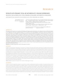
FIG. 3 Left: Anteroposterior radiograph made eight years after a total hip arthroplasty performed with use of a hydroxyapatite-coated femoral component. There is relative osteopenia (top arrow), bone condensation and hypertrophy (middle arrow), and radiolucency around the uncoated portion of the stem (bottom arrow). Right: Photograph showing the prosthesis (Osteonics, Allendale, New Jersey).
but greater hypertrophy of cancellous bone was noted proximally in association with the stems coated with hydroxyapatite. The second series involved a press-fit version of the same type of stem with and without a hydroxyapatite coating on the grooved, grit-blasted proximal surface5265'25. This prospective, randomized, multicenter trial revealed substantial differences between the two groups at a minimum of two and a half years. The Harris hip scores and the relief of pain were better for the hydroxyapatite-coated stems, and the prevalence of radiolucent lines in the coated region was approximately 1 per cent for that group compared with approximately 25 per cent for the press-fit stems. Clinical Experience with Hydroxy apatiteCoated Acetabular Components Most clinical trials of hydroxyapatite-coated implants have focused on the performance of the femoral component rather than that of the acetabular component. In one study, fully porous-coated hemispherical acetabular components supplemented with a hydroxyapatite coating demonstrated improved fixation radiographically at a minimum of three years as compared with partially porous-coated components without a hydroxyapatite coating12''. In the multicenter study of D'Antonio et al., three hydroxyapatite-coated acetabular cups in a series of 320 components had migrated measurably at two years but none had been revised for
aseptic loosening40. That study was unique because it included titanium acetabular components of three different designs: a microstructured, fully coated, dualradius hemispherical component designed to optimize the peripheral rim fit; a fully coated threaded cup; and a non-hydroxyapatite-coated porous-coated cup. At the five-year follow-up examination, the 165 non-threaded hemispherical hydroxyapatite-coated cups had a combined rate of radiographic and clinical failure of 19 per cent2238. This finding is in direct contrast to both the results for the 103 non-hydroxyapatite-coated porouscoated cups (a combined rate of failure of 6 per cent) and those for the hydroxyapatite-coated threaded cups from the same manufacturer in the two European trials (a combined rate of failure of 3 per cent of 118 and 0 per cent of 107, respectively). Additionally, Geesink and Hoefnagels reported a six-year survival rate of 99 per cent for 100 hydroxyapatite-coated threaded acetabular components74. It is paradoxical that these components yielded excellent results compared with those for hemispherical cups, as non-hydroxyapatite-coated threaded cups have been associated with poor results in other series63160184. Geesink and Hoefnagels found that fixation with screws, holes in the cup, the locking mechanism of the polyethylene, and the method of sterilization of the polyethylene did not contribute to the increased rate of failure of the microstructured cups74. Factors that were found to be associated with loosening were the use of a T H E J O U R N A L OF BONE AND JOIN T SURGERY
TOTAL HIP ARTHROPLASTY WITH HYDROXYAPATITE-COATED PROSTHESES
thirty-two-millimeter femoral head, female gender, and an age of forty-five years or less. The most important factor was believed to be the lack of any macrostructuring (as with threads, beads, or arc-deposited titanium) that would have allowed for a mechanical interlock 74 . Moilanen et al.,in a retrospective study, reported the two to three-year results for press-fit cobalt-chromium acetabular components with a central peg and two superolateral flanges, with or without a hydroxyapatite coating that was eighty to 120 micrometers thick132. T h e r e w e r e n o differences in the clinical results, but the hydroxyapatite-coated c o m p o n e n ts were associated with fewer radiolucent lines and less rotational and proximal migration. Adaptive Host-Bone Remodeling Hydroxy apatite-Coated Femoral
in Association Components
with
1925
There was no bone loss between two and seven years. In another study, proximal bon e loss in a matched group of eighty-seven patients was found to be greater, two to five years postoperatively, in patients who had a porouscoated, diaphysis-filling, cobalt-chromium stem than in those who had a proximally fitting hydroxyapatitecoated titanium stem157. The average bon e loss in the two proximal zones of G r u e n et al. was 29 per cent for the porous-coated implants and 14 per cent for the hydroxyapatite-coated implants. Analysis of hydroxyapatite-coated femoral components with use of dual-energy x-ray absorptiometry has revealed results that are favorable compared with those of studies, performed with use of the same modality, of stems inserted with cement and evaluated retrospectively 123145 , of p o r o u s - c o a t e d stems retrieved at autopsy59124169, and of porous-coated stems evaluated retrospectively 90106107145 . A paired, bilateral, dual-energy x-ray absorptiometry comparison of hydroxyapatitecoated and porous-coated stems of the same design revealed greater proximal bon e loss in association with the porous-coated components 155 . Titanium stems with a proximal hydroxyapatite coating have been associated with minimum, nonprogressive proximal loss of b o n e and with strong evidence of proximal biological fixation in densitometric and radiographic studies39-42'65•72'74•10,•114•130'155-157•166'175'l76'181. Non-hydroxyapatite-coated titanium stems also have been associated with minimum short-term decreases in bone-mineral density, although radiographic signs of biological fixation have b e e n lacking. Radiographic and dual-energy x-ray absorptiometry analyses have shown that porous-coated cobalt-chromium stems and stems inserted with cement caused greater resorption of femoral bon e than hydroxyapatite-coated stems and that biological fixation did not occur as consistently in association with porous-coated stems.
Proximally hydroxyapatite-coated femoral stems were associated with a characteristic radiographic remodeling patter n in multiple trials22'39-42'6572'74101"4166'75176. T h e c o m m o n findings were a high prevalence of radiolucent lines surrounding the distal, uncoated portion of the stem and a low prevalence of radiolucent lines adjacent to the proximal, coated portion of the stem3941'65'7274101'114'166175176; cancellous condensation at the distal extent of the coating39-41'7274126; and fusiform cortical h y p e r t r o p h y beginning at the transition between the coated and uncoated regions and extending distally39'427274101 (Fig. 3). In the largest study of radiographic remodeling of the femur after implantation of a hydroxyapatite-coated femoral stem of which we are aware, 68 per cent of 224 stems had radiolucent lines surrounding the distal, uncoated portion of the stem and 2 per cent had radiolucent lines around the proximal, coated region as seen on both anteroposterior and lateral radiographs 4 1 . These findings of new-bone formation and extensive proximal fixation were consistent and progressive throughout the seven-year follow-up period (Figs. 4-A through 4-D).
Analysis
Dual-energy x-ray absorptiometry has b e e n used to quantify the regional changes in bone-mineral density that occur in the femur adjacent to a component that has b e e n inserted without cement. In two prospective studies of proximally hydroxyapatite-coated femoral components, there was an 8 to 12 per cent decrease in bone-mineral density in the proximal part of the femur after eighteen to twenty-four months of follow-up130181. In a cross-sectional, retrospective study of eightyeight patients w h o were followed for seven years after implantation of a hydroxyapatite-coated femoral stem, dual-energy x-ray absorptiometry revealed a 25 per cent decrease in bone-mineral density proximally between six weeks and six months postoperatively 156 . Between six m o n t h s and two years, bone-mineral density increased proximally, with an average loss of 15 per cent, and it increased distally, returning to values equal to or greater than those before the operation.
Most retrieved hydroxyapatite-coated components have been femoral stems. There are two types of retrieval studies of hydroxyapatite-coated components, revision and autopsy, and they have demonstrated different results. Studies of implants retrieved at the time of a revision hip arthroplasty have revealed findings of concern, including dissolution and delamination of the coating. Examination of a patient who initially had a well fixed hydroxyapatite-coated titanium femoral component but who had severe pain in the thigh at two years did not reveal histological evidence of the original hydroxyapatite coating, but the prosthesis-host interface was covered predominantly with trabecular bone 19 . Bloebaum et al. reported on a series of fourteen different implants retrieved for a variety of reasons, including osteolysis, excessive wear of the polyethylene, infection, pain in the hip, trauma, loosening, and death 1316 . The implants had been in situ for an average of twenty-eight
VOL. 78-A, NO. 12, DECEMBER 1996
of Retrieved Hydroxyapatite-Coated
Implants
1926
W. L. JAFFE AND D. F. SCOTT
FIG.
4-A
Figs. 4-A through 4-D: Anteroposterior and lateral radiographs showing progressive femoral remodeling around a stem with a proximal hydroxyapatite coating. Fig. 4-A: Six months postoperatively.
months and were of six different designs. In addition to titanium and polyethylene debris, hydroxyapatite particulate material was discovered in the periprosthetic tissues and was found to be embedded in the surfaces of the polyethylene liner. In some areas, a ten to twenty-micrometer gap was evident between the hydroxyapatite and the metal substrate, suggesting delamination of the hydroxyapatite coating. Those authors noted that it was not possible to determine which of the three types of particles — titanium, polyethylene, or hydroxyapatite — had caused the osteolysis; how-
ever, they cited references suggesting that hydroxyapatite particulate material could cause an inflammatory tissue response and osteolysis1316. In order to assess indirectly the potential problem of hydroxyapatite particulate debris and resultant third-body wear, Bauer et al. used laser interference microscopy to measure the roughness of the surface of the modular cobalt-chromium femoral heads of fifteen clinically retrieved hydroxyapatite-coated components and to compare it with that of the heads of fifteen retrieved porous-coated components that had
FIG. 4-B One year postoperatively.
THE JOURNAL OF BONE AND JOINT SURGERY
TOTAL HIP A R T H R O P L A S TY WITH HYDROXYAPATITE-COATED
PROSTHESES
1927
FIG. 4-C Three years postoperatively.
been inserted without cement and also with that of the heads of fifteen components that had been inserted with cement9. They found increased median surface roughness as compared with the initial specifications of the manufacturer in all groups; however, the heads from the hydroxyapatite-coated components had less roughness and fewer deep scratches than those from either the porous-coated components or the components that had been inserted with cement. This suggests that the problem of third-body wear is not greater with hydroxyapatite-coated implants than with porouscoated implants or with implants inserted with cement. There also has been a number of investigations of hydroxyapatite-coated implants retrieved at autopsy. Bloebaum et al. examined retrieved bilateral femo-
ral components with identical porous-coated titanium stems, except that the stem retrieved from the right hip had a seventy to 100-micrometer proximal airplasma-sprayed hydroxyapatite coating'4. Backscattered electron analysis showed 50 per cent more bone in the implants with a hydroxyapatite coating. In another autopsy-retrieval study, Bloebaum et al. performed an analysis of hydroxyapatite-coated femoral and acetabular components retrieved three weeks after implantation15. Light microscopy showed the presence of osteoid on 20 per cent and 40 per cent of the femoral and acetabular regions, respectively. Bauer et al. performed a histological analysis of five autopsy-retrieved hydroxyapatite-coated femoral stems that had been in situ for an average of twelve months7. A uniform coating
FIG. 4-D Seven years postoperatively. VOL. 78-A, NO. 12, D E C E M B E R 1996
1928
W. L. JAFFE AND D. F. SCOTT
of hydroxyapatite was identified on each stem, with extensive direct apposition of bone. There was no evidence of delamination of the coating, formation of a fibrous membrane, inflammation, or necrosis. Five other reports concerning autopsy-retrieved components revealed early deposition of woven bone directly on the hydroxyapatite surface, with extensive formation of bone and without intervening fibrous tissue44678384"'3. Resorption of the hydroxyapatite coating was identified in one implant, with host bone in direct contact with the metal. The disparity between the findings from the two types of retrieval studies of hydroxyapatite-coated femoral components makes interpretation of the results difficult. The studies of components retrieved during revision revealed delamination of the coating as well as hydroxyapatite particulate debris, but the studies of components retrieved at autopsy did not corroborate these findings. It is possible that the stems that were retrieved at the time of revision had failed because of a direct complication associated with the hydroxyapatite coating, but it is equally possible that there was another primary cause of failure, such as infection (which reduces pH and therefore adversely affects the hydroxyapatite coating), malposition, or excessive wear of the polyethylene. Thus, the damage to the hydroxyapatite coating could have been secondary, and it could have been complicated by artefactual damage during removal of the component. We believe that additional retrieval studies are needed before clear conclusions can be made regarding the failure of the hydroxyapatite coating and its severity. Hydroxyapatite-coated acetabular components have been specifically examined in only a few retrieval studies. In one such study, apposition of bone was found to be most prominent in areas of load transmission around the periphery, and focal loss of hydroxyapatite without a histiocytic tissue response was observed in areas of low load transmission8. Another study of components that were revised early because of malposition revealed a thin membrane of connective tissue between the hydroxyapatite and the bone, without evidence of biological fixation152. A third study demonstrated the presence of osteoid on 40 per cent of a retrieved component" 5. It is revealing to compare the results of these reports concerning retrieved hydroxyapatite-coated prostheses with those of reports concerning retrieved porouscoated components. Several studies have demonstrated minimum bone ingrowth in association with porouscoated femoral and acetabular components17-26-272960-8696100. Optimum Design ofHydroxyapatite-Coated Components for Total Hip Arthroplasty The initial biological fixation of the femoral component depends on the stability of the implant, and longterm fixation depends on preservation of bone and minimization of particulate and ionic debris. We believe
that, in order to achieve these goals, hydroxyapatitecoated femoral stems designed for primary hip arthroplasty should be fabricated from titanium2590145157179, have excellent metaphyseal fit and fill, and have a coating that is limited to the proximal portion of the stem58-92. We also believe that these stems should be collarless24"8129 because an easily seated collar prevents fit and fill and a collar that is difficult to seat promotes fracture of the calcar. A macrostructured or porous substrate, or a threaded design, should be used for the acetabular component. The hydroxyapatite coating should be of the highest crystallinity and purity. The process of application should be closely monitored with strict qualitycontrol measures, and the thickness of the coating should be between fifty and seventy-five micrometers464775. Currently, air or vacuum plasma-spraying is the preferred method of application. Indications for the Use of Hydroxyapatite-Coated Components for Total Hip Arthroplasty The indications for the use of any type of femoral component without cement are unclear, and there are no specific indications for the use of hydroxyapatitecoated as opposed to porous-coated components. Historically, femoral components inserted with cement have demonstrated poor longevity in younger patients53103 but have performed well in older patients who place a lower demand on the implant69-"12-"'3-154. However, there have been recent reports of better performance of so-called second and third-generation femoral components inserted with cement in young, active patients6168. The indications for insertion of acetabular components with cement are better defined, and fixation without cement is preferred except in patients more than seventy years old6168. We believe that insertion of the femoral component with cement is still the best choice for patients who place a low or moderate demand on the implant. Stems that rely on biological fixation should be reserved for healthy, physically active patients less than sixty years old who have non-inflammatory osteoarthrosis, good bone quality, and a femoral geometry that is amenable to proximal fixation. It is unclear what role hydroxyapatite-coated components should have in revision procedures, but Geesink reported encouraging results73. Future Directions in the Development of Hydroxyapatite- Coated Components for Total Hip Arthroplasty Several recent developments may lead to improved performance of hydroxyapatite-coated hip components. So-called sandwich coatings, consisting of a composite of a deep layer of hydroxyapatite and a surface layer of rapidly dissolving biphasic calcium phosphates, and alternative types of ceramics, such as fluorapatite or bioactive polymer coatings, may improve initial osTHE JOURNAL OF BONE AND JOINT SURGERY
TOTAL HIP ARTHROPLASTY WITH HYDROXYAPATITE-COATED PROSTHESES
seointegration and long-term stability. Other methods of application of the coating, such as sputter-coating, which allow a uniform coating on an implant with a complex shape, and low-temperature precipitation processes, which permit the incorporation of bioactive substances into the hydroxyapatite coating, also hold promise. Osteoinductive agents8"401*'153 such as bone morphogenetic protein might accelerate biological fixation. Antibiotics adsorbed to the hydroxyapatite coating"3158186 could be released into the immediate periprosthetic area as prophylaxis against infection in patients having a primary hip arthroplasty or as therapeutic agents in patients having a revision hip arthroplasty because of infection. These improvements may be applicable primarily to the acetabular component, as the rate of mechanical failure of the femoral component has remained extremely low. Biomechanically optimum stem designs (for example, isoelastic polymer stems25-92'98'141 •142-15' or more radical designs such as short-stemmed components97134) may be developed. Hydroxyapatite coatings may make such designs more successful. Finite element modeling has suggested that application of hydroxyapatite in specific patterns (for example, stripes)91 could minimize proximal stress-shielding while maintaining biological fixation. Overview Except for hydroxyapatite-coated microtextured hemispherical acetabular shells, hydroxyapatite-coated
1929
components have proved to be equal to, and in most instances better than, non-hydroxyapatite-coated components with regard to the achievement of prompt biological fixation. Proximal hydroxyapatite coating enhances the early fixation of the femoral component, which has a positive effect on femoral remodeling and bone density. Radiographs of the femur have provided evidence of successful biological fixation of the proximal hydroxyapatite-coated zones, with stress transfer to the femur at the junction of the coated and uncoated regions of the stem. Hydroxyapatite-coated components permit a less intimate fit of the prosthesis than do other components designed to be inserted without cement because biological fixation may occur with larger gaps in the bone-prosthesis interface. There is no firm evidence of loosening caused by failure or delamination of the coating, although the long-term stability of hydroxyapatite coatings is unknown. If one accepts the premise that all bioactive ceramic coatings will eventually resorb or be replaced by host bone, hydroxyapatite appears to do so at a rate that does not result in a fibrous layer at the bone-prosthesis interface. The encouraging experimental evidence as well as the intermediate-term clinical results, coupled with the questionable long-term results associated with stems inserted with cement and with porous-coated stems in young patients who place a high functional demand on the implant, support the continued clinical use of hydroxyapatite-coated components in total hip arthroplasty for this population of patients.
References 1. Abcrman, H. M.; Jones, L. C ; Baines, D. P.; Villanueva, A. R.; Constant/, B. R.; Hungerford, D. S.; and Dumbleton, J. H.: Gap healing in a non-weight bearing dog model: effectiveness of a solution precipitated apatite coating. Orthop. Trans., 17: 777-778,1993-1994. 2. Akao, M.; Aoki, H.; and Kato, K.: Mechanical properties of sintered hydroxyapatite for prosthetic applications. J. Mater. ScL, 16: 809-812,1981. 3. Albee, F. H., and Morrison, H. F.: Studies in bone growth. Triple calcium phosphate as a stimulus to osteogenesis. Ann. Surg., 71: 32-39,1920. 4. American Society for Testing and Materials: Standard Specifications for Tricalcium Phosphate for Surgical Implants, F-1088-87. Philadelphia, American Society for Testing and Materials, 1987. 5. American Society for Testing and Materials: Standard Specifications for Ceramic Hydroxyapatite for Surgical Implants, F-1185-89. Philadelphia, American Society for Testing and Materials, 1989. 6. Ballard, W. T.; Callaghan, J. J.; Sullivan, P. M.; and Johnston, R. C: The results of improved cementing techniques for total hip arthroplasty in patients less than fifty years old. A ten-year follow-up study../. Bone and Joint Surg., 76-A: 959-964, July 1994. 7. Bauer, T. W.; Geesink, R. C. T.; Zimmerman, R.; and McMahon, J. T.: Hydroxyapatite-coated femoral stems. Histological analysis of components retrieved at autopsy. J. Bone and Joint Surg., 73-A: 1439-1452, Dec. 1991. 8. Bauer, T. W.; Stulberg, B. N.; Ming, J.; and Geesink, R. G.: Uncemented acetabular components. Histologic analysis of retrieved hydroxyapatite-coated and porous implants. J. Arthroplasty, 8:167-177,1993. 9. Bauer, T. W.; Taylor, S. K.; Jiang, M.; and Medendorp, S. V.: An indirect comparison of third-body wear in retrieved hydroxyapatitecoated, porous, and cemented femoral components. Clin. Orthop., 298:11-18,1994. 10. Ben-Nissar, B.; Chai, C; and Evans, L.: Crystallographic and spectroscopic characterization of morphology of biogenic and synthetic apatites. Part B. In Encyclopedic Handbook of Biomaterials and Bioengineering, p. 196. Edited by D. L. Wise. New York, Marcel Dekker, 1995. 11. Berry, J. L.; Geiger, J. M.; Moran, J. M.; Skraba, J. S.; and Greenwald, A. S.: Use of tricalcium phosphate or electrical stimulation to enhance the bone-porous implant interface../. Biomed. Mater. Res., 20: 65-77,1986. 12. Block, M. S., and Kent, J. N.: Long-term radiographic evaluation of hydroxylapatite-augmented mandibular alveolar ridges. J. Oral and Maxillofac. Surg., 42: 793-796,1984. 13. Bloebaum, R. D., and Dupont, J. A.: Osteolysis from a press-fit hydroxyapatite-coated implant. A case study. / Arthroplasty, 8: 195-202,1993. 14. Bloebaum, R. D.; Bachus, K. N.; Rubman, M. H.; and Dorr, L. D.: Postmortem comparative analysis of titanium and hydroxyapatite porous-coated femoral implants retrieved from the same patient. A case study. J. Arthroplasty, 8: 203-211,1993. 15. Bloebaum, R. D.; Merrell, M.; Gustke, K.; and Simmons, M.: Retrieval analysis of a hydroxyapatite-coated hip prosthesis. Clin. Orthop., 267:97-102,1991. VOL. 78-A, NO. 12, DECEMBER 1996
1930
W. L. JAFFE AND D. F. SCOTT
16. Bloebaum, R. D.; Beeks, D.; Dorr, L. D.; Savory, C. G.; Dupont, J. A.; and Hofmann, A. A.: Complications with hydroxyapatite particulate separation in total hip arthroplasty. Clin. Orthop., 298:19-26,1994. 17. Bobyn, J. D.; Engh, C. A.; and Classman, A. H.: Histologic analysis of a retrieved microporous-coated femoral prosthesis. A seven-year case report. Clin. Orthop., 224:303-310,1987. 18. Brossa, E; Cigada, A.; Chiesa, R.; Paracchini, L.; and Consonni, C: Adhesion properties of plasma sprayed hydroxylapatite coatings for orthopaedic prostheses. Bio-Med. Mater, and Eng., 3:127-136,1993. 19. Biima, P., and Gardeniers, J. W.: Tissue reactions around a hydroxyapatite-coated hip prosthesis. Case report of a retrieved specimen. / Arthroplasty, 10: 389-395,1995. 20. Butler, C. A.; Jones, L. C; and Hungerford, D. S.: Initial implant stability of porous coated total hip femoral components: a mechanical study of micromovement. Trans. Orthop. Res. Soc, 13: 549,1988. 21. Capello, W. N.: Hydroxyapatite in total hip arthroplasty: five-year clinical experience. Orthopedics, 17: 781-792,1994. 22. Capello, W. N.; D'Antonio, J. A.; and Feinberg, J. R.: Hydroxyapatite-coated stems in patients under 50 years old: clinical and radiographic results at five-year minimum follow-up. Orthop. Trans., 19:399,1995. 23. Capello, W. N.; D'Antonio, J. A.; Manley, M. T.; and Feinberg, J. R.: Hydroxyapatite versus porous coated cups in conjunction with hydroxyapatite-coated stems: five-year minimum follow-up. Read at the Annual Meeting of The American Academy of Orthopaedic Surgeons, Orlando, Florida, Feb. 20,1995. 24. Carlsson, A. S.; Rydberg, J.; and Onnerfalt, R.: A large collar increases neck resorption in total hip replacement. 204 hips evaluated during 5 years. Acta Orthop. Scandinavica, 66: 339-342,1995. 25. Cheal, E. J.; Spector, M.; and Hayes, W. C: Role of loads and prosthesis material properties on the mechanics of the proximal femur after total hip arthroplasty. J. Orthop. Res., 1.0: 405-422,1992. 26. Collier, J. P.; Mayor, M. B.; Chae, J. C; Surprenant, V. A.; and Surprenant, H. P.: Macroscopic and microscopic evidence of prosthetic fixation with porous-coated materials. Clin. Orthop., 235:173-180,1988. 27. Cook, S. D.; Thomas, K. A.; and Haddad, R. J., Jr.: Histologic analysis of retrieved human porous-coated total joint components. Clin. Orthop., 234:90-101,1988. 28. Cook, S. D.; Thomas, K. A.; and Kay, J. F.: Experimental coating defects in hydroxylapatite-coated implants. Clin. Orthop., 265: 280-290,1991. 29. Cook, S. D.; Barrack, R. L.; Thomas, K. A.; and Haddad, R. J., Jr.: Quantitative analysis of tissue growth into human porous total hip components. J. Arthroplasty, 3:249-262,1988. 30. Cook, S. D.; Kay, J. F.; Thomas, K. A.; and Jarcho, M.: Interface mechanics and histology of titanium and hydroxylapatite-coated titanium for dental implant applications. Internat. J. Oral and Maxillofac. Implants, 2:15-22,1987. 31. Cook, S. D.; Thomas, K. A.; Dalton, J. E.; and Kay, J. F.: Enhanced bone ingrowth and fixation strength with hydroxyapatite-coated porous implants. Sem. Arthroplasty, 4: 268,1991. 32. Cook, S. D.; Thomas, K. A.; Dalton, J. E.; Volkman, T.; and Kay, J. F.: Enhancement of bone ingrowth and fixation strength by hydroxylapatite coating porous implants. Trans. Orthop. Res. Soc, 16: 550,1991. 33. Cook, S. D.; Thomas, K. A.; Dalton, J. E.; Yolkman, T. K.; Whitecloud, T. S., Ill; and Kay, J. F.: Hydroxylapatite coating of porous implants improves bone ingrowth and interface attachment strength. J. Biomed. Mater. Res., 26: 989-1001,1992. 34. Cooley, D. R.; Van Dellen, A. F.; Burgess, J. O.; and Windeler, A. S.: The advantages of coated titanium implants prepared by radiofrequency sputtering from hydroxyapatite./ Prosthet. Dent., 67: 93-100,1992. 35. Cranin, A. N., and Satler, N. M.: Human mandibular alveolar ridge augmentation with HA: final report of a five-year investigation. Read at the Annual Meeting of the Society for Biomaterials, April 27-May 4,1984. 36. Dallemagnes, M. J., and Richelle, L. J.: Inorganic chemistry of bone. In Biological Mineralization, p. 23. Edited by I. Zipkin. New York, John Wiley and Sons, 1973. 37. Dalton, J. E.; Cook, S. D.; Thomas, K. A.; and Kay, J. E: The effect of operative fit and hydroxyapatite coating on the mechanical and biological response to porous implants./ Bone and Joint Surg., 77-A: 97-110, Jan. 1995. 38. D'Antonio, J. A.: Cementless acetabular reconstruction: a comparison of four designs. Read at the Annual Total Hip and Knee Replacement Symposium, Vail, Colorado, Jan. 29,1996. 39. D'Antonio, J. A., and Capello, W. N.: Radiographic remodeling around hydroxylapatite coated femoral components — five-year minimum follow-up. Orthop. Trans., 19: 399-400,1995. 40. D'Antonio, J. A.; Capello, W. N.; and Jaffe, W. L.: Hydroxylapatite-coated hip implants. Multicenter three-year clinical and roentgenographic results. Clin. Orthop., 285:102-115,1992. 41. D'Antonio, J. A.; Capello, W. N.; and Manley, M. T.: Remodeling of bone around hydroxyapatite-coated femoral stems. / Bone and Joint Surg., 78-A: 1226-1234, Aug. 1996. 42. D'Antonio, J. A.; Capello, W. N.; Crothers, O. D.; Jaffe, W. L.; and Manley, M. T.: Early clinical experience with hydroxyapatite-coated femoral implants. / Bone and Joint Surg., 74-A: 995-1008, Aug. 1992. 43. Davis, S. D.; Gibbons, D. E; Martin, R. L.; Levitt, S. R.; Smith, J.; and Harrington, R. V.: Biocompatibility of ceramic implants in soft tissue./ Biomed. Mater. Res., 6:425-449,1972. 44. de Bruijn, J. D.; Klein, C. P.; de Groot, K.; and van Blitterswijk, C. A.: The ultrastructure of the bone-hydroxyapatite interface in vitro. / Biomed. Mater. Res., 26:1365-1382,1992. 45. Deer, W. A.; Howie, R. A.; and Zussman, J.: An Introduction to the Rock Forming Minerals, pp. 504-509. Hong Kong, Longman, 1985. 46. de Groot, K.: HA coatings for implants in surgery. In High Tech Ceramics, pp. 381-386. Edited by P. Vincencini. Amsterdam, Elsevier Science, 1987. 47. de Groot, K.; Geesink, R.; Klein, C. P.; and Serekian, P.: Plasma sprayed coatings of hydroxylapatite. / Biomed. Mater. Res., 21: 1375-1381,1987. 48. de Groot, K.; Jansen, J. A.; Wolke, J. G. C; Klein, C. P. A. T.; and van Blitterswijk, C. A.: Developments in bioactive coatings. In Hydroxylapatite Coatings in Orthopaedic Surgery, pp. 49-61. Edited by R. G. T. Geesink and M. T. Manley. New York, Raven Press, 1993. 49. Dejong, W. E: La substance material dans les os. Rec. trav. chim., 45: 445-446,1926. 50. Dheit, W. J. A.; Klein, C. P. A. T.; and Wolke, J. G. C: Fluorapatite, magnesium-whitlockite, and hydroxyapatite-coated titanium plugs: mechanical bonding and the effect of different implant sites. In Ceramics in Substitutive and Reconstructive Surgery, pp. 385-390. Edited by P. Vincencini. Amsterdam, Elsevier Science, 1991.
THE JOURNAL OF BONE AND JOINT SURGERY
TOTAL HIP ARTHROPLASTY WITH HYDROXYAPATITE-COATED PROSTHESES
1931
51. Dorr, L. D.: Clinical total hip replacement with hydroxyapatite from 1984-1991. Sem. Arthroplasty, 2:289-294,1991. 52. Dorr, L. D., and Smith, C: Clinical results from the calcitite-coated press fit APR hip system. Dental Clin. North America, 36: 239246,1992. 53. Dorr, L. D.; Kane, T. J., Ill; and Conaty, J. P.: Long-term results of cemented total hip arthroplasty in patients 45 years old or younger. A 16-year follow-up study. J. Arthroplasty, 9: 453-456,1994. 54. Driessens, F. C : Physiology of hard tissues in comparison with the solubility of synthetic calcium phosphates. Ann. New York Acad. Sci., 523:131-136,1988. 55. Driessens, F. C; van Dijk, J. W.; and Borggreven, J. M.: Biological calcium phosphates and their role in the physiology of bone and dental tissues I. Composition and solubility of calcium phosphates. Calcif. Tissue Res., 26:127-137,1978. 56. Dumbleton, J. H.: Personal communication, 1996. 57. el Deeb, M.; Tompach, P. C ; Morstad, A. T.; and Kwon, P.: Long-term follow-up of the use of nonporous hydroxyapatite for augmentation of the alveolar ridge. J. Oral and Maxillofac. Surg., 49: 257-261,1991. 58. Engh, C. A., and Bobyn, J. D.: The influence of stem size and extent of porous coating on femoral bone resorption after primary cementless hip arthroplasty. Clin. Orthop., 231:7-28,1988. 59. Engh, C. A.; McGovern, T. F.; Bobyn, J. D.; and Harris, W. H.: A quantitative evaluation of periprOsthetic bone-remodeling after cementless total hip arthroplasty. J. Bone and Joint Surg., 74-A: 1009-1020, Aug. 1992. 60. Engh, C. A.; Hooten, J. P., Jr.; Zettl-Schaffer, K. F.; Ghaffarpour, M.; McGovern, T. F.; and Bobyn, J. D.: Evaluation of bone ingrowth in proximally and extensively porous-coated anatomic medullary locking prostheses retrieved at autopsy. J. Bone and Joint Surg., 11-A: 903-910, June 1995. 61. Epinette, J.-A.: Hydroxylapatite-coated implants for total hip replacement: clinical experience in France. In Hydroxylapatite Coatings in Orthopaedic Surgery, pp. 227-248. Edited by R. G. T. Geesink and M. T. Manley. New York, Raven Press, 1993. 62. Fiiiaggi, M. J.; Coombs, N. A.; and Pilliar, R. M.: Characterization of the interface in the plasma-sprayed HA coating/Ti-6Al-4V implant system./. Biomed. Mater. Res., 25:1211-1229,1991. 63. Fox, G. M.; McBeath, A. A.; and Heiner, J. P.: Hip replacement with a threaded acetabular cup. A follow-up study. J. Bone and Joint Surg., 16-A: 195-201, Feb. 1994. 64. Franck, T.; Rondia, J.; Gillet, P.; and Lemaire, R.: Etude radiologique a 5 ans de l'interface os-implant dans deux series de cupules R.M. non cimentees, avec ou sans revetement d'hydroxyapatite. Acta Orthop. Belgica, 59 (Supplement 1): 335-338,1993. 65. Friedman, R. J.: Advances in biomaterials and factors affecting implant fixation. In Instructional Course Lectures, The American Academy of Orthopaedic Surgeons. Vol. 41, pp. 127-136. Park Ridge, Illinois, The American Academy of Orthopaedic Surgeons, 1992. 66. Furlong, R.: Six years use of the unmodified Furlong hydroxyapatite ceramic coated total hip replacement. Acta Orthop. Belgica, 59 (Supplement 1): 323-325,1993. 67. Furlong, R. J., and Osborn, J. F.: Fixation of hip prostheses by hydroxyapatite ceramic coatings. J. Bone and Joint Surg., 73-B(3): 741-745,1991. 68. Galgut, P. N.; Verrier, J.; Waite, I. M.; Linney, A.; and Cornick, D. E.: Computerized densitometric analysis of interproximal bone levels in a controlled clinical study into the treatment of periodontal bone defects with ceramic hydroxyapatite implant material. J. Periodontal., 62:44-50,1991. 69. Garellick, G.; Herberts, P.; Stromberg, C ; and Malchau, H.: Long-term results of Charnley arthroplasty. A 12-16-year follow-up study. J. Arthroplasty, 9: 333-340,1994. 70. Geesink, R. G.: Experimental and clinical experience with hydroxyapatite-coated hip implants. Orthopedics, 12:1239-1242,1989. 71. Geesink, R. G.: Clinical, radiological and human histological experience with hydroxyapatite coatings in orthopaedic surgery. Acta Orthop. Belgica, 59 (Supplement 1): 160-164,1993. 72. Geesink, R. G. T.: Hydroxyapatite-coated total hip prostheses. Two-year clinical and roentgenographic results of 100 cases. Clin. Orthop., 261:39-58,1990. 73. Geesink, R. G. T.: Revision arthroplasty with hydroxylapatite-coated implants. In Hydroxylapatite Coatings in Orthopaedic Surgery, pp. 271-286. Edited by R. G. T. Geesink and M. T. Manley. New York, Raven Press, 1993. 74. Geesink, R. G. T., and Hoefnagels, N. H. M.: Six-year results of hydroxyapatite-coated total hip replacement. J. Bone and Joint Surg., 77-B(4): 534-547,1995. 75. Geesink, R. G. T.; de Grool, K.; and Klein, C. P. A. T.: Chemical implant fixation using hydroxyl-apatite coatings. The development of a human total hip prosthesis for chemical fixation to bone using hydroxyl-apatite coatings on titanium substrates. Clin. Orthop., 225: 147-170,1987. 76. Geesink, R. G. T.; de Groot, K.; and Klein, C. P. A. T.: Bonding of bone to apatite-coated implants. /. Bone and Joint Surg., 70-B(l): 17-22,1988. 77. Golec, T. S., and Krauser, J. T.: Long-term retrospective studies on hydroxyapatite coated endosteal and subperiosteal implants. Dental Clin. North America, 36: 39-65,1992. 78. Gottlander, M., and Albrektsson, T.: Histomorphometric studies of hydroxylapatite-coated and uncoated CP titanium threaded implants in bone. Internat. J. Oral and Maxillofac. Implants, 6: 399-404,1991. 79. Griss, P.; Werner, E.; Heimke, G.; and Raute-Krenisen, U.: Vergleic bonde experimental^ mit bioglas beschichteter A1203-Keramik. Arch. Orthop. and Trauma Surg., 92:199-210,1978. 80. Haldeman, K. O., and Moore, J. M.: Influence of a local excess of calcium and phosphorus on the healing of fractures. An experimental study. Arch. Surg., 29: 385-396,1934. 81. Haman, J. D.; Lucas, L. C ; and Crawmer, D.: Characterization of high velocity oxy-fuel combustion sprayed hydroxyapatite. Biomaterials, 16:229-237,1995. 82. Hamon, G.; Vanhoye, M.; Brabants, K.; Conix, B.; Delporte, P.; and de Knop, E.: Prothese totale de hanche A.B.G. + hydroxyapatite. Resultats de deux annees d'experience. Acta Orthop. Belgica, 59 (Supplement 1): 339-342,1993. 83. Hardy, D. C. R.; Frayssinet, P.; and Delince, P. E.: Devenir a deux ans de l'interface os-hydroxyapatite. Etude histologique de 10 hemiarthroplasties femorales. Acta Orthop. Belgica, 59 (Supplement 1): 372-373,1993. 84. Hardy, D. C. R.; Frayssinet, P.; Guilhem, A.; Lafontaine, M. A.; and Delince, P. E.: Bonding of hydroxyapatite-coated femoral prostheses. Histopathology of specimens from four cases. / Bone and Joint Surg., 73-B(5): 732-740,1991. 85. Hawkins, M. V.; Bauer, T. W.; Friedman, R. J.; Zimmerman, M. C; Ricci, J. L.; and Jaffe, W. L.: Evaluation of an arc-deposited CPTI VOL. 78-A, NO. 12, DECEMBER 1996
1932
86. 87. 88. 89. 90.
91. 92. 93. 94. 95. 96. 97. 98. 99. 100. 101.
102. 103. 104. 105. 106. 107. 108. 109. 110. 111. 112. 113. 114. 115. 116. 117. 118. 119. 120. 121.
W. L. JAFFE AND D. F. SCOTT
surface with and without hydroxylapatite coating in three in vivo studies. Presented as a poster exhibit at the Annual Meeting of The American Academy of Orthopaedic Surgeons, Orlando, Florida, Feb. 17,1995. Heikkila, J. T.; Aho, A. J.; Yli-Urpo, A.; Andersson, O. H.; Aho, H. J.; and Happonen, R. P.: Bioactive glass versus hydroxylapatite in reconstruction of osteochondral defects in the rabbit. Acta Orthop. Scandinavica, 64:678-682,1993. Heintke, G., and Griss, P.: Ceramic implant materials. Med. and Biol. Eng. and Comput., 18:503,1980. Heling, I.; Heindel, H.; and Merin, B.: Calcium-fluorapatite. A new material for bone implants. / Oral lmplantol, 9:548-555,1981. Horisaka, Y.; Okamoto, Y.; Matsumoto, N.; Yoshimura, Y.; Kawada, J.; Yamashita, K.; and Takagi, T.: Subperiosteal implantation of bone morphogenetic protein adsorbed to hydroxyapatite. Clin. Orthop., 268: 303-312,1991. Hughes, S. S.; Furia, J. P.; Smith, P.; and Pellegrini, V. D., Jr.: Atrophy of the proximal part of the femur after total hip arthroplasty without cement. A quantitative comparison of cobalt-chromium and titanium femoral stems with use of dual x-ray absorptiometry. J. Bone and Joint Surg., 11-A: 231-239, Feb. 1995. Huiskes, R., and van Rietbergen, B.: Preclinical testing of total hip stems. The effects of coating placement. Clin. Orthop., 319: 64-76,1995. Huiskes, R.; Weinans, H.; and Dalstra, M.: Adaptive bone remodeling and biomechanical design considerations for noncemented total hip arthroplasty. Orthopedics, 12:1255-1267,1989. Jaffe, W. L., and Scott, D. E: Rationale and clinical application of hydroxyapatite coatings in pressfit total hip arthroplasty. Sem. Arthroplasty, 4:159-166,1993. Jansen, J. A.; Wolke, J. G.; Swann, S.; Van der Waerden, J. P.; and de Groot, K.: Application of magnetron sputtering for producing ceramic coatings on implant materials. Clin. Oral Implants Res., 4:28-34,1993. Jarcho, M.: Calcium phosphate ceramics as hard tissue prosthetics. Clin. Orthop., 157: 259-278,1981. Jasty, M.; Bragdon, C ; Jiranek, W.; Chandler, H.; Maloney, W.; and Harris, W. H.: Etiology of osteolysis around porous-coated cementless total hip arthroplasties. Clin. Orthop., 308:111-126,1994. Jasty, M.; Krushell, R.; Zalenski, E.; O'Connor, D.; Sedlacek, R.; and Harris, W.: The contribution of the nonporous distal stem to the stability of proximally porous-coated canine femoral components../. Arthroplasty, 8:33-41,1993. Jasty, M.; Bragdon, C. R.; Maloney, W. J.; Mulroy, R., Jr.; Haire, T.; Crowninshield, R. D.; and Harris, W. H.: Bone ingrowth into a low-modulus composite plastic porous-coated canine femoral component. ]. A rthroplasty, 7:253-259,1992. Ji, H., and Marquis, P. M.: Effect of heat treatment on the microstructure of plasma-sprayed hydroxyapatite coating. Biomaterials, 14: 64-68,1993. Kamerer, D. B.; Hirsch, B. E.; Snyderman, C. H.; Costantino, P.; and Friedman, C. D.: Hydroxyapatite cement: a new method for achieving watertight closure in transtemporal surgery. Am. J. Otol, 15: 47-49,1994. Karrholm, J.; Malchau, H.; Snorrason, F.; and Herberts, P.: Micromotion of femoral stems in total hip arthroplasty. A randomized study of cemented, hydroxyapatite-coated, and porous-coated stems with roentgen stereophotogrammetric analysis. J. Bone and Joint Surg., 76-A: 1692-1705, Nov. 1994. Katz, R. P.; Callaghan, J. J.; Sullivan, P. M.; and Johnston, R. C: Results of cemented femoral revision total hip arthroplasty using improved cementing techniques. Clin. Orthop., 319:178-183,1995. Kavanagh, B. F.; Wallrichs, S.; Dewitz, M.; Berry, D.; Currier, B.; and Ilstrup, D.: Charnley low-friction arthroplasty of the hip. Twenty-year results with cement. /. Arthroplasty, 9:229-234,1994. Kay, J. F.: A new concept for noncemented fixation of orthopaedic devices. Tech. Orthop., 2:1,1987. Kester, M. A.; Manley, M. T.; Taylor, S. K.; and Cohen, R. C: Influence of thickness on the mechanical properties and bond strength of HA coatings applied to orthopaedic implants. Trans. Orthop. Res. Soc, 16: 95,1991. Kilgus, D. J.; Shimaoka, E. E.; Seeger, L.; and Eberle, R. W.: Femoral bone remodeling after total hip arthroplasty. Sem. Arthroplasty, 4: 277-287,1993. Kilgus, D. J.; Shimaoka, E. E.; Tipton, J. S.; and Eberle, R. W.: Dual-energy x-ray absorptiometry measurement of bone mineral density around porous-coated cementless femoral implants. Methods and preliminary results./. Bone and Joint Surg., 75-B(2): 279-287,1993. Kitsugi, T.; Yamamuro, T.; Takeuchi, H.; and Ono, M.: Bonding behavior of three types of hydroxyapatite with different sintering temperatures implanted in bone. Clin. Orthop., 234: 280-290,1988. Klein, C. P.; de Groot, K.; Driessen, A. A.; and Van der Lubbe, H. B.: A comparative study of different beta-whitlockite ceramics in rabbit cortical bone with regard to their biodegradation behaviour. Biomaterials, 7:144-146,1986. Klein, C. P.; Wolke, J. G.; de Blieck-Hogervorst, J. M.; and de Groot, K.: Calcium phosphate plasma-sprayed coatings and their stability: an in vivo study. / Biomed. Mater. Res, 28: 909-917,1994. Klein, C. P.; Wolke, J. G.; de Blieck-Hogervorst, J. M.; and de Groot, K.: Features of calcium phosphate plasma-sprayed coatings: an in vitro study./ Biomed. Mater. Res, 28: 961-967,1994. Koch, F. W.; Mepier, H. H.; Wagner, U.; and Meyer, H. J.: Kurzfristige Ergebnisse (2 bis 5 Jahre) der Hydroxylapatit-beschichteten Hiiftendoprothesen vom Typ Furlong. Zcitschr. Orthop. Grenzgeb., 131: 562-567,1993. Korkusuz, E; Uchida, A.; Shinto, Y.; Araki, N.; Inoue, K.; and Ono, K.: Experimental implant-related osteomyelitis treated by antibioticcalcium hydroxyapatite ceramic composites. / Bone and Joint Surg., 75-B(l): 111-114,1993. Kroon, P.-O., and Freeman, M. A. R.: Hydroxyapatite coating of hip prostheses. Effect on migration into the femur. / Bone and Joint Surg., 74-B(4): 518-522,1992. Lacout, J. L.; Assarane, J.; and Trombe, J. C: Sur la fixation du titane paries mineraux phosphates. C R.Acad. Sci, 29:173,1984. Legeros, R. Z.: Biodegradation and bioresorption of calcium phosphate ceramics. Clin. Mater, 14: 65-88,1993. Legeros, R. Z., and Orly, I.: Substrate surface dissolution and interfacial biological mineralization. In The Bone-Biomaterial Interface, pp. 76-88. Edited by J. E. Davies. Toronto, University of Toronto Press, 1991. Ling, R. S. M.: The use of a collar and precoating on cemented femoral stems is unnecessary and detrimental. Clin. Orthop., 285: 73-83,1992. Livesley, P. J.; Srivastiva, V. M.; Needoff, M.; Prince, H. G.; and Moulton, A. M.: Use of a hydroxyapatite-coated hemiarthroplasty in the management of subcapital fractures of the femur. Injury, 24: 236-240,1993. Locardi, B.; Pazzaglia, U. E.; Gabbi, C; and Profilo, B.: Thermal behaviour of hydroxyapatite intended for medical applications. Biomaterials, 14: 437-441,1993. Lugschreider, E.; Weber, T. H.; and Krepper, M.: Production of biocompatible coatings of hydroxyapatite and fluorapatite. Read at the National Thermal Spray Conference, Cincinnati, Ohio, 1988.
THE JOURNAL OF BONE AND JOINT SURGERY
TOTAL HIP ARTHROPLASTY WITH HYDROXYAPATITE-COATED
PROSTHESE S
1933
122. Lum, L. B.; Beirne, O. R.; and Curtis, D. A.: Histologic evaluation of hydroxylapatite-coated versus uncoated titanium blade implants in delayed and immediately loaded applications. Intemat. J. Oral and Maxillofac. Implants, 6: 456-462,1991. 123. McCarthy, C. K.; Steinberg, G. G.; Agren, M.; Leahey, D.; Wyman, E.; and Baran, D. T.: Quantifying bone loss from the proximal femur after total hip arthroplasty. J. Bone and Joint Surg., 73-B(5): 774-778,1991. 124. McGovern, T. E; Engh, C. A.; Zettl-Schaffer, K.; and Hooten, J. P., Jr.: Cortical bone density of the proximal femur following cementless total hip arthroplasty. Clin. Orthop., 306:145-154,1994. 125. McPherson, E. J.; Friedman, R. J.; and Dorr, L. D.: The APR-1 experience with hydroxylapatite. In Hydroxylapatite Coatings in Orthopaedic Surgery, pp. 249-261. Edited by R. G. T. Geesink and M. T. Manley. New York, Raven Press, 1993. 126. McPherson, E. J.; Dorr, L. D.; Gruen, T. A.; and Saberi, M. T.: Hydroxyapatite-coated proximal ingrowth femoral stems. A matched pair control study. Clin. Orthop., 315: 223-230,1995. 127. Manley, M. T., and Koch, R.: Clinical results with the hydroxyapatite-coated Omnifit hip stem. Dental Clin. North America, 36: 257-262,1992. 128. Maquet, P.: Reaction biologique a la prothese converte d'hydroxyapatite. Acta Orthop. Belgica, 59 (Supplement 1): 326-328,1993. 129. Markolf, K. L.; Amstutz, H. C; and Hirschoveitz, D. L.: The effect of calcar contact on femoral component micromovement. A mechanical study../. Bone and Joint Surg., 62-A: 1315-1323, Dec. 1980. 130. Massari, L.; Bagni, B.; Biscione, R.; and Traina, G. C: Periprosthetic bone density in uncemented femoral implants with proximal hydroxylapatite coating. Bull. Hosp. Joint Dis., 54: 206-210,1996. 131. Mikic, S., and Pfirrmann, R. W.: Die Hydroxylapatite-beschichtete Totalprothese der Hiifte. III. Generation Htiftprothesen. Zentral. Chir., 115:695-705,1990. 132. Moilanen, T.; Stocks, G. W.; Freeman, M. A. R.; Scott, G.; Goodier, W. D.; and Evans, S. J. W.: Hydroxyapatite coating of an acetabular prosthesis. Effect on stability../. Bone and Joint Surg., 78-B(2): 200-205,1996. 133. Monroe, E. A.; Votava, W.; Bass, D. B.; and McMullen, J.: New calcium phosphate ceramic material for bone and tooth implants. J. Dental Res., 50: 860-861,1971. 134. Morrey, B. F.: Short-stemmed uncemented femoral component for primary hip arthroplasty. Clin. Orthop., 249:169-175,1989. 135. Nagahara, K.; Isogai, M.; Shibata, K.; and Meenaghan, M. A.: Osteogenesis of hydroxyapatite and tricalcium phosphate used as a bone substitute. Intemat. J. Oral and Maxillofac. Implants, 7: 72-79,1992. 136. Nagase, M.; Abe, Y.; Chigira, M.; and Udagawa, E.: Toxicity of silica-containing calcium phosphate glasses demonstrated in mice. Biomaterials, 13:172-175,1992. 137. Najjar, T. A.; Lerdrit, W.; and Parsons, J. R.: Enhanced osseointegration of hydroxylapatite implant material. Oral Surg., Oral Med., Oral Pathol., 71:9-15,1991. 138. Nishino, T.: [Experimental study on hydroxyapatite soaked sodium fluoride — with special reference to bone formation] [Japanese], / Japanese Orthop. Assn., 65:1199-1210,1991. 139. Ogino, M.; Ohuchi, F.; and Hench, L. L.: Compositional dependence of the formation of calcium phosphate films on bioglass. /. Biomed. Mater. Res., 14: 55-64,1980. 140. Ono, I.; Ohura, T.; Murata, M.; Yamaguchi, H.; Ohnuma, Y.; and Kuboki, Y.: A study on bone induction in hydroxyapatite combined with bone morphogenetic protein. Plast. and Reconstr. Surg., 90: 870-879,1992. 141. Otani, T.; Whiteside, L. A.; and White, S. E.: Strain distribution in the proximal femur with flexible composite and metallic femoral components under axial and torsional loads. J. Biomed. Mater. Res., 27: 575-585,1993. 142. Otani, T.; Whiteside, L. A.; White, S. E.; and McCarthy, D. S.: Effects of femoral component material properties on cementless fixation in total hip arthroplasty. A comparison study between carbon composite, titanium alloy, and stainless steel. ./. Arthroplasty, 8: 6774,1993. 143. Pereira, D. S.; Ricci, J. L.; Scott, D. F.; Casar, R. S.; Jaffe, W. L.; Hawkins, M. V.; Oh, Y. H.; and Alexander, H.: Effects of macroroughness and hydroxyapatite coating on osteointegration and osteoconduction. Read at the Annual Meeting of the Society for Biomaterials, San Francisco, California, March 19,1995. 144. Poser, R. D.; Kay, J. F.; Magee, F. P.; and Hadday, A. K.: Preclinical evaluation of the effect of hydroxyapatite on the "comprehensive" bone-implant interface. In Hydroxylapatite Coatings in Orthopaedic Surgery, pp. 137-149. Edited by R. G. T. Geesink and M. T. Manley. New York, Raven Press, 1993. 145. Pritchett, J. W.: Femoral bone loss following hip replacement. A comparative study. Clin. Orthop., 314:156-161,1995. 146. Ragni, P., and Lindhohn, T. S.: Interaction of allogeneic demineralized bone matrix and porous hydroxyapatite bioceramics in lumbar interbody fusion in rabbits. Clin. Orthop., 272: 292-299,1991. 147. Ray, D. R., and Ward, A. A.: A preliminary report of studies of basic calcium phosphate in bone replacement. In Surgical Forum. Vol. • 51, p. 429. Philadelphia, W.B.Saunders, 1952. 148. Ricci, J. L.; Spivak, J. M.; Alexander, H.; Blumenthal, N. C; and Parsons, J. R.: Hydroxyapatite ceramics and the nature of the bone-ceramic interface. Bull. Hosp. Joint Dis., 49:178-191,1989. 149. Rivero, D. P.; Fox, J.; Skipor, A. K.; Urban, R. M.; and Galante, .1. O.: Calcium phosphate-coated porous titanium implants for enhanced skeletal fixation./ Biomed. Mater. Res., 22:191-201,1988. 150. Rossi, P.; Sibelli, P.; Fumero, S.; and Crua, E.: Short-term results of hydroxyapatite-coated primary total hip arthroplasty. Clin. Orthop., 318:98-102,1995. 151. Rothman, R. H.; Hozack, W. J.; Ranawat, A.; and Moriarty, L.: Hydroxyapatite-coated femoral stems. A matched-pair analysis of coated and uncoated implants./ Bone and Joint Surg., 78-A: 319-324,March 1996. 152. Santavirta, S.; Nordstrom, D.; Ylinen, P.; Konttinen, Y. T.; Silvennoinen, T.; and Rokkanen, P.: Biocompatibility of hydroxyapatitecoated hip prostheses. A rch. Orthop. and Trauma Surg., 110: 288-292,1991. 153. Sato, T.; Kawamura, M.; Sato, K.; Iwata, H.; and Miura. T.: Bone morphogenesis of rabbit bone morphogenetic protein-bound hydroxyapatite-fibrin composite. Clin. Orthop., 263: 254-262,1991. 154. Schulte, K. R.; Callaghan, .1. J.; Kelley, S. S.; and Johnston, R. C: The outcome of Charnley total hip arthroplasty with cement after a minimum twenty-year follow-up. The results of one surgeon. / Bone and Joint Surg., 75-A: 961 -975, July 1993. 155. Scott, D. F., and Jaffe, W. L.: Host bone response to porous-coated cobalt-chrome and hydroxyapatite-coated titanium femoral components in hip arthroplasty. J. Arthroplasty, 11: 429-437,1996. 156. Scott, D. F.; Stein, K. S.; Jaffe, W. L.; and Geesink, R. G.: Seven year radiographic and densitometric results of hydroxyapatite-coated VOL. 78-A, NO. 12, D E C E M B E R 1996
1934
157. 158. 159. 160. 161. 162. 163. 164. 165. 166. 167. 168. 169. 170. 171. 172. 173.
174.
175. 176. 177. 178. 179. 180. 181.
182. 183. 184. 185.
186. 187.
W. L. JAFFE A N D D. F. SCOTT
femoral components. Presented as a poster exhibit at the Annual Meeting of The American Academy of Orthopaedic Surgeons, Orlando, Florida, Feb. 17,1995. Scott, D. F.; Stein, K. S.; Jaffe, W. L.; and Jaffe, R: Dual energy xray absorptiometry analysis of five-year femoral remodeling of uncemented hip prostheses. Orthop. Trans., 19: 402,1995. Shinto, Y.; Uchida, A.; Korkusuz, E; Araki, N.; and Ono, K.: Calcium hydroxyapatile ceramic used as a delivery system for antibiotics. J. Bone and Joint Surg., 74-B(4): 600-604,1992. Shirandami, R., and Esat, 1.1.: New design of hip prosthesis using carbon fibre reinforced composite. J. Biomed. Eng., 12:19-22,1990. Snorrason, F., and Karrholm, J.: Primary migration of fully-threaded acetabular prostheses. A roentgen stereophotogrammetric analysis. /. Bone and Joint Surg., 72-B(4): 647-652,1990. Soballe, K.; Brockstedt-Rasmussen, H.; Hansen, E. S.; and Biinger, C: Hydroxyapatile coating modifies implant membrane formation. Controlled micromotion studied in dogs. Acta Orthop. Scandinavica, 63:128-140,1992. Soballe, K.; Hansen, E. S.; Brockstedt-Rasmussen, H.; and Biinger, C: Hydroxyapatite coating converts fibrous tissue to bone around loaded implants. / Bone and Joint Surg., 75-B(2): 270-278,1993. Soballe, K.; Gotfredsen, K.; Brockstedt-Rasmussen, H.; Nielsen, P. T.; and Rechnagel, K.: Histologic analysis of a retrieved hydroxyapatite-coated femoral prosthesis. Clin. Orthop., 272: 255-258,1991. Soballe, K.; Hansen, E. S.; B.-Rasmussen, H.; Jorgensen, P. H.; and Biinger, C : Tissue ingrowth into titanium and hydroxyapatitecoated implants during stable and unstable mechanical conditions./ Orthop. Res., 10: 285-299,1992. Soballe, K.; Hansen, E. S.; Brockstedt-Rasmussen, H.; Hjortdal, V. E.; Juhl, G. I.; Pedersen, C. M.; Hvid, I.; and Biinger, C: Gap healing enhanced by hydroxyapatite coating in dogs. Clin. Orthop., 272: 300-307,1991. Soballe, K.; Toksvig-Larsen, S.; Gelineck, J.; Fruensgaard, S.; Hansen, E. S.; Ryd, L.; Lucht, U.; and Biinger, C: Migration of hydroxyapatite coated femoral prostheses. A roentgen stereophotogrammetric study. / Bone and Joint Surg., 75-B(5): 681-687,1993. Stephenson, P. K.; Freeman, M. A.; Revell, P. A.; Germain, J.; Tuke, M.; and Pirie, C. J.: The effect of hydroxyapatite coating on ingrowth of bone into cavities in an implant. J. Arthroplasty, 6: 51-58,1991. Sullivan, P. M.; MacKenzie, J. R.; Callaghan, J. J.; and Johnston, R. C : Total hip arthroplasty with cement in patients who are less than fifty years old. A sixteen to twenty-two-year follow-up study. /. Bone and Joint Surg., 76-A: 863-869, June 1994. Sychterz, C. J., and Engh, C. A.: The influence of clinical factors on periprosthetic bone remodeling. Clin. Orthop., 322:285-292,1996. Thomas, K. A.; Kay, J. F.; Cook, S. D.; and Jarcho, M.: The effect of surface macrotexture and hydroxylapatite coating on the mechanical strengths and histologic profiles of titanium implant materials. J. Biomed. Mater. Res., 21:1395-1414,1987. Tonino, A. J.; Romanini, L.; Rossi, P.; Borroni, M.; Greco, F.; Garcia-Araujo, C; Garcia-Dihinx, L.; Murcia-Mazon, A.; Hein, W.; and Anderson, J.: Hydroxyapatite-coated hip prostheses. Early results from an international study. Clin. Orthop., 312: 211-225,1995. Urist, M. R. [editor]: Bone transplants and implants. In Fundamental and Clinical Bone Physiology, pp. 361-368. Philadelphia, J. B. Lippincott, 1980. van Blitterswijk, C. A.; Bovell, Y. P.; Flach, J. S.; Leenders, B. S.; van den Brink, I.; and de Bruijn, J. D.: Variations in hydroxyapatite crystallinity: effects on interface reactions. In Hydroxylapatite Coatings in Orthopaedic Surgery, pp. 33-45. Edited by R. G. T. Geesink and M. T. Manley. New York, Raven Press, 1993. van Dijk, K.; Schaeken, H. G.; Wolke, J. C; Maree, C. H.; Habraken, F. H.; Verhoeven, J.; and Jansen, J. A.: Influence of discharge power level on the properties of hydroxyapatite films deposited on Ti6A14V with RF magnetron sputtering. J. Biomed. Mater. Res., 29: 269-276,1995. Vaughn, B. K.; Lombardi, A. V.; and Mallory, T. H.: Clinical and radiographic experience with a hydroxyapatite-coated titanium plasma-sprayed porous implant. Sem. Arthroplasty, 2: 309-316,1991. Vaughn, B. K.; Lombardi, A. V., Jr.; and Mallory, T. H.: Clinical and radiographic experience with a hydroxyapatite-coated titanium plasma-sprayed porous implant. Dental Clin. North America, 36: 263-272,1992. Vermeire, D.: The centrometaphyseal femoral hip prosthesis: 8-year follow-up. Acta Orthop. Belgica, 59 (Supplement 1): 294-296,1993. Vidalain, J. P.: La prothese Corail.5 annees d'experience du groupe ARTRO. Acta Orthop. Belgica, 59 (Supplement 1): 165-169,1993. Weinans, H.; Huiskes, R.; and Grootenboer, H. J.: Effects of material properties of femoral hip components on bone remodeling. J. Orthop. Res., 10: 845-853,1992. Wicki, O.; Mikic, S.; and Gerber, P.: Die mit Hydroxylapatit beschichtete Totalprosthese der Hiifte. Helvetica Chir. Acta, 57: 107115,1990. Wixson, R. L.; Stulberg, S. D.; Van Flandern, G.; and Puri, L.: Maintenance of proximal bone mass with a proximally porous coated, close fitting uncemented femoral hip prosthesis: analysis with DEXA and radiographs. Presented as a scientific exhibit at the Annual Meeting of The American Academy of Orthopaedic Surgeons, Orlando, Florida, Feb. 16-21,1995. Wolke, J. G. C; de Groot, K.; and Kraak, T. G.: The characterization of HA coatings sprayed with VPS, APS, and DJ systems. In Thermal Spray Coatings: Properties, Processes, and Applications, pp. 481-490. Materials Park, Ohio, ASM International, 1991. Wolke, J. G. C; Klein, C. P. A. T.; and de Groot, K.: Bioceramics for maxillofacial applications. In Bioceramics for the Human Body, pp. 166-180. Edited by A. Ravaglioli and A.Krajewski. Amsterdam, Elsevier Science, 1991. Yahiro, M. A.; Gantenberg, J. B.; Nelson, R.; Lu, H. T.; and Mishra, N. K.: Comparison of the results of cemented, porous-ingrowth, and threaded acetabular cup fixation. A meta-analysis of the orthopaedic literature. J. A rthroplasty, 10:339-350,1995. Yankee, S. J.; Platka, B. J.; Luckey, H. A.; and Johnson, W. A.: Process for fabricating HA coatings for biomedical applications, thermal spray research and applications. In Proceedings of the Third Annual Thermal Spray Conference, May 20-25, 1990, pp. 433-438. Long Beach, California, 1990. Yu, D.; Wong, J.; Matsuda, Y.; Fox, J. L.; Higuchi, W. I.; and Otsuka, M.: Self-setting hydroxyapatite cement: a novel skeletal drugdelivery system for antibiotics./ Pharmaceut. 5c/., 81: 529-531,1992. Zyman, Z.; Weng, J.; Liu, X.; Zhang, X.; and Ma, Z.: Amorphous phase and morphological structure of hydroxyapatite plasma coatings. Biomaterials, 14: 225-228,1993.
THE JOURNAL OF BONE AND JOINT SURGERY