A PET/CT Directed, 3D Ultrasound-Guided Biopsy System for Prostate Cancer Baowei Fei 1,2,5,*, Viraj Master3, Peter Nieh3, Hamed Akbari1, Xiaofeng Yang1, Aaron Fenster4, and David Schuster1 1
Department of Radiology and Imaging Sciences 2 Winship Cancer Institute 3 Department of Urology, Emory University, Atlanta, GA 30329 4 Imaging Research Laboratories, Robarts Research Institute, London, Ontario, Canada, N6A 5K8 5 Department of Biomedical Engineering, Emory University and Georgia Institute of Technology, Atlanta, GA 30329
[email protected]
Abstract. Prostate cancer affects 1 in 6 men in the USA. Systematic transrectal ultrasound (TRUS)-guided biopsy is the standard method for a definitive diagnosis of prostate cancer. However, this "blind" biopsy approach can miss at least 20% of prostate cancers. In this study, we are developing a PET/CT directed, 3D ultrasound image-guided biopsy system for improved detection of prostate cancer. In order to plan biopsy in three dimensions, we developed an automatic segmentation method based wavelet transform for 3D TRUS images of the prostate. The segmentation was tested in five patients with a DICE overlap ratio of more than 91%. In order to incorporate PET/CT images into ultrasound-guided biopsy, we developed a nonrigid registration algorithm for TRUS and PET/CT images. The registration method has been tested in a prostate phantom with a target registration error (TRE) of less than 0.4 mm. The segmentation and registration methods are two key components of the multimodality molecular image-guided biopsy system. Keywords: Molecular imaging, PET/CT, image segmentation, nonrigid image registration, wavelet transform, 3D ultrasound imaging, prostate cancer, imageguided biopsy.
1
Introduction
Systematic transrectal ultrasound (TRUS)-guided prostate biopsy is considered as the standard method for prostate cancer detection. The current biopsy technique has a significant sampling error and can miss at least 20% of cancers [1]. As a result, a patient may be informed of a negative biopsy result but may in fact be harboring an occult early-stage cancer. It is a difficult challenge for physicians to manage patients *
Corresponding author.
A. Madabhushi et al. (Eds.): Prostate Cancer Imaging 2011, LNCS 6963, pp. 100–108, 2011. © Springer-Verlag Berlin Heidelberg 2011
A PET/CT Directed, 3D Ultrasound-Guided Biopsy System for Prostate Cancer
101
with false negative biopsies who, in fact, harbor curable prostate cancer as indicated by biochemical measurements such as rising prostate specific antigen (PSA), as well as patients diagnosed with early-stage disease. Although ultrasound imaging is a preferred method for image-guided biopsy because it is performed in real time and because it is portable and cost effective, current ultrasound imaging technology has difficulty to differentiate carcinoma from benign prostate tissue. MR spectroscopic imaging (MRSI) is playing an increasing role in prostate cancer management [2-3]. Various PET imaging agents have been developed for prostate cancer detection and staging, these include 18F-FDG [4], 11Ccholine [5], 18F-fluorocholine [6], 11C-acetate [7], 11C-methionine [8], and other PET agents. 18F-FDG is widely used in cancer applications. However, it has low sensitivity in the primary staging of prostate cancer and poor detection of abdominal-pelvic nodes because of excretion of tracers in the ureters, bladder, and bowel. PET imaging with new molecular imaging tracers such as FACBC has shown promising results for detecting and localizing prostate cancer in humans [9]. FACBC PET images show focal uptake at the tumor and thus could be ideal information to direct targeted biopsy. By combining PET/CT with 3D ultrasound images, multimodality imageguided targeted biopsy may be able to improve the detection of prostate cancer.
2
Multimodality Molecular Image-Guided Biopsy System
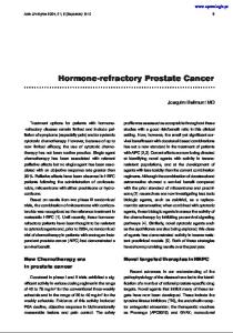
We focus on a PET/CT directed, 3D ultrasound-guided biopsy system (Fig. 1). The steps of targeted prostate biopsy are as follows. (1) Before undergoing prostate biopsy, the patient undergoes a PET/CT scan with FACBC as part of the examinations. The anatomic CT images will be combined with PET images for improved localization of the prostate and suspicious tumors. (2) During biopsy, 3D ultrasound images are acquired immediately before needle insertion. The 3D ultrasound images are registered with the PET/CT data for biopsy planning. Threedimensional visualization tools guide the biopsy needle to a suspicious lesion. (3) At the end of each core biopsy, the needle tip position is recorded on the real-time ultrasound images. The location information of biopsy cores is saved and then restored in the re-biopsy procedure. This allows the physician to re-biopsy the same area for a follow-up examination. The location information of the biopsy cores can also be used to guide additional biopsy to different locations if the original biopsy was negative.
102
B. Fei et al.
Molecular Imaging with PET/CT and/or MRI/MRSI
PET/CT
Deformable Registration
MRI/MRSI
Image Fusion
3D Visualization 2D/3D/4D Visualization
Real-time 3D ultrasound-guided biopsy and/or therapy
3D Ultrasound
Needle planning
Prostate Biopsy
Fig. 1. Molecular image-directed, ultrasound-guided system for targeted biopsy. Top: The PET/CT and MRI/MRS were acquired from the same patient at our institution. PET/CT with FACBC shows a focal lesion within the prostate (white arrow). MR images also show the suspicious lesion in the gland. The 3D visualization of the pelvis and prostate can aid the insertion of the biopsy needle into a suspicious tumor target. Bottom: A mechanically assisted navigation device was developed to acquire 3D TRUS images from the patient. The prostate boundaries are segmented from each slice and are then used to generate a 3D model of the prostate. Real-time TRUS images are acquired and registered to the 3D model in order to guide the biopsy. To incorporate PET/CT into ultrasound-guided procedures, deformable registration, segmentation, fusion, and visualization are the key technologies.
3
Automatic Segmentation of 3D Prostate Ultrasound Images
Many methods for semi-automatic or automatic segmentation of the prostate in TRUS images have been proposed. Active shape models (ASM) was proposed to segment the prostate [10]. Knoll et al. proposed a deformable segmentation model that uses one-dimensional wavelet transform as a multi-scale contour parameterization tool to constrain the shape of the prostate model [11]. Our proposed method consists of the training and application stages. Two training TRUS images were used for wavelet features training and ten patients are used to make a predefined model. The prostate boundaries have been manually defined by specialists. A prostate shape model is created based on the allowable models of shape variations and its probability. This model is employed to modify the prostate boundaries. The prostate textures are locally captured by training the locally placed Wavelet-based support vector machines (W-SVMs). With integrating local texture features and geometrical data, W-SVMs can robustly differentiate the prostate tissue
A PET/CT Directed, 3D Ultrasound-Guided Biopsy System for Prostate Cancer
103
from the adjacent tissues. The trained W-SVMs are employed to tentatively label the respective voxels around the surface into prostate and non-prostate tissues based on their texture features from different Wavelet filters. Subsequently, after an affine transformation of the shape model to the pre-defined prostate region that optimally matches with the texture features of the prostate boundary, the surface of the model is driven to the boundary between the tentatively labeled prostate and non-prostate tissues based on defined weighting functions and labeled voxels. TRUS image textures can provide important features for accurately defining the prostate, especially for the regions where prostate boundaries are not clear. Biorthogonal wavelets 1.3, 1.5, and 4.4 are employed to extract the texture features of the prostate. Designing biorthogonal wavelets allows more degrees of freedom comparing to orthogonal wavelets. One additional degree of freedom is the possibility to construct symmetric wavelet functions. A number of W-SVMs on different regions of the surface model are placed and trained to adaptively label the tissue based on its texture and location. Each W-SVM is composed of 5 wavelet filter banks, voxel coordinates, and a Kernel Support Vector Machine (KSVM). The wavelet filters are employed to extract texture features from TRUS images, and the KSVM is used to nonlinearly classify the Wavelet texture features for tissue differentiation. Each W-SVM corresponds to an individual sub-region in order to characterize and differentiate image textures locally and adaptively. All W-SVMs are trained to differentiate the texture features around its corresponding sub-regions in the training set. The trained W-SVMs are employed to tentatively label each voxel into prostate and non-prostate tissues in the application stage. To find more accurate segmentation, the set of W-SVMs is trained and applied in 3 planes (sagittal, coronal, transverse). Three sets of 2-D Wavelet filters were located at three orthogonal planes and were trained in each plane. Therefore, each voxel was tentatively labeled in three planes as prostate or non-prostate voxel. Fig. 2 shows the algorithm flowchart of the segmentation method.
Fig. 2. The flowchart of the wavelet-based segmentation algorithm
104
B. Fei et al.
A prostate probability model was used for modifying the segmentation. To build the prostate model, ten segmented prostates were registered using an affine transformation. Other registration methods can also be used to register the prostate of different patients [12-14]. In this study, we used the registration approach that is based on the principal axis transformation. This method was chosen because of its computational properties, speed and simplicity. The prostate volume was translated and rotated with respect to each other. The principal axis transformation is known from the classical theory of rigid bodies. A rigid body is uniquely located by knowledge of the position of its center of mass and its orientation (rotation) with respect to its center of mass. The center of mass, inertia matrix, and principal axes can be determined for any rigid body. For simple geometric shapes, the principal axes coincide with the axes of symmetry. In general, an orthogonal coordinate system is set up with their origin at the center of mass. When computed in the principal axis coordinate system, the inertia matrix is diagonal. The basic parameters that were used for registration of the prostate are the position of the center of mass and rotation of the prostate about the center of mass, and the lengths of the principle axes. These properties uniquely determine the location and geometry of the prostate in threedimensional space. After overlaying these 10 registered volumes, a probability model was created for each voxel based on how many prostate models are labeled as a voxel of the prostate at that region.
4
Nonrigid Registration of TRUS and CT Images
Our non-rigid registration method includes three terms: (1) surface landmark matching, (2) internal landmark matching, (3) volume overlap matching. Let xiCT and
yiUS are surface landmarks of the prostate from the segmented CT and TRUS images, respectively, ukCT and vlUS are internal landmarks e.g. urethra and calcification within the prostate on the CT and TRUS images, respectively. BCT and BUS represent the bladder neck region on the CT and TRUS images, respectively. Inspired by [16-18], we design an overall similarity function to integrate the similarities between same type of landmarks and add smoothness constraints on the estimated transformation between segmented CT and TRUS images. The transformation between CT and TRUS images are represented by a general function, which can be modeled by various function basis. In our study, we choose B-splines as the transformation basis. The similarity function is written as: CT US E ( f ) = α ESS ( f {xiCT , yUS j }) + β EIS ( f {uk , vl }) + γ EVM ( f {BCT , BUS }) + λ ES ( f )
Where I
J
I
J
I
J
CT ESS ( f ) = pij ( yUS j − f ( xi ) ) + δ pij log( pij ) − ξ pij 2
i =1 j =1
K
L
i =1 j =1
i =1 j =1
K
K
L
L
EIS ( f ) = qkl vlUS − f (ukCT ) + τ qkl log(qkl ) − η qkl k =1 l =1
2
k =1 l =1
k =1 l =1
A PET/CT Directed, 3D Ultrasound-Guided Biopsy System for Prostate Cancer
EVM ( f ) = 1 −
2 * ( f ( BCT ) BUS ) f ( BCT ) + BUS
∂2 f ∂2 f ∂2 f ES ( f ) = ( 2 + 2 + 2 ) 2 dxdydz ( x , y , z )∈Ω M ∂x ∂y ∂z
α , β ,γ ,
λ
105
2
ESS is the similarity for surface landmarks, and EIS is the similarity for internal landmarks. EVM is the energy term for the bladder-neck volume matching; and ES is the smoothness constraint term. δ and τ are called the temperature parameter and its weighted term is an entropy term comes from the deterministic annealing technique [28]. ξ and η are the weight for the outlier rejection term. Matrixes pij and qkl are the fuzzy correspondence matrixes [25]. f denotes the transformation between CT and and
are the weights for each energy term.
TRUS images, which is B-spline transformation in our method. The overall similarity function can be minimized by an alternating optimization algorithm that successively updates the correspondences matrixes
pij
and qkl , and
the transformation function f. First, with the fixed transformation f, the correspondence matrixes between landmarks are updated by minimizing E ( f ) . The updated correspondence matrixes are then treated as the temporary correspondences between landmarks. Second, with the fixed temporary correspondence matrixes
pij
and qkl , the transformation function f is updated. The two steps are alternatively repeated until there are no updates of the correspondence matrixes.
5
Results
We developed a 3D ultrasound-guided biopsy system for the prostate. The system uses: (1) Passive mechanical components for guiding, tracking, and stabilizing the position of a commercially available, end-firing, transrectal ultrasound probe; (2) Software components for acquiring, storing, and reconstructing real- time, a series of 2D TRUS images into a 3D image; and (3) Software that displays a model of the 3D scene to guide a biopsy needle in three dimensions. The system allows real-time tracking and recording of the 3D position and orientation of the biopsy needle as a physician manipulates the ultrasound transducer. The segmentation method was evaluated by five patient data sets of 3-D TRUS. Fig. 3 shows sample segmentation and its comparison with the corresponding gold standard. Quantitative performance assessment of the method was done by comparing the results with the corresponding gold standard data from manual segmentation. The Dice similarity and Sensitivity were used as performance assessment metrics in prostate classification algorithm. The numerical results of these evaluation criteria are
106
B. Fei et al.
shown in Table 1. The dice d ratio is between 88.7%-95.0% among five prostate volumes. The mean and staandard deviation of the dice function are 90.7% and 2.55%, respectively.
Fig. 3. 2-D segmentation resu ults in different planes: red lines are the gold standard boundaaries and green lines are the segm mentation boundaries. (a) Coronal plane (b) Sagittal planee (c) Transverse plane. Table 1. Quantittative evaluation results of the segmentation method Patients
1
2
3
4
5
Mean
STD D
DICE
90.2%
88 8.7%
88.9%
95.0%
90.8%
90.7%
2.5% %
Sensitivity
91.2%
95 5.2%
82.3%
93.6%
91.1%
90.7%
4.9% %
Prostate Pro ostate
Urethra
Urethra
Lesion Lesion
Fig. 4. An ultrasound image (lleft) is registered with an MR image (middle) of the same prosstate phantom. The 3D visualizatio on shows the relative location of the ultrasound plane withinn the 3D MR image (right) where grreen is the prostate and red is the lesion.
Fig. 4 shows the registraation of ultrasound and MR images of a prostate phantoom. The registration works well in the phantom experiment. We also evaluated the registration method using five f sets of pre- and post-biopsy TRUS data of the saame patients. The size of TRUS S data is 244×244×175 voxels and the spatial resolutionn is 0.38×0.38×0.39 mm3. Wee used pre-biopsy images as the reference images and
A PET/CT Directed, 3D Ultrasound-Guided Biopsy System for Prostate Cancer
107
registered the post-biopsy images of the same patient. For five sets of patient data, the target registration error (TRE) was 0.88±0.16 mm and the maximum TRE is 1.08±0.21 mm.
6
Discussion and Conclusion
We developed a PET/CT directed, 3D ultrasound-guided biopsy system for the prostate. In order to include other imaging modality such as PET/CT into 3D ultrasound-guided biopsy, we developed a 3D non-rigid registration method that combines point-based registration and volume overlap matching methods. The registration method was evaluated for TRUS and MR images. The registration method was also used to register 3D TRUS images acquired at different time points and thus can be used for potential use in TRUS-guided prostate re-biopsy. Our next step is to apply this registration method to CT and TRUS images and then incorporate PET/CT images into ultrasound image-guided targeted biopsy of the prostate in human patients. In order to build a 3D model of the prostate, a set of Wavelet-based support vector machines and a shape model are developed and evaluated for automatic segmentation of the prostate TRUS images. Wavelet transform was employed for prostate texture extraction. A probability prostate model was incorporated into the approach to improve the robustness of the segmentation. With the model, even if the prostate has diverse appearance in different parts and weak boundary near bladder or rectum, the method is able to produce a relatively accurate segmentation in 3-D TRUS images. Acknowledgement. This research is supported in part by NIH grant R01CA156775 (PI: Fei), Coulter Translational Research Grant (PI: Fei), Georgia Cancer Coalition Distinguished Clinicians and Scientists Award (PI: Fei), Emory Molecular and Translational Imaging Center (NIH P50CA128301), SPORE in Head and Neck Cancer (NIH P50CA128613), and Atlanta Clinical and Translational Science Institute (ACTSI) that is supported by the PHS Grant UL1 RR025008 from the Clinical and Translational Science Award program.
References 1. Roehl, K.A., Antenor, J.A., Catalona, W.J.: Serial biopsy results in prostate cancer screening study. J. Urol. 167(6), 2435–2439 (2002) 2. Jambor, I., Borra, R., Kemppainen, J., Lepomäki, V., Parkkola, R., Dean, K., Alanen, K., Arponen, E., Nurmi, M., Aronen, H.J., Minn, H.: Functional imaging of localized prostate cancer aggressiveness using 11C-acetate PET/CT and 1H-MR spectroscopy. J. Nucl. Med. 51(11), 1676–1683 (2010) 3. Carlani, M., Mancino, S., Bonanno, E., Finazzi Agrò, E., Simonetti, G.: Combined morphological, [1H]-MR spectroscopic and contrast-enhanced imaging of human prostate cancer with a 3-Tesla scanner: preliminary experience. Radiol Med. 113(5), 670–688 (2008)
108
B. Fei et al.
4. Schöder, H., Herrmann, K., Gönen, M., Hricak, H., Eberhard, S., Scardino, P., Scher, H.I., Larson, S.M.: 2-[18F]fluoro-2-deoxyglucose positron emission tomography for the detection of disease in patients with prostate-specific antigen relapse after radical prostatectomy. Clin. Cancer Res. 11(13), 4761–4769 (2005) 5. Schilling, D., Schlemmer, H.P., Wagner, P.H., Böttcher, P., Merseburger, A.S., Aschoff, P., Bares, R., Pfannenberg, C., Ganswindt, U., Corvin, S., Stenzl, A.: Histological verification of 11C-choline-positron emission/computed tomography-positive lymph nodes in patients with biochemical failure after treatment for localized prostate cancer. BJU Int. 102(4), 446–451 (2008) 6. DeGrado, T.R., Coleman, R.E., Wang, S., Baldwin, S.W., Orr, M.D., Robertson, C.N., Polascik, T.J., Price, D.: Synthesis and evaluation of 18F-labeled choline as an oncologic tracer for positron emission tomography: initial findings in prostate cancer. Cancer Res. 61(1), 110–117 (2001) 7. Oyama, N., Akino, H., Kanamaru, H., Suzuki, Y., Muramoto, S., Yonekura, Y., Sadato, N., Yamamoto, K., Okada, K.: 11C-acetate PET imaging of prostate cancer. J. Nucl. Med. 43(2), 181–186 (2002) 8. Nuñez, R., Macapinlac, H.A., Yeung, H.W., Akhurst, T., Cai, S., Osman, I., Gonen, M., Riedel, E., Scher, H.I., Larson, S.: Combined 18F-FDG and 11C-methionine PET scans in patients with newly progressive metastatic prostate cancer. J. Nucl. Med. 43(1), 46–55 (2002) 9. Schuster, D.M., Votaw, J.R., Nieh, P.T., Yu, W., Nye, J.A., Master, V., Bowman, F.D., Issa, M.M., Goodman, M.: Initial experience with the radiotracer anti-1-amino-3-18Ffluorocyclobutane-1-carboxylic acid with PET/CT in prostate carcinoma. J. Nucl. Med. 48(1), 56–63 (2007) 10. Hodge, A.C., Fenster, A., Downey, D.B., Ladak, H.M.: Prostate boundary segmentation from ultrasound images using 2D active shape models: Optimisation and extension to 3D. Computer Methods and Programs in Biomedicine 84, 99–113 (2006) 11. Knoll, C., Alcaniz, M., Grau, V., Monserrat, C., Juan, M.C.: Outlining of the prostate using snakes with shape restrictions based on the wavelet transform. Pattern Recognit. 32, 1767–1781 (1999) 12. Fei, B., Duerk, J.L., Sodee, D.B., Wilson, D.L.: Semiautomatic nonrigid registration for the prostate and pelvic MR volumes. Academic Radiology 12, 815–824 (2005) 13. Fei, B., Lee, Z., Duerk, J.L., Lewin, J.S., Sodee, D.B., Wilson, D.L.: Registration and Fusion of SPECT, High Resolution MRI, and interventional MRI for Thermal Ablation of the Prostate Cancer. IEEE Transactions on Nuclear Science 51(1), 177–183 (2004) 14. Yang, X., Akbari, H., Halig, L., Fei, B.: 3D non-rigid registration using surface and local salient features for transrectal ultrasound image-guided prostate biopsy. In: Proc. SPIE 7964 (2011) 15. Alpert, N.M., Bradshaw, J.F., Kennedy, D., Correia, J.A.: The principal axes transformation a method for image registration. The Journal of Nuclear Medicine 31(10), 1717–1722 (1990) 16. Chui, H., Rangarajan, A.: A new point matching algorithm for non-rigid registration. Computer Vision and Image Understanding 89(2-3), 114–141 (2002) 17. Zhan, Y., Ou, Y., Feldman, M., Tomaszeweski, J., Davatzikos, C., Shen, D.: Registering histologic and MR images of prostate for image-based cancer detection. Acad. Radiol. 14, 1367–1381 (2007) 18. Yang, J., Blum, R.S., Williams, J.P., Sun, Y., Xu, C.: Non-rigid Image Registration Using Geometric Features and Local Salient Region Features. In: CVPR, pp. 1825–1832 (2006)
Fei B, Master V, Nieh P, Akbari H, Yang X, Fenster A, Schuster D. A PET/CT directed, 3D ultrasoundguided biopsy system for prostate cancer. Workshop on Prostate Cancer Imaging, The Annual Meeting of the Society of Medical Imaging Computing and Image Assisted Interventions (MICCAI), Toronto, Canada, September 18-22, 2011. A. Madabhushi et al. (Eds.): Prostate Cancer Imaging 2011, LNCS 6963, pp. 100–108, 2011
A. Madabhushi et al. (Eds.): Prostate Cancer Imaging 2011, LNCS 6963, pp. 100–108, 2011.© SpringerVerlag Berlin Heidelberg 2011. One print or electronic copy may be made for personal use only. Systematic reproduction and distribution, duplication of any material in this paper for a fee or for commercial purposes, or modification of the content of the paper are prohibited.