CHAPTER 3
Control of breathing P.M.A. Calverley Correspondence: P.M.A. Calverley, University Hospital Aintree, Liverpool, UK.
Most respiratory clinicians recognise that the control of breathing is a complex and multi-factorial process that appears to be rather mysterious. Everyone would agree that the optimum matching of ventilation and perfusion within the lungs is necessary to maintain appropriate homeostasis for the arterial blood gases and hence for oxygen delivery to the tissues. How this is achieved continues to elude a simple and comprehensive explanation. For instance, the mechanisms whereby ventilation increases appropriately and almost immediately at the start of exercise continue to be controversial with advocates of both peripheral and central mechanisms [1]. Given this lack of a simple understanding of ventilatory control in some of the commonest physiological situations, it is not surprising that when disease intervenes it is hard to develop tools which are clinically useful. Many diseases derange blood gas tensions and these remain one of the most useful practical ways of assessing the ability of the respiratory control system to meet the demands placed upon it. In practice, most clinicians think about changes in blood gases in terms of the structural processes that have produced them, rather than as manifestations of deranged ventilatory control. Likewise, it is much easier to measure directly, or to infer, abnormalities of lung mechanics than it is to evaluate the role that they play in disordered control mechanisms. With a few well-recognised exceptions, abnormal ventilatory control is not the primary reason why patients present to the respiratory physician. Nonetheless, an understanding of the factors that influence the control of breathing and the way in which it can modify the patients behaviour is helpful. However, the clinical need to request the tests described briefly below are rather small. In this chapter some of the considerations relevant to an understanding of ventilatory control in health will be examined, followed by a review of some of the approaches that have been used to evaluate respiratory control mechanisms. Finally, some of the diseases where abnormal ventilatory control plays a role will be mentioned.
Ventilatory control in health Conventionally, the neural mechanisms that regulate ventilation are considered to be a hierarchy, with more primitive processes regulated by the higher centres [2]. There is still disagreement about whether the output of the control system is regulated to optimise ventilation or breathing pattern [3], although systems control approaches emphasise the advantages of reducing total respiratory system work for any overall level of activity [4]. Inevitably, most of the data regarding the mechanisms regulating these processes have come from animal studies with parts of the system damaged or stimulated under anaesthesia. These are very unphysiological conditions and drawing detailed interpretations about the function of the ventilatory control system from them may be misleading. Nonetheless, a general picture has emerged about how this system is organised. Eur Respir Mon, 2005, 31, 44–56. Printed in UK - all rights reserved. Copyright ERS Journals Ltd 2005.
44
CONTROL OF BREATHING
The central feature of this scheme is the respiratory pattern generator, which is believed to lie within the medulla oblongata [5]. Here, at least three groups of neurones receive inputs from important chemoreceptors and mechanoreceptors. Those going to the dorsal respiratory group are predominately inspiratory but are not influenced by stretch receptor inputs. Those in the ventral respiratory group are also thought to be inspiratory, at least in the upper part of this complex, but activate expiratory muscles in the lower part. Finally, neurones in the pontine respiratory group appear to cover the period during late inspiration and early expiration and are important in terms of respiratory rhythm generation. A basic rhythmic oscillatory pattern is developed between these neuronal groups, which acts as a form of respiratory "pacemaker" but this is greatly modulated by the nature and severity of the inputs from the other peripheral sensors [6]. Mechanical and chemical inputs arise from changes in chest-wall movement and/or blood gas tensions provide the feedback, which modify the intensity and timing of the respiratory neural outputs [7, 8]. This has a number of practical consequences: 1. If the ribcage and diaphragm are unable to produce adequate ventilation then the mechanoreceptors in the joints and intercostals muscles will increase their output to the respiratory centre and this leads to a change in the timing and amplitude of the neural stimulus passing through the phrenic nerve [2]. 2. If alveolar ventilation is inadequate and arterial oxygen tension (Pa,O2) falls then the peripheral chemoreceptors at the junction of the external and common carotid arteries increase their discharge rate [7]. One of the most consistent findings in animal studies is the relationship between the Pa,O2 and the carotid sinus nerve traffic and this clearly shows that these receptors are only significantly activated once the Pa,O2 falls below 8.0 kPa [9]. However, some tonic activity does persist as this can be abolished by oxygen therapy and this may be particularly important during exercise. 3. Increases in arterial CO2 tension (Pa,CO2) stimulates both peripheral, but especially central chemoreceptor, firing rate [10]. It is still difficult in humans to distinguish the effect of CO2 on the central chemoreceptors from those on the peripheral chemoreceptors and brief stimulation, as used in many tests of ventilatory control, may not give an appropriate index of more sustained hypercapnia, as happens in disease [11]. 4. The degree of acid base compensation within the cerebrospinal fluid is an important determinant of the effects of raised carbon dioxide and this is relevant for those individuals who develop persistent hypercapnia or slowly worsening hypercapnia in the course of an episode of respiratory failure [12]. 5. All of the above are influenced significantly by the higher centres, as is demonstrated by the change in ventilatory control that occurs with the onset of sleep. In these circumstances chemical and mechanical stimulation produces less in the way of increased ventilation and this appears to be related to the depth of sleep, with stages three and four being those in which the patient is least responsive [13, 14]. There is continuing debate about which part of rapid eye movement sleep is associated with stimulation and which with depression of ventilation, although in general periods of tonic rapid eye movement sleep has a lower responsivity than when the eye movements are most brisk. Thus, the integrated function of the respiratory system can be modified significantly by changes in Pa,O2, Pa,CO2, alterations in the impedance of the respiratory system and changes in conscious level. In health, the system appears to be regulated so as to minimise the overall energy expenditure of the respiratory system [4]. In practice this may involve a trade-off between the increased mechanical impedance and the disordered blood gas 45
P.M.A. CALVERLEY
tensions. One further consequence of this is the close relationship between the situation of conflict and the sensation of breathlessness. This promotes behavioural changes, which are often the most effective way of decreasing ventilatory demand and hence the burden on the respiratory system [15]. For reasons of muscle energetics it is not sensible to push the activation of the inspiratory or expiratory muscles to the point where muscle fatigue develops and most patients either voluntarily or involuntarily adopt a breathing pattern and a pattern of behaviour that prevents this happening [16, 17].
Assessing ventilatory control There is no simple single method of assessing how breathing is controlled. The investigator either opens the "closed loop" of physiological control, for example by adding carbon dioxide to the inspired gas, or measures the breathing pattern under resting or challenged conditions and compares this with the pattern seen in normal subjects in similar situations. In both cases it is assumed that a brief test lasting only a few minutes to half an hour will give information that explains how ventilation is controlled over much longer periods. This is clearly not the case and longer exposures to abnormal gas mixtures and mechanical loads produce different and sometimes contradictory effects to those seen during the acute test [18]. Thus, most tests of ventilatory control give insights into the immediate response to changes in breathing conditions but are unreliable guides to the long-term control of breathing. The testing of ventilatory control has gone through several phases. In the 1950s and 1960s attention focused on abnormalities in the blood gas tensions and investigators studied the response to raised carbon dioxide and lowered oxygen in the inspired gas. In the 1970s studies of the way in which ventilation changed in the face of added respiratory loads similar to, or greater than, those seen in disease became an important way of exploring control mechanisms and at this stage studies of respiratory muscle fatigue became popular. In the 1980s longer term exposures to abnormal gas mixtures were also investigated and highlighted the contradictory finding that increases in ventilation with chronic hypoxia decline in intensity with more prolonged exposure, although in most studies to date chronic hypoxia produces a level of ventilation that is greater than that at rest. A simple approach advocated in some laboratories is the use of the breath-holding time, although this has limitations, which will be outlined below. With the exception of the breath-holding time, all the tests relate an external stimulus e.g. loading (change in inspired gases) to measures that directly or indirectly reflect respiratory centre output. The most widely used of these is minute ventilation, although some authors have advocated reporting changes in tidal volume and respiratory frequency separately. Unlike other tests of respiratory function it has been difficult to establish a normal range among younger, let alone older adults. This significantly restricts the clinical usefulness of these tests.
Measuring the output Minute ventilation and breathing pattern Minute ventilation can be measured directly using a Douglas bag or gas meter or can be derived from integration of the flow signal using a flow sensor at the mouth. The presence of a noseclip and mouthpiece influences resting ventilation and tends to increase it possibly as a result of the added dead space [19]. This has stimulated research on 46
CONTROL OF BREATHING
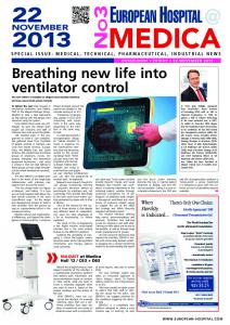
TV camera
Motion analyser Markers position
Geometrical models
Flow Volume computing
Poes Pga
Chest wall compartmental volume changes (Vrc, Vab, Vcw) Fig. 1. – Schematic presentation of a data acquisition system used for the noninvasive measurement of lung volume by optoelectronic plethysmography. With the subject seated with the arms supported away from the chest light from 89 reflective markers positioned around the chest wall is collected by the television cameras and processed to derive the volume of the space enclosed by a series of triangles constructed by the computer from the markers position on the chest. This allows the absolute lung volume and the volume enclosed by the rib cage and abdominal compartments of the chest wall to be calculated. Poes: oesophageal pressure; Pga: gastric pressure; Vrc: volume of the rib cage; Vab: volume of the abdomen; Vcw: volume of the chest wall.
noninvasive methods of measuring ventilation and for a period the magnetometer approach of quantifying the excursion of the ribcage and the abdomen became popular [20]. Commercial devices incorporating impedance coils into bands placed around the chest and abdomen were used to simplify the application of the system. Unfortunately, it proved difficult to calibrate this system and changes in position of even a minor degree significantly affected the reliability of the measurement. More accurate noninvasive measurements are now available using optoelectronic plethysmography [21, 22]. To date these have only been used in a research setting (fig. 1).
Analysis of breathing patterns Breathing patterns can be characterised simply from the combination of tidal volume and respiratory frequency but can be further separated into mean inspiratory flow (tidal volume divided by inspiratory time) and the duty cycle (tidal volume divided by total cycle duration) [23]. The characteristics can also be used to define minute ventilation. In general, the mean inspiratory flow is the variable that responds to external stimulation, although when mechanically limited this can decline in the face of a rising neural drive. In this context it can be more useful to measure the mouth occlusion pressure (the pressure recorded 100 ms after the onset of inspiration against a closed airway). In this brief period the muscles should shorten simply in response to the pattern of impulses in the phrenic nerve [24]. Unfortunately, technical considerations related to the shape of the diaphragm and to identifying reliably the onset of the occlusion of pressure waveform make this technique less robust in disease than it is in healthy subjects. Nonetheless, it is a significant improvement on simply reporting tidal volume in patients with abnormal 47
P.M.A. CALVERLEY
resting lung mechanics. An alternative tactic that is less well validated is to express the tidal volume or minute ventilation as a per cent of that predicted [25]. Formulae exist for deriving the predicted maximal minute ventilation and some data suggest that this approach may be useful [26]. However, the lack of widespread normal values limits its application. A further, more direct but unfortunately more invasive, method involves the use of a balloon catheter system placed in the oesophagus and stomach from which the transdiaphragmatic pressure can be calculated. Even if this is simplified to just an oesophageal balloon it is an uncomfortable procedure that is rather time consuming. The advantage is that this should indicate the point at which inspiration begins and from which the occlusion pressure waveform can be calculated. Unfortunately, this has problems in patients with chronic obstructive pulmonary disease (COPD) where there is a degree of intrinsic positive end-expiratory pressure, which makes it hard to be sure at which point the neural inspiration has begun [27]. A yet more invasive approach is to monitor diaphragmatic electromyogram, which gives a measure of the electrical activation [28]. Unfortunately, relating the size of the electrical signal to either the pressure developed or the mechanical change it produces can be very difficult [28].
Chemosensitivity responses to hypoxia and hypercapnia As noted previously, the arterial blood gas tensions at rest often provide the simplest indicator of the adequacy of ventilatory control. The role of abnormal gas exchange is considered elsewhere in this Monograph. Nonetheless, in many circumstances, particularly those where there is little or no mechanical abnormality, an elevation in the carbon dioxide tension is a good pointer of inadequate ventilation and impaired control mechanisms. Protocols for stimulation either by increasing carbon dioxide or reducing the inspired oxygen concentration normally involve the subject rebreathing from a sealed anaesthetic bag, which containsy6 L of premixed gas, and then recording the change in gas tensions at the mouth and relating it to either ventilation or occlusion pressure [29, 30]. Fortunately, the relationships between a fall in oxygen saturation and ventilation are themselves linear and hence peripheral oxygen saturation can be measured as a useful surrogate for Pa,O2. Linearised in this way makes the slope of the ventilation saturation relationship, which is an inverse one, as useful a guide to chemosensitivity as is the slope of the ventilation–CO2 relationship [30]. In health, wide ranges exist for both values and it is important to specify the hypoxic response in terms of the CO2 tension maintained during the trial. Isocapnic testing is essential in studies of hypoxic response and is normally achieved by re-circulating the expired gas through a carbon dioxide scrubber circuit [30]. Likewise, oxygen tensions during the CO2 re-breathing are an important practical consideration and patients normally begin with a gas mixture containing 93% oxygen and 7% CO2.
Mechanical loading This is a more specialised and largely research technology, which might yet prove useful if developed as a clinical tool. A variety of loaded breathing circuits have been developed involving either exposure to a single load and observing the effect on the first loaded breath relative to the preceding breaths or studying the effects of stimulated 48
CONTROL OF BREATHING
breathing with hypoxia or hypercapnia during a fixed resistive load [25]. It is more difficult to apply sustained elastic loads and so most data derives from studies during resistive loading. Although it would be intellectually interesting to understand the effects of stiffening of the chest wall, such studies would be unlikely to produce different data from that during resistive breathing. Again, the nature of the disease to be studied affects the results obtained and in this context ventilatory restriction due to mechanical impairment of the chest wall or the effect of intrinsic positive end expiratory pressure in COPD can complicate the interpretation of these tests [31]. When resistive loading is used it is important to know the linearity of the resistance as higher levels of ventilation may expose the patient to substantially greater inspiratory loads than is initially anticipated. While minute ventilation is normally depressed in the face of inspiratory resistive loading the degree that subjects defend their ventilation varies significantly [32]. Likewise, individuals will adopt rather different breathing patterns when acutely loaded. The relevance of the breathing pattern adopted to more chronic loading is less clear and in general loaded breathing produces a reduction in tidal volume and a shortening of respiratory cycle duration i.e. a rapid, shallow breathing pattern [33–34]. This seems to be true whether the load is predominantly resistive or elastic. Increasing knowledge of the abnormalities of lung mechanics in many conditions suggests that in life the loaded breathing is a combination of the two.
Breath-holding time This simple test requires only a noseclip and a stop-watch. The subject exhales to residual volume, inhales to total lung capacity and then holds the breath for as long as possible. Analysis of the expired gas allows the stimulus to changing gas tensions to be related to the breath-holding time. The test can be repeated until reproducible results are obtained or subject exhaustion intervenes [34]. Like many other tests of ventilatory control there are a number of other important confounders that interfere with the interpretation of this test. Specifically, respiratory muscle strength and the geometry of the chest wall are very relevant to the patient’s ability to hold the breath [35, 36]. If this test is to be used clinically it is necessary that the laboratory establish its own normal range so that some comment on when the breath-holding time is abnormal can be made.
Ventilatory control and disease Problems of interpretation A variety of factors interfere with the interpretation of ventilatory control and these are well illustrated from studies in COPD, which is one of the most examined conditions. In general, these can be summarised as: 1. Impaired gas mixing: in COPD and chronic asthma, lung gas stores are increased and mixing of fresh and resident gases is not homogeneous. Slowly ventilated areas of the lungs may not achieve the same gas concentrations as the well ventilated ones, with a discrepancy resulting between measurements made on end tidal expiratory breaths and the true gas tensions within the alveoli. 2. Mechanical inequalities: abnormal time constants throughout the respiratory system (the product of resistance and compliance) can delay the equilibration of pressure applied within the chest and that recorded at the mouth. This is a particular problem 49
P.M.A. CALVERLEY
for obstructive lung disease, but is not an issue for interstitial lung disease where the time constants are short. 3. Changes in the respiratory muscles: changes in the configuration of the respiratory muscles can greatly influence their ability to develop pressure for a given neural stimulus [37]. Thus, patients with COPD who have a flattened diaphragm, which shortens this muscle, will be unable to develop the same pressure for an equivalent stimulus as would someone with interstitial lung disease where the diaphragm has a normal configuration. This is very relevant when indices, like mouth occlusion pressure, are used as the surrogate for respiratory centre output. Moreover, changes in muscle structure occur in chronically shorted muscle, like the flattened diaphragm in patients where lung volume is increased [38], although how relevant these changes are to human disease is uncertain. 4. Mechanical loading by preventing muscle shortening can itself reduce outputs such as minute ventilation, irrespective of the stimulus being applied to the respiratory muscles. This problem has been noted previously.
Specific diseases Primary disorders of ventilatory control The assessment of ventilatory control can be a useful adjunct to the investigation of patients in a number of clinical settings. The two most important of these are in patients who either under breathe (hypoventilate) or over breathe (hyperventilate). In each case the investigation has been mainly used as a research tool rather than one required to make a clinical diagnosis. In the relatively rare primary alveolar hypoventilation syndrome, which is seen mainly in children, there is an absence of chemoreceptor responsiveness [39]. This is compensated for during the day by the wakefulness-related drive to breathe, as commented on previously. But at night when this influence declines, so does the minute ventilation with profound falls in Pa,O2 and an increase in Pa,CO2. These subjects show a reduced response to CO2 and hypoxic stimuli even when awake, and this becomes much more dramatic when they are asleep. Why this condition arises is still problematic, but it commonly presents in childhood or early adolescence with failure to thrive, intellectual impairment, daytime tiredness or even light heart failure and pulmonary hypertension. Appropriate therapy with nocturnal ventilation can dramatically improve the well being of these patients and permits adequate correction of their blood gas tensions [40]. The problems of disproportionate breathlessness associated with individuals who appear to develop a larger ventilatory response than required during exercise, have been known for many years. More recently, careful scientific study has shown that these patients have abnormal ventilatory control [41]. They can usually be identified either by a low Pa,CO2 and a high Pa,O2 at rest and will commonly have larger tidal volumes and higher respiratory frequencies than would be predicted during progressive exercise testing. They exhibit ventilatory limitation in this setting, and some who are not hypocapnic at rest will become so during the exercise, although this tends to resolve as their mechanical inability to sustain high ventilations eventually limits their capacity to continue. Why these patients behave in this way is unclear, but their ability to sustain the hyperventilation for extended periods during the daytime and overnight suggest that there is a primary problem with ventilatory control, rather than simply a secondary psychological abnormality. Undoubtedly, the chronicity of their problems produces psychological difficulties and it has been difficult to disentangle cause and effect in these circumstances. 50
CONTROL OF BREATHING
A third situation when ventilation will be abnormal arises with periodic breathing. This is now known to be a relatively frequent finding during sleep in patients with chronic congestive heart failure, having been originally recognised only during the daytime in patients with gross congestive heart failure, or major strokes. The waxing and waning of ventilation with the system "hunting" to find a stable CO2 tension is characteristic of this form of breathing. Changes in CO2 threshold, which occurred during sleep, appear to be important for initiating this form of respiration, although the arousal responses that commonly accompany it are also relevant [42]. At present, periodic breathing appears to be a marker of other diseases, rather than a primary abnormality of ventilatory control, which itself produces ill health.
Chronic obstructive pulmonary disease This is the most widely studied condition from a research perspective and is reviewed in detail elsewhere [43]. Attempts to distinguish patients who developed hypoxia and hypercapnoea from those who maintained their CO2 tensions have tracked the history of this research methodology. The initial observation that suggested that those with hypoxia had lower ventilatory responses has been hard to substantiate. In general, the worse the mechanical impairment in COPD the lower the ventilatory response to altered gas tensions. The same mechanical abnormalities limit the interpretation of occlusion pressure responses to either mechanical or chemical loading [44, 45]. The most consistent findings have been in the breathing pattern, with patients who develop carbon dioxide retention also having a smaller tidal volume and reduced inspiratory time [46]. Effectively, the smaller tidal volume in the face of the fixed dead space increases the dead space tidal volume ratio and promotes carbon dioxide retention as predicted from steady state analysis of gas exchange. Researchers continue to try and disentangle the cause and effect nature of both respiratory sensory abnormalities and the mechanical impairment that characterises these patients. Nonetheless, many of us who have studied these problems are left with the feeling that there is a difference in the basic responsiveness of patients who will readily allow themselves to become hypoxaemic and hypercapnic [47] and this is an issue that still awaits adequate resolution. A further controversy in the field of ventilatory control on COPD has been the effect of high inspired oxygen concentrations. This issue has been reviewed on several occasions [48, 49] and it is clear that some of the conflicting data reflects the severity of the disease in which the measurements have been made. In very severe COPD, with marked hypoxaemia and possibly associated haemodynamic instability, exposure to a high concentration of oxygen produces CO2 retention largely by a predictable effect on ventilation-perfusion matching within the lung [50]. In contrast, in nonventilated patients breathing high oxygen concentrations during an exacerbation, a proportion will develop CO2 retention because of a degree of ventilatory depression, perhaps secondary to a reduction in chemoreceptor activation. The possibility that there has also been an effect of the change of gas tensions on airway calibre must also be considered [51]. In practice, it is clinically desirable to avoid this and this is still best done with controlled oxygen therapy as described by Campbell over 40 years ago.
Bronchial asthma No specific ventilatory control abnormalities have been identified in most patients with bronchial asthma. However, it is clear that some asthmatics deteriorate acutely and there is increasing data that these patients perceive changes in chemical stimuli relatively poorly [52]. This occurs despite the fact that some appear to have reasonably normal lung 51
P.M.A. CALVERLEY
mechanics between attacks and suggest that there is either an inherent or acquired defect in ventilatory control, which when combined with the onset of severe asthma exposes these patients to particular risk. Early studies showed that the ventilatory response to CO2 was reduced in some asthmatics during the recovery from a severe exacerbation [53] and where there is chronic loading both respiratory perception and ventilatory responses to chemical stimuli were impaired [54].
Interstitial lung disease This heterogeneous group of conditions is associated with the increasing elastic work of breathing and as noted previously, the breathing pattern in response to this tends to be rapid and shallow. Arterial blood gas tensions are usually normal at rest in these conditions but there is marked desaturation during exercise as ventilation fails to match the increased perfusion. Typically, occlusion pressure responses are increased in these patients, but it is unlikely that they are mounting a greater ventilatory response for a given mechanical load than other subjects.
Chest wall and neuromuscular diseases During the 1970s careful examination of well-characterised patients with kyphoscoliosis showed that the reduction in response to CO2, which would be anticipated in these patients, was related to the reduction of the compliance of the respiratory system and particularly the change in the chest wall compliance [55]. Again, the breathing pattern was rapid and shallow, as is seen in patients with other disorders associated with impaired lung mechanics. The management of these patients has been revolutionised by the introduction of nocturnal positive pressure ventilation treatment but good studies examining ventilatory responsiveness before and after the introduction of this therapy are lacking. There is a continuing suspicion that alterations in chest wall mechanics may explain why Pa,CO2 falls during the day, although a resetting of the chemoreceptors as a result of better nocturnal ventilation is also a possibility. Patients with neuromuscular disease normally show similar abnormalities in their response to CO2 and oxygen tension perturbation as do other subjects who are unable to mount an adequate ventilatory response [56]. These patients are particularly dependent upon the diaphragm function, which decreases during rapid eye movement and often the earliest sign of future ventilatory impairment is the development of oxygen desaturation in this sleep stage. However, their overall respiratory function as assessed by their vital capacity is a better predictor of prognosis than the presence of this isolated ventilatory control problem [57, 58].
Sleep and breathing disorders Most attention has focused on the anatomical abnormalities, which determine the occurrence of upper airway obstruction during sleep, rather than changes in ventilatory control. Most clinicians accept that there must be some individual variation in response, and alterations in ventilatory control may explain some of the variation in the severity of nocturnal oxygen desaturation and duration of the apnoeic periods [59]. As yet, no simple way of assessing this has been developed. The large number of complex and changing variables, which characterise upper airway obstruction during sleep, make the simple elucidation of this problem unlikely in the near future. 52
CONTROL OF BREATHING
Conclusion The regulation of ventilatory control is abnormal in some relatively rare disease states but its major role in most cases is to modify the clinical presentation and subsequent progress of patients with many different forms of respiratory disease. Although major steps forward have been made in understanding some of the complex interactions between altered blood gas tensions, lung mechanics and the central nervous system processing of these signals, it is difficult to turn this information into tools that modify clinical decision making. It seems likely that quite different approaches to understand ventilatory control, perhaps coming from areas of systems control theory, will ultimately give better ways of explaining what is happening. Until such "high tech" solutions are available, an awareness of the additional impact of altered ventilatory control is helpful when considering patient management.
Summary The maintenance of blood gas homeostasis is dependent on the balance between respiratory drive and peripheral, mechanical and chemoreceptor responses. No single measurement encapsulates all aspects of this complex control system. Most investigators and clinical tests rely on relatively short-term changes in inspired gas concentrations and/or additional predominantly inspiratory mechanical loading to determine how the control system responds. Usually ventilation or an index of neural drive, such as mouth occlusion pressure, is used as the output measurement. Changes in the mechanical properties of the lungs make interpretation of these tests difficult and in common diseases such as chronic obstructive pulmonary disease, asthma and interstitial lung diseases the usual index of ventilatory control abnormality is a change in the arterial blood gas tension. In some conditions, e.g. hypo- or hyperventilation syndromes, investigation of respiratory control mechanisms may be useful. Studies of disordered respiratory control have helped understanding of the pathophysiology of disease and continue to inform current clinical practice, e.g. in the prescription of highflow oxygen. Future developments using modern computerised methods to analyse breathing pattern and relate this to neural activation may offer more appropriate clinical tools. Keywords: Chemoreceptor, chronic obstructive pulmonary disease, hypercapnoea, hypoxia, sleep disorders.
References 1.
2.
3.
Wasserman KB, Whipp BJ, Casaburi R. Respiratory control during exercise. In: Cherniack NS, Widdicombe JG, eds. Handbook of Physiology; Control of Breathing. Vol 2, Part 2, Section 3. Bethesda, American Physiology Society, 1986; pp. 595–619. Euler C von. Brain stem mechanisms for generation and control of breathing pattern. In: Cherniack NS, Widdicombe JG, eds. Handbook of Physiology; Control of Breathing. Vol. 2, Part 2, Section 3. Bethesda, American Physiological Society, 1986; pp. 1–67. Mead J. Control of respiratory frequency. J Appl Physiol 1960; 15: 325–336.
53
P.M.A. CALVERLEY
4. 5. 6. 7. 8.
9. 10. 11. 12.
13. 14. 15. 16. 17. 18. 19. 20. 21.
22. 23. 24. 25. 26.
27. 28.
Poon CS. Effects of inspiratory resistive load on respiratory control in hypercapnia and exercise. J Appl Physiol 1989; 66: 2391–2399. Richter DW. Generation and maintenance of the respiratory rhythm. J Exp Biol 1982; 100: 93– 107. Remmers JE. Central neural control of breathing. In: Altose MD, Kawakami Y, eds. Control of Breathing in Health and Disease. New York, Marcel Dekker, 1999; pp. 1–35. Bisgard GE, Neubauer JA. Peripheral and central effects of hypoxia. In: Dempsey JA, Pack AI, eds. Regulation of Breathing. 2nd Edn. New York, Marcel Dekker, 1995; pp. 617–668. Coleridge HM, Coleridge JCG. Reflexes evoked from tracheobronchial tree and lungs. In: Cherniack NS, Widdicombe JG, eds. Handbook of Physiology; Control of breathing. Vol 2, Part 2, Section 3. Bethesda, American Physiology Society, 1986; pp. 395–430. Biscoe TJ, Bradley GW, Purves MJ. The relation between carotid body chemoreceptor discharge, carotid sinus pressure and carotid body venous flow. J Physiol (London) 1970; 208: 99–120. Bruce EN, Cherniack NS. Central chemoreceptors. J Appl Physiol 1987; 62: 389–402. Easton PA, Slykerman LJ, Anthonisen NR. Ventilatory response to sustained hypoxia in normal adults. J Appl Physiol 1986; 61: 906–911. Bledsoe SW, Hornbein TF. Central chemoreceptors and the regulation of their chemical environment. In: Hornbein TF, ed. Regulation of Breathing. New York, Marcel Dekker, 1981: 347–406. Douglas NJ, White DP, Weil JV, Pickett CK, Zwillich CW. Hypercapnic ventilatory response in sleeping adults. Am Rev Respir Dis 1982; 126: 758–762. White DP, Douglas NJ, Pickett CK, Weil JV, Zwillich CW. Hypoxic ventilatory response during sleep in normal premenopausal women. Am Rev Respir Dis 1982; 126: 530–533. Cherniack NS. Respiratory sensation as a respiratory controller. In: Adams L, Guz A, eds. Respiratory Sensation. New York, Marcel Dekker, 1996; pp. 213–230. Bellemare F, Wight D, Lavigne CM, Grassino A. Effect of tension and timing of contraction on the blood flow of the diaphragm. J Appl Physiol 1983; 54: 1597–1606. Bellemare F, Grassino A. Effect of pressure and timing of contraction on human diaphragm fatigue. J Appl Physiol 1982; 53: 1190–1195. Easton PA, Slykerman LJ, Anthonisen NR. Ventilatory response to sustained hypoxia after pretreatment with aminophylline. J Appl Physiol 1988; 64: 1445–1450. Maxwell DL, Cover D, Hughes JMB. Effect of respiratory apparatus on timing and depth of breathing in man. Respir Physiol 1985; 61: 255–264. Konno K, Mead J. Measurement of the separate volume changes of rib cage and abdomen during breathing. J Appl Physiol 1967; 22: 407–422. Aliverti A, Dellaca R, Pelosi P, Chiumello D, Gattinoni L, Pedotti A. Compartmental analysis of breathing in the supine and prone positions by Opto-electronic Plethysmography. Ann Biomed Eng 2001; 29: 60–70. Cala SJ, Kenyon CM, Ferrigno G, et al. Chest wall and lung volume estimation by optical reflectance motion analysis. J Appl Physiol 1996; 81: 2680–2689. Clark FJ, von Euler C. On the regulation of depth and rate of breathing. J Physiol (London) 1972; 222: 267–295. Whitelaw WA, Derenne JP. Airway occlusion pressure. J Appl Physiol 1993; 74: 1475–1483. Cherniack NS, Altose MD. Respiratory responses to loading. In: Hornbein TK, ed. The Regulation of Breathing Part II. New York, Marcel Dekker Inc, 1981; pp. 905–964. Dillard TA, Hnatiuk OW, McCumber TR. Maximum voluntary ventilation: spirometric determinants in chronic obstructive pulmonary disease patients and normal subjects. American Review of Respiratory Disease 1993; 147: 870–875. Pride NB, Milic-Emili J. Lung mechanics. In: Calverley PMA, MacNee W, Pride NB, Rennard SI, eds. Chronic Obstructive Pulmonary Disease. London, Arnold, 2003; pp 151–174. Lopata M, Onal E, Cromydas G. Respiratory load compensation in chronic airway obstruction. J Appl Physiol 1985; 59: 1947–1954.
54
CONTROL OF BREATHING
29. 30. 31. 32. 33. 34.
35. 36. 37. 38. 39. 40. 41.
42.
43. 44.
45.
46. 47. 48. 49. 50.
51.
Read DJC. A clinical method for assessing the ventilatory response to carbon dioxide. Australa Ann Med 1967; 16: 20–32. Rebuck AS, Campbell EJM. A clinical method for assessing the ventilatory response to hypoxia. Am Rev Respir Dis 1974; 109: 345–350. Cherniack NS, Milic-Emili J. Mechanical aspects of loaded breathing. In: Roussos C, Macklem PT, eds. The Thorax. New York, Marcel Dekker, 1985; pp. 751–786. Clague JE, Carter J, Pearson MG, Calverley PM. Effort sensation, chemoresponsiveness, and breathing pattern during inspiratory resistive loading. J Appl Physiol 1992; 73: 440–445. Loveridge B, West P, Anthonisen NR, Kryger MH. Breathing pattern in patients with chronic obstructive pulmonary disease. Am Rev Respir Dis 1984; 130: 730–733. Stanley NN, Cunningham EL, Altose MD, Kelsen SG, Levinson RS, Cherniack NS. Evaluation of breath holding in hypercapnia as a simple clinical test of respiratory chemosensitivity. Thorax 1975; 30: 337–343. Stanley NN, Altose MD, Kelsen SG, Ward CF, Cherniack NS. Changing effect of lung volume on respiratory drive in man. J Appl Physiol 1975; 38: 768–773. Whitelaw WA, Derenne J, Noble S, McBride B. Similarities between behavior of respiratory muscles in breath-holding and in elastic loading. Respir Physiol 1988; 72: 151–161. Similowski T, Yan S, Gauthier AP, Macklem PT, Bellemare F. Contractile properties of the human diaphragm during chronic hyperinflation. N Engl J Med 1991; 325: 917–923. Farkas GA, Roussos C. Diaphragm in emphysematous hamsters: Sarcomere adaptability. J Appl Physiol 1983; 54: 1635–1640. Shea SA, Andres LE, Shannon DC, Banzett RB. Ventilatory responses to exercise in humans lacking ventilatory chemosensitivity. J Physiol (Lond) 1993; 468: 623–640. American Thoracic Society. Idiopathic congenital central hypoventilation syndrome: diagnosis and management. Am J Respir Crit Care Med 1999; 160: 368–373. Jack S, Rossiter HB, Pearson MG, Ward SA, Warburton CJ, Whipp BJ. Ventilatory responses to inhaled carbon dioxide, hypoxia, and exercise in idiopathic hyperventilation. Am J Respir Crit Care Med 2004; 170: 118–125. Davies RJ, Bennet LS, Barbour C, Tarassenko L, Stradling JR. Second by second patterns in cortical electroencephalograph and systolic blood pressure during Cheyne-Stokes. Eur Respir J 1999; 14: 940–945. Calverley PMA. Ventilatory control and dyspnea. In: Calverley PMA, Pride NB, eds. Chronic Obstructive Pulmonary Disease. London, Chapman and Hall, 1995; pp. 205–242. Oliven A, Kelsen SG, Deal EC, Cherniack NS. Mechanisms of CO2 retention during flowresistive loading in patients with chronic obstructive pulmonary disease. J Clin Invest 1983; 71: 1442–1249. Gorini M, Spinelli A, Ginanni R, Duranti R, Gigliotti F, Scano G. Neural respiratory drive and neuromuscular coupling in patients with chronic obstructive pulmonary disease. Chest 1990; 98: 1179–1186. Gorini M, Misuri G, Corrado A, et al. Breathing pattern and carbon dioxide retention in severe chronic obstructive pulmonary disease. Thorax 1996; 51: 677–683. Mountain R, Zwillich CW, Weil J. Hypoventilation in obstructive lung disease. The role of familial factors. N Engl J Med 1978; 298: 521–525. Stradling JR. Hypercapnia during oxygen therapy in airways obstruction: a reappraisal. Thorax 1986; 41: 897–902. Calverley PMA. Oxygen-induced hypercapnia revisited. Lancet 2000; 356: 1538–1539. Aubier M, Murciano D, Milic-Emili J, et al. Effects of the administration of O2 on ventilation and blood gases in patients with chronic obstructive pulmonary disease during acute respiratory failure. Am Rev Respir Dis 1980; 122: 747–754. Robinson TD, Freiberg DB, Regnis JA, Young IH. The role of hypoventilation and ventilationperfusion redistribution in oxygen-induced hypercapnia during acute exacerbations of chronic obstructive pulmonary disease. Am J Respir Crit Care Med 2000; 161: 1524–1529.
55
P.M.A. CALVERLEY
52. 53. 54.
55. 56. 57.
58. 59.
Kikuchi Y, Okabe S, Tamura G, et al. Chemosensitivity and perception of dyspnea in patients with a history of near-fatal asthma. N Engl J Med 1994; 330: 1329–1343. Rebuck AS, Read J. Patterns of ventilatory response to carbon dioxide during recovery from severe asthma. Clin Sci 1971; 41: 13–21. Altose MD, McCauley WC, Kelsen SG, Cherniack NS. Effects of hypercapnia and inspiratory flow resistive loading on respiratory activity in chronic airways obstruction. J Clin Invest 1987; 59: 500–507. Kafer E. Idiopathic scoliosis. Mechanical properties of the respiratory system and the ventilatory response to carbon dioxide. J Clin Invest 1975; 55: 1153–1163. Gigliotti F, Pizzi A, Duranti R, Gorini M, Iandelli I, Scano G. Control of breathing in patients with limb girdle dystrophy: a controlled study. Thorax 1995; 50: 962–968. Phillips MF, Quinlivan RCM, Edwards RHT, Calverley PMA. Changes in spirometry over time as a prognostic marker in patients with duchenne muscular dystrophy. Am J Respir Crit Care Med 2001; 164: 2191–2194. Phillips MF, Smith PE, Carroll N, Edwards RH, Calverley PM. Nocturnal oxygenation and prognosis in duchenne muscular dystrophy. Am J Respir Crit Care Med 1999; 160: 198–202. Stradling JR. Handbook of Sleep Related Breathing Disorders. Oxford, Oxford University Press, 1993: pp. 13–21.
56