10 Conservative Management of Anterior Knee Pain: The McConnell Program Jenny McConnell and Kim Bennell
Introduction Traditionally, conservative management of patellofemoral pain syndrome (PFPS) involved pain-relieving techniques and standard quadriceps strengthening in non-weight-bearing positions. In 1986, an Australian physiotherapist, Jenny McConnell, proposed an innovative management program based on the premise that abnormal patellar tracking plays a key role in the etiology of PFPS.56 Passive, active, and neural factors predisposing to abnormal patellar tracking were to be identified via a thorough assessment of the patient. Based on the assessment findings, the treatment program aimed first to unload abnormally stressed soft tissue around the patellofemoral joint by optimizing the patellar position, and second to improve the lower limb mechanics. The program included vastus medialis obliquus (VMO) retraining in functional weight-bearing positions combined with patellar taping, patellar mobilization, correction of foot mechanics, and stretching to reduce pain and enhance VMO activation. The McConnell program is now used routinely in Australia and increasingly around the world. There has been much research on the effects of patellar taping but less investigating the efficacy of the overall program. Case series and a recent randomized controlled trial indicate that the program is effective in treating PFPS. This chapter will focus on the McConnell program for conservatively managing PFPS. It will describe factors predisposing to PFPS as a theoretical
rationale for the program and include details of assessment and treatment.
Factors Predisposing to Patellofemoral Pain Individuals with patellofemoral pain tend to demonstrate a failure of the intricate balance of the soft tissue structures around the joint. This may alter the pressure distribution from the patella to the femur. However, the mechanism of pain production in patellofemoral pain is not fully understood. Patellofemoral pain is most likely due to either tension or compression of the soft tissue structures. Patellofemoral pain may therefore be classified by area of pain as this usually indicates the compromised structure and the possible mechanism for the compromise. For example, lateral pain may be indicative of adaptive shortening of the lateral retinaculum. Those with lateral pain will have chronically tilted patellae (excessive lateral pressure syndrome) and there is often evidence of small nerve injury in the lateral retinaculum when the retinaculum is sectioned histologically.29 Inferior patellar pain is likely to implicate the infrapatellar fat pad, one of the most pain-sensitive structures in the knee.2,23 A patient with a recurrently subluxing patella often presents with medial patellofemoral pain because the medial retinaculum is chronically overstretched. It is unusual for this type of patient to have tight lateral structures as the
167
168
patella is generally mobile in all directions and the VMO is poorly developed. It is postulated that in individuals who complain of a deep ache in the knee, the articular cartilage has failed such that the load is now borne on the richly innervated underlying subchondral bone.29 These patients often have the classic chondromalacia patellae where softening and fissuring is present on the undersurface of the patella.
Biomechanical Faults Although a direct blow or a traumatic dislocation of the patella may precipitate patellofemoral pain, suboptimal mechanics of the patella from biomechanical faults is thought to be the major contributory factor.29,43,52,68,81 The biomechanical faults may be divided into structural and nonstructural. Structural causes of malalignment may be divided into intrinsic and extrinsic causes and may be quite subtle. The extrinsic factors are more common and magnify the effect of the nonstructural faults. Intrinsic structural factors relate to dysplasia of the patella or femoral trochlea and the position of the patella relative to the trochlea. Although uncommon, developmental abnormalities such as patella or trochlea dysplasia will create patellofemoral incongruence with resultant instability of the patella.68,81 Extrinsic structural faults are reported to cause a lateral tracking of the patella.43,49,52 The extrinsic factors include increased Q angle and tightness of the hamstrings and gastrocnemius muscles. The Q angle has been used to estimate the angle of pull of the quadriceps muscle group.52 It forms a valgus vector particularly in extension. The outer limit of Q angle for females is 15°, for males 12°.49,52 The Q angle varies dynamically, decreasing with knee flexion and increasing with knee extension due to the external rotation of the tibia which occurs during the screw home mechanism to allow full extension to occur.29,49 Increased femoral anteversion, external tibial torsion, or a lateral displacement of the tibial tubercle can cause an increase in Q angle.54 Often individuals with an increased Q angle have “squinting” patellae. These individuals usually present with an anteversion of the femur.54
Soft Tissue Tightness Soft tissue tightness is particularly prevalent during the adolescent growth spurt where the long bones are growing faster than the surrounding
Etiopathogenic Bases and Therapeutic Implications
soft tissues.60 This leads not only to problems with lack of flexibility and alteration of stress through joints but also to muscle control problems where the motor program is no longer able to appropriately control the limb. A decrease in extensibility of the lateral retinaculum, a reduction in the flexibility of the tensor fasciae latae, hamstrings, gastrocnemius, or rectus femoris will adversely affect patellar tracking. When the knee flexes, a shortened lateral retinaculum will come under excessive stress as the patella is drawn into the trochlea and the iliotibial band pulls posteriorly on the already shortened lateral retinaculum.28,29 This will cause a lateral tracking and tilting of the patella and often a weakness of the medial retinaculum.29 Additionally, a tight TFL, through its attachment into the iliotibial band, will cause a lateral tracking of the patella, particularly at 20° of knee flexion when the band is at its shortest. Hamstrings and gastrocnemius tightness also cause a lateral tracking of the patella, by increasing the dynamic Q angle.5,70,77 During running, tight hamstrings will lead to increased knee flexion when the foot lands. Because the knee cannot straighten, an increased amount of dorsiflexion is required to position the body over the planted foot. If the range of full dorsiflexion has already occurred at the talocrural joint, further range is achieved by pronating the foot, particularly at the subtalar joint. This causes an increase in the valgus vector force and hence increases the dynamic Q angle.77
Muscle Imbalance While it would seem that the control and timing of the lower limb muscles, in particular VMO and vastus lateralis (VL), are critical to the smooth functioning of the patellofemoral joint, this is still a controversial area. Voight and Weider82 found that the reflex response time of the VMO was earlier than the VL in an asymptomatic group, but in a symptomatic patellofemoral group there was a reversal of the pattern. These findings were recently confirmed by Witvrouw and colleagues,83 but curiously these investigators found that there was a shorter reflex response time in the PF group relative to the control group.83 Dynamically this issue has been supported by the work of Koh et al.,48 who examined isokinetic knee extension at 250°s−1, following hamstrings preactivation, finding that the VMO activated 5.6 ms earlier than VL. Even though this finding was statistically significant, these authors questioned
Conservative Management of Anterior Knee Pain: The McConnell Program
the functional relevance. Our research group has shown that the EMG onset of VMO is delayed relative to VL during both stair stepping9 and postural perturbation tasks11 in patients with patellofemoral pain compared with asymptomatic controls. However, in the asymptomatic group, VMO onset occurred at the same time as VL and not before. Others have found that the VMO did not fire earlier than the VL in the asymptomatic group and that the VMO was not delayed in the symptomatic group.30,45,64,65 There is also contention about whether there is a difference in the ratio of VMO and VL activity.55,64 Part of the conflict might relate to the problem of normalization of EMG data. Normalization involves obtaining a ratio of the recorded muscle activity and muscle activity from the maximal voluntary contraction (MVC), which then enables comparison of the ratio of one muscle relative to its maximal with another muscle relative to its maximal. There has been some discussion that normalization is affected by the presence of pain that will mask differences as there may be error in the MVC that may appear in the error of the recorded EMG.74 Furthermore, there is debate about the reliability of the maximal contraction, throwing some concern on the normalization process.42,85 Where does this leave the clinician and what is the best method of facilitating recovery in a patient with patellofemoral pain? This issue will be addressed in the section concerned with muscle training.
Altered Foot Biomechanics Altered foot biomechanics such as excessive, prolonged, or late pronation will alter the tibial rotation at varying times through range, thus having an effect on patellofemoral joint mechanics.70,77 It is essential for the therapist to realize that the foot may be mobile or stiff and that if a foot problem is discovered, orthotics may not necessarily be the only course of action – joint mobilization and muscle training can be extremely effective, particularly if the foot is stiff.
Clinical Examination In the history, the clinician needs to elicit the location of the pain, the aggravating activities, the history of the pain, its behavior, and any other associated symptoms such as giving way or swelling. Simple outcome measures that are valid and reliable should also be obtained from the patient so that the effectiveness of treatment can be evaluated. These measures include a
169
visual analogue scale for overall usual or worst pain in the past week or the anterior knee pain scale.16 A change of more than 2 cm out of 10 on the visual analogue scale is needed to represent a clinically important change. The clinical examination is important to establish the diagnosis and to determine the underlying causative factors of the patient’s symptoms so the appropriate treatment can be implemented. The patient is initially examined in standing for assessment of lower extremity alignment. Biomechanical faults are noted so that the clinician has a reasonable indication of how the patient will move. Of particular interest is femoral position, which is easier to see when the patient has the feet together. Internal femoral rotation is a common finding in patients with patellofemoral pain (Figure 10.1). The term internal femoral rotation is preferred to femoral anteversion, because the term rotation implies not only the bony position, but also the soft tissue adaptation that occurs as a result of the femoral anteversion. The soft tissue changes are quite amenable to change by conservative management. The internal femoral rotation often causes a squinting of the patellae but if the lateral structures of the patellofemoral joint are tight, the patella may appear straight. The clinician is interested in the presence of an enlarged fat pad, which indicates that the patient is standing in hyperextension or a “locked back” knee position. The muscle bulk of the VMO is observed and compared with the other side. The VL and ITB are palpated to determine the resting tension. Presence of varus/valgus and/or torsion of the tibia is noted. The talus is palpated on the medial and lateral sides to check for symmetry of position. In relaxed standing, the patient should be in midstance position, so ideally, the subtalar joint should be in mid-position.51,70 If the talus is more prominent medially, then the patient’s subtalar joint is pronated. The shape of medial and lateral longitudinal arches is noted. If, for example, the medial longitudinal arch is flattened, then the patient will exhibit a prolonged amount of pronation during walking. The great toe and first metatarsal are examined for callus formation as well as position. If the patient has callus on the medial aspect of the first metatarsal or the great toe, or has a hallux valgus, then the therapist should expect the patient to have an unstable push-off in gait. When examined prone this patient will have a forefoot deformity.
170
Etiopathogenic Bases and Therapeutic Implications
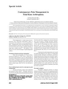
Figure 10.1. Common biomechanical presentation: internal rotation of the femurs.
From the side, the clinician can check pelvic position, to determine whether there is an anterior tilt, posterior tilt, or a sway back posture.47 Position of hyperextension or lock back knees can be verified looking from the side. From behind, the level of the PSIS is checked, gluteal bulk is assessed, and the position of the calcaneum is observed. If the therapist finds that the calcaneum is in a relatively neutral or inverted position and the talus is more prominent on the medial side, then the therapist could probably expect that the patient would have a stiff subtalar joint. Thus, from a person’s static alignment, the clinician can have a reasonable idea of the dynamic picture. Any deviations from the anticipated gives a great deal of information about the muscle control of the activity.
Dynamic Examination The aim of the dynamic examination is not only to evaluate the effect of muscle action on the static mechanics, but also to reproduce the patient’s symptoms so the clinician has an objective reassessment activity to evaluate the effectiveness of the treatment. The least stressful activity of walking is examined first. For example, individuals with patellofemoral pain who stand in hyperextension will not exhibit the nec-
essary shock absorption at the knee, at heel strike. Consequently, the femur will internally rotate and the quadriceps will not function well in inner range due to lack of practice. If the patient’s symptoms are not provoked in walking, then evaluation of more stressful activities, such as stair climbing, is performed. If symptoms are still not provoked then squat and oneleg squat may be examined and used as a reassessment activity. For the athlete, the clinician will, in many cases, be evaluating the control of the one-leg squat as symptom production in the clinic may be difficult.
Supine Lying Examination With the patient in supine lying, the clinician gains an appreciation of the soft tissue structures and begins to confirm the diagnosis. Gentle, but careful palpation should be performed on the soft tissue structures around the patella. First, the joint lines are palpated to exclude obvious intrarticular pathology. Second, palpation of the retinacular tissues determine which parts of the retinaculum are under chronic recurrent stress. If pain is elicited in the infrapatellar region on palpation, the clinician should shorten the fat pad by lifting it toward the patella. If on further palpation, the
Conservative Management of Anterior Knee Pain: The McConnell Program
pain is gone, then the clinician can be relatively certain that the patient has a fat pad irritation. If the pain remains, then patellar tendonosis is the most likely diagnosis. The knee is passively flexed and extended with overpressure applied so the clinician has an appreciation of the quality of the end feel. If any of these maneuvers reproduce pain, they can be used as a reassessment sign;53 for example, the symptoms of fat pad irritation can often be produced with an extension overpressure maneuver. The hamstrings, iliopsoas, rectus femoris, tensor fascia latae, gastrocnemius, and soleus muscles are tested for length. Tightness of any of these muscles has an adverse effect on patellofemoral joint mechanics and will have to be addressed in treatment. The iliopsoas, rectus femoris, and tensor fascia latae may be tested using the Thomas test.41,46 Hamstrings flexibility may be examined by a passive straight leg raise, once the lumbar spine is flattened on the plinth and the pelvis is stable.46 Normal-length hamstrings should allow 80–85° of hip flexion when the knee is extended and the lumbar spine is flattened.46 An essential part of patellofemoral evaluation in supine is assessment of the orientation of the patella relative to the femur. In order to maximize the area of contact of the patella with the femur, the patellar position should be optimal before the patella enters the trochlea. The clinician needs to consider the patellar position not with respect to the normal, but with respect to the optimal, because articular cartilage is nourished and maintained by evenly distributed, intermittent compression.4,37,61 An optimal patellar position is one where the patella is parallel to the femur in the frontal and the sagittal planes, and the patella is midway between the two condyles when the knee is flexed to 20°.56,57 The position of the patella is determined by examining four discrete components; glide, lateral tilt, anteroposterior tilt, and rotation, in a static and dynamic manner. Determination of the glide component involves measuring the distance from the midpole of the patella to the medial and lateral femoral epicondyles (Figure 10.2). The patella should be sitting equidistant (+/−5 mm) from each epicondyle when the knee is flexed 20°. A 5 mm lateral displacement of the patella causes a 50% decrease in VMO tension.1 In some instances, the patella may sit equidistant to the condyles, but moves lateral, out of the line of the femur,
171
Figure 10.2. Assessment of patellar glide.
when the quadriceps contracts, indicating a dynamic problem. The dynamic glide examines both the effect of the quadriceps contraction on patellar position as well as the timing of the activity of the different heads of quadriceps. If the passive lateral structures are too tight, then the patella will tilt so that the medial border of the patella will be higher than the lateral border and the posterior edge of the lateral border will be difficult to palpate. This is a lateral tilt and, if severe, can lead to excessive lateral pressure syndrome.29 When the patella is moved in a medial direction, it should initially remain parallel to the femur. If the medial border rides anteriorly, the patella has a dynamic tilt problem that indicates that the deep lateral retinacular fibers are too tight, affecting the seating of the patella in the trochlea. An optimal position also involves the patella being parallel to the femur in the sagittal plane. A most common finding is a posterior displacement of the inferior pole of the patella (Figure 10.3). This will result in fat pad irritation and often manifests itself as inferior patella pain that
172
Etiopathogenic Bases and Therapeutic Implications
Figure 10.3. Assessment of posterior tilt of the inferior pole of the patella.
is exacerbated by extension maneuvers of the knee.58 A dynamic posterior tilt problem can be determined during an active contraction of the quadriceps muscle as the inferior pole is pulled posteriorly, particularly in patients who hyperextend. To complete the ideal position, the long axis of the patella should be parallel to the long axis of the femur. In other words, if a line was drawn between the most medial and most lateral aspects of the patella, it should be perpendicular to the long axis of the femur (Figure 10.4). If the inferior pole is sitting lateral to the long axis of the femur, the patient has an externally rotated patella. If the inferior pole is sitting medial to the long axis of the femur, then the patient has an internally rotated patella. The presence of a rotation component indicates that a particular part of the retinaculum is tight. Tightness in the retinacular tissue compromises the tissue and can be a potent source of the symptoms.29
Side Lying The retinacular tissue can be specifically tested for tightness with the patient in side lying and the knee flexed to 20°. The therapist moves the patella in a medial direction, so the lateral femoral condyle is readily exposed. If the lateral femoral condyle is not readily exposed, the superficial retinacular fibers are tight. To test the deep fibers, the therapist places his or her hand on the middle of the patella, takes up the slack of the glide, and applies an anteriorposterior pressure on the medial border of the patella. The lateral should move freely away from the femur, and on palpation the tension in
Figure 10.4. Assessment of rotation of the patella.
the retinacular fibers should be similar along the length of the patella. This test procedure can also be used as a treatment technique. Iliotibial band tightness may be confirmed further by Ober’s test.41
Prone In prone, the clinician may examine the foot to determine whether the patient has a primary foot deformity that is contributing to the patient’s patellofemoral symptoms. The deformity will need to be addressed with orthotics or specific muscle training. In the prone position, the clinician is also able to evaluate the flexibility of the anterior hip structures, by examining the patient in a figure of four position, with the underneath foot at the level of the tibial tubercle (Figure 10.5). This position tests the available extension and external rotation at the hip, which is often limited because of chronic adaptive shortening of the anterior structures as a result of the underlying femoral anteversion. The distance of the ASIS from the plinth is measured, so the clinician has an objective measure of change.
Conservative Management of Anterior Knee Pain: The McConnell Program
173
Figure 10.5. Assessment of the flexibility of the anterior hip structures.
A modification of the test position can also be used as a treatment technique. A lumbar spine palpation can be performed at this stage of the examination, if the clinician feels that the knee symptoms have been referred from a primary pathology in the lumbar spine. A summary of the examination process is listed in Table 10.1. Once the patellofemoral joint has been thoroughly examined, and the primary problems have been identified, appropriate treatment can be instigated.
Treatment Conservative Most patellofemoral conditions may be successfully managed with physical therapy. Treatment aims to optimize the patellar position and improve lower limb mechanics and thus decrease the patient’s symptoms. Stretching the tight lateral structures and changing the activation pattern of the VMO may decrease the tendency for the patella to track laterally and should enhance the position of the patella. Stretching the tight lateral structures can be facilitated passively by the therapist mobilizing and massaging the lateral retinaculum and the iliotibial band, as well as the patient performing a self-stretch on the retinacular tissue. However, the most effective stretch to the adaptively shortened retinacular tissue may be obtained by a sustained low load, using tape, to facilitate a permanent elongation
of the tissues. This utilizes the creep phenomenon, which occurs in viscoelastic material when a constant low load is applied. It has been widely documented that the length of soft tissues can be increased with sustained stretching.27,38,40,59,80 The magnitude of the increase in displacement is dependent on the duration of the applied stretch.59,80 If the tape can be maintained for a prolonged period of time, then this, plus training of the VMO to actively change the patellar position, should have a significant effect on patellofemoral mechanics. However, there is some debate as to whether tape actually changes the position of the patella. Some investigators have found that tape changes PF angle and lateral patellar displacement, but congruence angle is not changed.69 Others have concurred, finding no change in congruence angle when the patella is taped, but congruence angle is measured at 45° knee flexion, so subtle changes in patellar position may have occurred before this.8 A study of asymptomatic subjects found that medial glide tape was effective in moving the patella medially but ineffective in maintaining the position after vigorous exercise. However, tape seems to prevent the lateral shift of the patella that occurred with exercise.3 The issue for a therapist, however, is not whether the tape changes the patellar position on x-ray, but whether the therapist can decrease the patient’s symptoms by at least 50%, so the patient can exercise and train in a pain-free manner.
174
Etiopathogenic Bases and Therapeutic Implications
Table 10.1. Examination checklist PATIENT STANDING: Examine for biomechanical abnormalities Observe alignment from: 1. In front Normal standing position of the feet with respect to the legs Q angle tibial valgum/varum tibial torsion talar dome position navicular position Morton’s toe hallux valgus Feet together squinting patellae VMO bulk VL tension 2. Side pelvic position: tilt hyperextension of the knees 3. Behind PSIS position gluteal bulk calf bulk calcaneal position DYNAMIC EVALUATION: Evaluate the effect of the bony alignment and soft tissue on dynamic activities 1. Walking, if no pain 2. Steps, if no pain 3. Squat, if no pain 4. One leg squat ASSESSMENT IN SUPINE LYING: Determine the causative factors of the symptoms and formulate a diagnosis 1. Palpation of the tibiofemoral joint line and soft tissue structures of the patellofemoral joint 2. Tibiofemoral tests 3. Meniscal tests 4. Ligament tests 5. Thomas’s test: psoas, rectus femoris, tensor fascia lata 6. Tests for hamstrings, gastrocs 7. Slump test for dural length, particularly indicated if the patient complains of lateral knee pain when sitting with the legs out straight. 8. Hip tests (if applicable) 9. Orientation of the patella glide, dynamic glide lateral tilt anteroposterior tilt rotation ASSESSMENT IN SIDELYING POSITION: Tests for tightness of the lateral structures 1. Medial glide: tests superficial lateral structures 2. Medial tilt: tests deep lateral structures 3. Ober’s test for iliotibial band tightness ASSESSMENT IN THE PRONE POSITION: 1. Lumbar spine palpation (only if applicable, i.e., if dural test positive) 2. Foot assessment 3. Hip rotation 4. Femoral nerve mobility ●
● ● ● ● ● ● ● ●
●
● ● ●
● ●
● ● ● ●
● ● ● ●
Patellar Taping Patellar taping is based on the assessment of the patellar position. The component(s) corrected, the order of correction, and the tension of the tape is tailored for each individual (Figures 10.6 and 10.7). After each piece of tape is applied, the symptom producing activity should be reassessed. The tape should always immediately improve a patient’s symptoms by at least 50%. If it does not, then the order in which the tape has been applied or the components corrected should be reexamined. In most cases, hypoallergenic tape is placed underneath the rigid sports tape to provide a protective layer for the skin and if there seems to be additional skin problems a plastic coating, either a spray or a roll-on, may be applied to the skin prior to the tape application. The patient must be taught how to position the tape on him- or herself. The patient should be in long sitting with the leg out straight and the quadriceps relaxed. If a posterior tilt problem has been ascertained on assessment, it must be corrected first, as taping over the inferior pole of the patella will aggravate the fat pad and exacerbate the patient’s pain. The posterior component is corrected together with a glide or a lateral tilt with the nonstretch tape being placed on the superior aspect of the patella, either on the lateral border to correct lateral glide or in the middle of the patella to correct lateral tilt. This positioning of the tape will lift the inferior pole out of the fat pad and prevent irritation of the fat pad. If there is no posterior tilt problem, the glide may be corrected by placing tape from the lateral patellar border to the medial femoral condyle. At the same time the soft tissue on the medial aspect of the knee is lifted toward the patella to create a tuck or fold in the skin. The skin lift helps anchor the tape more effectively and minimizes the friction rub (friction between the tape and the skin), which can occur when a patient has extremely tight lateral structures. The mediolateral tilt component is corrected by placing a piece of tape firmly from the middle of the patella to the medial femoral condyle. The object is to shift the lateral border away from the femur so that the patella becomes parallel with the femur in the frontal plane. Again, the soft tissue on the medial aspect of the knee is lifted toward the patella. External rotation is the most common rotation problem and to correct this the tape is positioned at the inferior pole and pulled upward
Conservative Management of Anterior Knee Pain: The McConnell Program
175
Figure 10.6. Taping components: (a) medial glide, (b) medial tilt, (c) internal rotation, (d) anterior tilt.
and medially toward the opposite shoulder while the superior pole is rotated laterally. Care must be taken so that the inferior pole is not displaced into the fat pad. Internal rotation, on the other hand, is corrected by taping from the superior pole downward and medially.
the fat pad, with the point of the V at the tibial tubercle coming wide to the medial and lateral joint lines (Figures 10.7D and 10.7E). As the tape is being pulled toward the joint line, the skin is lifted toward the patella, thus shortening the fat pad.
Unloading
Principles of Using Tape to Correct the Patella
The principle of unloading is based on the premise that inflamed soft tissue does not respond well to stretch. For example, if a patient presents with a sprained medial collateral ligament, applying a valgus stress to the knee will aggravate the condition, whereas a varus stress will decrease the symptoms. The same principle applies for patients with an inflamed fat pad, an irritated iliotibial band or a pes anserinus bursitis. The inflamed tissue needs to be shortened or unloaded. To unload an inflamed fat pad, for example a “V” tape is placed below
The tape is kept on all day, every day, until the patient has learned how to activate his or her VMO at the right time; that is, the tape is like trainer wheels on a bicycle and can be discontinued once the skill is established. The tape is removed with care in the evening allowing the skin time to recover. The tape can cause a breakdown in the skin either through a friction rub or as a consequence of an allergic reaction. Preparation of the skin and skin care advice is essential.
176
Etiopathogenic Bases and Therapeutic Implications
The patient should never train with or through pain or effusion as it has been shown quite conclusively in the literature that pain and effusion have an inhibitory effect on muscle activity.19,63,75,76 If the patient experiences a return of the pain, the tape should be
readjusted. If the activity is still painful, the patient must cease the activity immediately. The tape will loosen quickly if the lateral structures are extremely tight or the patient’s job or sport requires extreme amounts of knee flexion.
Figure 10.7. (a) Tape to correct lateral glide. (b) Tuck or fold in the skin.
Conservative Management of Anterior Knee Pain: The McConnell Program
177
Figure 10.7. (continued) (c) Tape in internal rotation to correct external rotation of the patella. (d,e) Unloading fat pad with tape, lift the soft tissue toward the patella.
178
Studies Investigating the Effects of Tape It has been fairly well established that taping the patella relieves pain,8,34,39 but the mechanism of the effect is still being debated in the literature. This topic has been well reviewed by Crossley et al.14 As mentioned previously, there is some evidence to show that taping can improve the radiographic position of the patella.50,69,73 Others have assessed the effect of tape on quadriceps function.8,34,72 Using isokinetic dynamometry, two studies found that tape significantly increased the quadriceps torque.8,34 However, the increase in muscle torque with tape did not necessarily correlate with pain reduction. Ernst et al.26 showed greater knee extensor moments and power during a vertical jump and lateral step-up in a taped condition compared with placebo and no tape conditions in PFPS subjects. It has also been found that during gait, individuals with PFPS decrease the amount of knee flexion in early stance to reduce the patellofemoral joint reaction force.17,20,32,67 Patellar taping results in small but significant increases in loading response knee flexion in a variety of gait conditions indicating a greater ability to load the knee joint with confidence.66 It has been suggested that patellar tape could influence the magnitude of VMO and VL activation, although most studies do not necessarily support this contention.6,18,39,62,72 Similarly there is conflict with regard to the effect of tape on onset timing of VMO and VL with some showing earlier timing of VMO. Taping the patella of symptomatic individuals such that the pain was decreased by 50% resulted in an earlier activation of the VMO relative to the VL on both step up and step down. Stepping down in particular caused an 8.3° differential between the knee angle at onset of VMO and VL as not only was the VMO activating earlier than the pre-taped condition, but the VL was significantly delayed in the taped condition.30 Our research group has also found that tape leads to a change in the onset timing of VMO relative to VL compared with placebo tape and no tape.12
Muscle Training There is currently debate about the best type of quadriceps strengthening for rehabilitating the PF joint. Powers concludes that because there is no difference in the activation pattern of the VMO and VL in symptomatic individuals and the ratio of the two muscles is the same, generalized quadriceps strengthening is all that is required.65 However, this is at odds with our recent research, which demonstrated that a McConnell-based
Etiopathogenic Bases and Therapeutic Implications
physiotherapy treatment regime (taping, functional training with biofeedback on VMO and VL) for PFPS alters the motor control of VMO relative to VL in both a functional task11 and a postural perturbation task.13 What types of exercises are most appropriate in training? From the current evidence available, it seems that closed chain exercise (when the foot is on the ground) is the preferred method of training, not only because closed kinetic training has been shown to improve patellar congruence, but muscle training has been found to be specific to limb position.21 In a group of patients with lateral patellar compression syndrome, it was found that open chain exercise with isometric quadriceps sets at 10° intervals with 3 kg weight resulted in more lateral patellar tilt and glide from 0–20° on CT scan. Closed chain exercise by pushing a foot-plate with resistance cords attached to provide 18 kg resistance led to improved congruence from 0–20°.21 Another study showed that in closed chain exercises, there is more selective VMO activation than in open chain exercises.79 However, there is still debate in the literature. Recently a 5-year follow-up of patients in a clinical trial found good maintenance of subjective and functional outcomes in both the open and closed kinetic chain exercise groups.84
Specificity of Training Before examining the issue of exercise prescription for the patellofemoral patient, some discussion on the different philosophies of strength training is required. The traditional strengthening view holds that strength gained in nonspecific muscle training can be harnessed for use in performance, that is, the engine (muscles) is built in the strength training room; learning how to turn the engine on (neural control) is acquired on the field.71 Strength is therefore increased by utilizing the overload principle, meaning exercising at least 60% of maximal.31 However, the muscles around the PF joint are stabilizing muscles and need to be endurance trained, so working at 20–30% of maximal is more appropriate. A more recent interpretation of how to facilitate strength is based on the premise that the engine (muscles) and how it is turned on (neural control) should both be built in the strength training room.71 Training should therefore simulate movement in terms of anatomical movement pattern, velocity, type and force of contraction. Thus, with training the neuromuscular system will tend to become better at generating tension for actions that resemble the muscle actions employed in training,
Conservative Management of Anterior Knee Pain: The McConnell Program
but not necessarily for actions that are dissimilar to those used in training. If the desired outcome of treatment is for the patient to be pain-free on weight-bearing activities, then the therapist must give the patient appropriate weight-bearing training. At no stage should the patient’s recovery be compromised by training into pain. A useful starting exercise is small-range knee flexion and extension movements (the first 30°) with the patient in stance position, where the feet are facing forward and positioned at the width of the pelvis. It is preferable that the patient has a dual channel biofeedback with electrodes on the VMO and the VL so the patient can monitor the timing of the contraction and the amount of activity. This is particularly important for those patients who have trouble activating the VMO. The patient is instructed to squeeze the gluteals and slowly flex the knees to 30° and slowly return to full extension without locking the knees back. The patient is aiming for the VMO to be activated before the VL and remain more than the VL during the activity. This clinical interpretation of the use of EMG biofeedback is at odds with the
179
research application, insofar as the activity of the VMO and the VL has not been normalized. Furthermore, the limited research to date does not necessarily show additional benefit from the incorporation of biofeedback.22 Progression of training involves simulation of the knee during the stance phase of walking, so the patient is in a walk-stance position. In this position VMO recruitment is usually poor and the seating of the patella in the trochlea is critical. Again, small-amplitude movements need to be practiced. Again, emphasis should be given to the timing and intensity of the VMO contraction relative to the VL. For a patient who is having difficulty contracting the VMO, muscle stimulation may be used to facilitate the contraction. Further progression of treatment can be implemented by introducing step training. The patients need to practise stepping down from a small height initially. This should be performed slowly, in front of a mirror, so that changes in limb alignment can be observed and deviations can be corrected (Figure 10.8). Specific work on the hip musculature may be necessary to improve
Figure 10.8. (a) Stepping down with correct limb alignment. (b) Stepping down with incorrect limb alignment.
180
the limb alignment. Some patients may only be able to do one repetition before the leg deviates. This is sufficient for them to start with, as inappropriate practice can be detrimental to learning. The number of repetitions should be increased as the skill level improves. It is therefore preferable for the therapist to emphasize quality, not quantity. Initially small numbers of exercises should be performed frequently throughout the day. The aim is to achieve a carryover from functional exercises to functional activities. Later, the patient can move to a larger step, initially decreasing the number of contractions and slowly increasing them again. As the control improves, the patient can alter the speed of the stepping activity and vary the place on descent where the stepping action is stopped. Weights may be introduced in the hands or in a backpack. Again, the number of repetitions and the speed of the movement should be decreased initially and built back up again. Training should be applicable to the patient’s activities/sport, so a jumping athlete, for example, should have jumping incorporated in his program. Figure-eight running, bounding jumping off boxes, jumping and turning, and other plyometric routines are particularly appropriate for the high-performance athlete. However, the patient’s VMO needs to be monitored at all times for timing and level of contraction relative to the VL. The number of repetitions performed by the patient at a training session will depend on the onset of muscle fatigue. The aim would be to increase the number of repetitions before the onset of fatigue. Patients should be taught to recognize muscle fatigue or quivering, so that they do not train through the fatigue and risk exacerbating their symptoms.
Improving Lower Limb Mechanics A stable pelvis will minimize unnecessary stress on the knee. Training of the gluteus medius (posterior fibers) to decrease hip internal rotation and the consequent valgus vector force that occurs at the knee is necessary to improve pelvic stability. Weakness of the hip abductors and external rotators has been documented in women with patellofemoral pain compared with pain-free controls.44 The posterior gluteus medius may be trained in weight bearing with the patient standing side-on to a wall. The leg closest to the wall is flexed at the knee so the foot is off the ground. The hip is in line with the standing hip. The patient should have all the
Etiopathogenic Bases and Therapeutic Implications
weight back through the heel of the standing leg, which is slightly flexed. The patient externally rotates the standing leg without turning the foot, the pelvis, or the shoulders. The patient should sustain the contraction for 20 seconds, so a burning can be felt in the gluteus medius region. If this exercise is difficult for a patient to coordinate, then rubber tubing may be used around the ankles to provide resistance as the patient stands on the affected leg while pushing the other leg back diagonally at 45°. The training may be progressed to standing on one leg where the pelvis is kept level and the lower abdominals and the glutei are worked together while the other leg is swinging back and forward, simulating the activity of the stance phase of gait. If the patient has marked internal femoral rotation stretching of the anterior hip structures, to increase the available external rotation may be required. The patient lies prone with the hip to be stretched in an abducted, externally rotated and extended position. The other leg is extended and lies on top of the bent leg. The malleolus of the underneath leg is at the level of the tibial tubercle. The patient attempts to flatten the abducted and rotated hip by pushing along the length of the thigh and holding the stretch for 5 seconds. This action activates gluteals in inner range. Although it is not functional, it may facilitate gluteus medius activity in someone who is finding it difficult to activate the muscle in weight bearing.
Muscle Stretching Appropriate flexibility exercises must be included in the treatment regime. The involved muscles may include hamstrings, gastrocnemius, rectus femoris, and TFL/ITB. A tight gastrocnemius will increase the amount of subtalar joint pronation exhibited in mid-stance phase of gait, so after the stretching, appropriate foot muscle training will be required.
Consideration of Foot Problems The supinators of the foot, specifically tibialis posterior, should be trained if the patient demonstrates prolonged pronation during the midstance in gait. With the foot supinated, the base of the first metatarsal is higher than the cuboid, which will allow the peroneus longus to work more efficiently to increase the stability of the first metatarsal complex for push-off. The therapist can train this action to improve the efficiency
Conservative Management of Anterior Knee Pain: The McConnell Program
of push-off. The position of training is in midstance, the patient is instructed to lift the arch while keeping the first metatarsal head on the floor, and then pushing the first metatarsal and great toe into the floor. If the patient is unable to keep the first metatarso-phalangeal joint on the ground when the arch is lifted, then the foot deformity is too large to correct with training alone and orthotics will be necessary to control the excessive pronation. The addition of orthotics to a physiotherapy program in a group of PFPS patients with documented rearfoot varus has been studied. The results showed less knee pain during aggravating activities after 8 weeks when compared with patients issued with a placebo foot insole.25 Another study showed that patellofemoral pain patients who responded best to off-the-shelf orthotics were those with forefoot valgus of 2°, passive great toe extension of 78°, or navicular drop of 3 mm.78 Gross and Foxworth33 provide a review of the role of foot orthoses as an intervention in this condition.
Evaluation of the McConnell Program Few clinical trials have evaluated the effectiveness of a McConnell-type program for PFPS.7,24,35 These and other physical interventions for PFPS have been reviewed by Crossley et al.14 and Heintjes et al.36 Harrison et al.35 performed a randomized, blinded, controlled trial investigating three physiotherapy treatment options, one of which best reflects the protocol designed by McConnell.56 At the end of the one-month intervention period, the subjects in the McConnell-based program showed significant improvements in pain and function compared with a group who had supervised exercises, but did not differ from the group given a home exercises program only. However, the sample size was only sufficient to detect a large effect between the groups. The large dropout rate (up to 48%) at 12 months may have affected the results at this time point, especially since a significantly greater number of subjects in the intervention group who showed substantial improvement were lost to follow-up. The authors concluded that any of the treatments could provide long-term improvements in pain and function. Clark et al.7 found that proprioceptive muscle stretching and strengthening aspects of physiotherapy have a beneficial effect at three months sufficient to permit discharge from physiotherapy. These benefits were maintained at three months. While they noted that taping did not influence the outcome, the numbers in each group
181
may not have been large enough to detect independent effects of taping. We conducted a randomized, double-blind, placebo-controlled trial of the McConnell program in 71 PFPS patients.10,15 Standardized treatment consisted of 6 treatment sessions, once weekly for both the physiotherapy and placebo groups. Sixty-seven (33 physiotherapy; 34 placebo) subjects completed the trial. The physiotherapy group demonstrated significantly better response to treatment and greater improvements in pain and functional activities than the placebo group. The physiotherapy treatment also changed the onset timing of VMO relative to VL measured using surface electromyography during stair stepping and postural perturbation tasks. At baseline in both groups, VMO came on significantly later than VL. Following treatment, there was no change in muscle onset timing of the placebo group. However, in the physiotherapy group, the onset of VMO and VL occurred simultaneously (concentric) or VMO actually preceded VL (eccentric).10,13 This study demonstrates that a McConnell-based physiotherapy program significantly improves pain and function and can alter EMG onset of VMO relative to VL compared with placebo treatment.
Conclusion Management of patellofemoral pain is no longer a conundrum if the therapist can determine the underlying causative factors and address those factors in treatment. It is imperative that the patient’s symptoms are significantly reduced. This is often achieved by taping the patella, which not only decreases the pain, but also promotes an earlier activation of the VMO and increases quadriceps torque. Management will need to include specific VMO training, gluteal control work, stretching tight lateral structures, and appropriate advice regarding the foot, be it orthotics, training, or taping.
References 1. Ahmed, A, S Shi, A Hyder et al. The effect of quadriceps tension characteristics on the patellar tracking pattern. Transactions of the 34th Orthopaedic Research Society, Atlanta, 1988; 280. 2. Bennell, K, P Hodges, R Mellor, C Bexander, and T Souvlis. The nature of anterior knee pain following injection of hypertonic saline into the infrapatellar fat pad. J Orthopaed Res 2004; 22: 116–121. 3. Bockrath, K, C Wooden, T Worrell et al. Effects of patella taping on patella position and perceived pain. Med Sci Sports Exer 1993; 25(9): 989–992.
182 4. Brandt, K. Pathogenesis of osteoarthritis. In Kelley, Harris, Ruddy, Sledge, eds., Textbook of Rheumatology. Philadelphia: W.B. Saunders, 1981, Chap. 88. 5. Buchbinder, R, N Naparo, and E Bizzo. The relationship of abnormal pronation to chondromalacia patellae in distance runners. J Am Pod Assoc 1979; 69(2): 159–161. 6. Cerny, K. Vastus medialis oblique/vastus lateralis muscle activity ratios for selected exercises in persons with and without patellofemoral pain syndrome. Phys Ther 1995; 75(8): 672–682. 7. Clark, DI, N Downing, J Mitchell et al. Physiotherapy for anterior knee pain: A randomised controlled trial. Ann Rheum Disease 2000; 59: 700–704. 8. Conway, A, T Malone, and P Conway. Patellar alignment/ tracking Alteration: Effect on force output and perceived pain. Isokin Exer Sci 1992; 2(1): 9–17. 9. Cowan, SM, KL Bennell, PW Hodges, KM Crossley, and J McConnell. Delayed electromyographic onset of vastus medialis obliquus and vastus lateralis in subjects with patellofemoral pain syndrome. Arch Phys Med Rehabil 2001; 82: 183–189. 10. Cowan, S, K Bennell, K Crossley, P Hodges, and J McConnell. Physical therapy treatment changes electromyographic onset timing of vastus medialis obliquus relative to vastus lateralis in subjects with patellofemoral pain syndrome. Med Sci Sport Exer 2002; 34: 1879–1885. 11. Cowan, SM, PW Hodges, KL Bennell, and KM Crossley. Altered vastii recruitment when people with patellofemoral pain syndrome complete a postural task. Arch Phys Med Rehabil 2002; 83: 989–995. 12. Cowan, S, K Bennell, and P Hodges. Therapeutic patellar taping changes the timing of vastii muscle activation in people with patellofemoral pain syndrome. Clin J Sport Med 2002; 12: 339–347. 13. Cowan, SM, PW Hodges, KL Bennell, KM Crossley, and J McConnell. Simultaneous feedforward recruitment of the vastii in untrained postural tasks can be restored by specific training. J Orthopaed Res 2003; 21: 553–558. 14. Crossley, K, S Cowan, J McConnell et al. Patellar taping: Is clinical success supported by scientific evidence? Manual Ther 2000; 5: 142–150. 15. Crossley, K, K Bennell, S Green, S Cowan, and J McConnell. Conservative management of patellofemoral pain: A randomised, double-blind controlled trial. Am J Sport Med 2002; 30: 857–865. 16. Crossley, K, K Bennell, S Green, and S Cowan. Analysis of outcome measures for persons with patellofemoral pain: Which outcome measures for individuals with patellofemoral pain are reliable and valid? Arch Phys Med Rehabil 2004; 85: 815–822. 17. Crossley, KM, SM Cowan, KL Bennell, and J McConnell. Knee flexion during stair ambulation is altered in individuals with patellofemoral pain. J Orthopaed Res 2004; 22: 267–274. 18. Christou, EA. Patellar taping increases vastus medialis oblique activing in the presence of patellofemoral pain. J Electromyogr Kinesiol 2004; 14: 495–504. 19. de Andrade, J, C Grant, and A Dixon. Joint distension and reflex muscle inhibition in the knee. J Bone Jt Surg 1965; 47A: 313. 20. Dillon, P, W Updyke and W Allen. Gait analysis with reference to chondromalacia patellae. J Orthop Sports Phys Ther 1983; 5(3): 127–131.
Etiopathogenic Bases and Therapeutic Implications 21. Doucette, S, and D Child. The effect of open and closed chain exercise and knee joint position on patellar tracking in lateral patellar compression syndrome. J Orthop Sports Phys Ther 1996; 23(2): 104–110. 22. Dursun, N, E Dursun, and Z Kilic. Electromyographic biofeedback-controlled exercise versus conservative care for patellofemoral pain syndrome. Arch Phys Med Rehab 2001; 82: 1692–1695. 23. Dye, S. The knee as a biologic transmission with an envelope of function. Clin Orthop Rel Res 1996; 325: 10–18. 24. Eburne, J, and G Bannister. The McConnell regimen versus isometric quadriceps exercises in the management of anterior knee pain: A randomised prospective controlled trial. Knee 1996; 3: 151–153. 25. Eng, J, and M Pierrynowski. Evaluation of soft foot orthotics in the treatment of patellofemoral pain syndrome. Phys Ther 1993; 73(2): 62–70. 26. Ernst, GP, J Kawaguchi, and E Saliba. Effect of patellar taping on knee kinetics of patients with patellofemoral pain syndrome. J Orthop Sports Phys Ther 1999; 29(11): 661–667. 27. Frankel, VH, and M Nordin. Basic Biomechanics of the Skeletal System. Philadelphia: Lea and Febiger, 1980. 28. Fulkerson, JP. Awareness of the retinaculum in evaluating patellofemoral pain. Am J Sports Med 1982; 10(3): 147–149. 29. Fulkerson, J, and D Hungerford. Disorders of the Patellofemoral Joint, 2nd ed. Baltimore: Williams & Wilkins, 1990. 30. Gilleard, W, J McConnell, and D Parsons. The effect of patellar taping on the onset of vastus medialis obliquus and vastus lateralis muscle activity in persons with patellofemoral pain. Phys Ther 1998; 78(1): 25–32. 31. Grabiner, MD, TJ Koh, and LF Draganich. Neuromechanics of the patellofemoral joint. Med Sci Sports Exer 1994; 26(1): 10–21. 32. Greenwald, AE, AM Bagley, FP France et al. A biomechanical and clinical evaluation of a patellofemoral knee brace. Clin Orthop Rel Res 1996; 324: 187–195. 33. Gross, MT, and JL Foxworth. The role of foot orthoses as an intervention for patellofemoral pain. J Orthopaed Sports Phys Ther 2003; 33: 661–670. 34. Handfield, T, and J Kramer. Effect of McConnell taping on perceived pain and knee extensor torques during isokinetic exercise performed by patients with patellofemoral pain. Physioth Can Winter 2000; 39–44. 35. Harrison, EL, MS Sheppard, and AM McQuarrie. A randomized controlled trial of physical therapy treatment programs in patellofemoral pain syndrome. Physioth Can Spring 1999; 93–106. 36. Heintjes, E, MY Berger, SM Bierma-Zeinstra, RM Bernsen, JA Verhaar, and BW Koes. Exercise therapy for patellofemoral pain syndrome. Cochrane Database of Systematic Reviews 2003; CD003472. 37. Helminen, H, I Kiviranta, M Tammi et al. Joint Loading. London: Butterworths, 1987. 38. Herbert, R. Preventing and treating stiff joints. In Crosbie, J, and J McConnell, eds., Key Issues in Musculoskeletal Physiotherapy. Oxford: ButterworthHeinemann, 1993. 39. Herrington, L, and CJ Payton. Effects of corrective taping of the patella on patients with patellofemoral pain. Physioth 1997; 83(11): 566–572. 40. Hooley, C, N McCrum, and R Cohen. The visco-elastic deformation of the tendon. J Biomech 1980; 13: 521.
Conservative Management of Anterior Knee Pain: The McConnell Program 41. Hoppenfeld, S. Physical Examination of the Spine and Extremities. New York: Appleton-Century-Crofts, 1976. 42. Howard, J, and R Enoka. Maximum bilateral contractions are modified by neurally mediated interlimb effects. J Appl Physiol 1991; 70: 306–316. 43. Insall, J. Chondromalacia patellae: Patellar malalignment syndrome. Orthopaed Clin North Am 1979; 10: 117–125. 44. Ireland, ML, JD Willson, BT Ballantyne, and IM Davis. Hip strength in females with and without patellofemoral pain. J Orthopaed Sports Phys Ther 2003; 33: 671–676. 45. Karst, GM, and GM Willett. Onset timing of electromyographic activity in the vastus medialis oblique and vastus lateralis muscles in subjects with and without patellofemoral pain syndrome. Phys Ther 1995; 75(9): 813–823. 46. Kendall, F, and L McCreary. Muscle Testing and Function. Baltimore: Williams and Wilkins, 1983. 47. Kendall, HD, FP Kendall, and DA Boynton. Posture and Pain. Baltimore: Williams and Wilkinson, 1952. 48. Koh, T, M Grabiner, and R DeSwart. In vivo tracking of the human patella. J Biomech 1992; 25(6): 637–643. 49. Kramer, PG. Patella malalignment syndrome: Rationale to reduce excessive lateral pressure. J Orthop Sports Phys Ther 1986; 8(6): 301–308. 50. Larsen, BE, A Adreasen, MR Urfer et al. Patellar taping: A radiographic examination of the medial glide technique. Am J Sports Med 1995; 23(4): 465–471. 51. Lutter, L. The knee and running. Clin Sports Med 1985; 4(4): 685–698. 52. Lyon, L, L Benzl, K Johnson et al. Q angle – A factor: Peak torque occurrence in isokinetic knee extension. J Orthop Sports Phys Ther 1988; 9(7): 250–253. 53. Maitland, GD. Vertebral Manipulation. London: Butterworths, 1986. 54. Malek, M, and R Mangine. Patellofemoral pain syndromes: A comprehensive and conservative approach. J Orthop Sports Phys Ther 1981; 2(3): 108–116. 55. Mariani, P, and I Caruso. An electromyographic investigation of subluxation of the patella. J Bone Jt Surg 1979; 61, 169–171. 56. McConnell, J. The management of chondromalacia patellae: A long-term solution. Aust J Physioth 1986; 32(4), 215–223. 57. McConnell, J. Training the vastus medialis oblique in the management of patellofemoral pain. Proceedings Tenth Congress of the World Confederation for Physical Therapy, Sydney, May 1987. 58. McConnell, J. Fat pad irritation: A mistaken patellar tendonitis. Sport Health 1991; 9(4): 7–9. 59. McKay-Lyons, M. Low-load, prolonged stretch in treatment of elbow flexion contractures secondary to head trauma: a case report. Phys Ther 1989; 69: 292. 60. Micheli, L, J Slater, E Woods et al. Patella alta and the adolescent growth spurt. Clin Orthop Rel Res 1986; 213: 159–162. 61. Mow, V, J Eisenfeld, and I Redler. Some surface characteristics of articular cartilage: II. On the stability of articular surface and a possible biomechanical factor in aetiology of chondrodegeneration. J Biomech 1974; 7: 457–467. 62. Ng, GY, and JM Cheng. The effects of patellar taping on pain and neuromuscular performance in subjects with patellofemoral pain. Clin Rehab 2002; 16: 821–827. 63. On, AY, B Uludag, E Taskirran, and C Eertekin. Differential corticomotor control of a muscle adjacent to a painful joint. Neurorehab & Neural Rep 2004; 18: 127–133.
183 64. Owings, TM, and MD Grabiner. Motor control of the vastus medialis oblique and vastus laterales is disrupted during eccentric contractions in subjects with patellofemoral pain. Am J Sports Med 2002; 30: 483–487. 65. Powers, CM, RF Landel, and J Perry. Timing and intensity of vastus muscle activity during functional activities in subjects with and without patellofemoral pain. Phys Ther 1996; 76: 946–955. 66. Powers, CM, R Landel, T Sosnick et al. The effects of patellar taping on stride characteristics and joint motion in subjects with patellofemoral pain. J Orthop Sports Phys Ther 1997; 26(6): 286–291. 67. Powers, CM, S Mortenson, D Nishimoto et al. Criterionrelated validity of a clinical measurement to determine the medial/lateral component of patellar orientation. J Orthop Sports Phys Ther 1999; 29(7): 372–377. 68. Radin, E. A rational approach to treatment of patellofemoral pain. Clin Orthop Rel Res 1979; Oct., 144: 107–109. 69. Roberts, JM. The effect of taping on patellofemoral alignment: A radiological pilot study. Proceedings Sixth Biennial Conference of the Manipulative Therapists Association of Australia, 1989, pp. 146–151. 70. Root, M, W Orien, and J Weed. Clinical Biomechanics, Vol. II. Los Angeles: Clinical Biomechanics Corp., 1977. 71. Sale, D, and D MacDougall. Specificity of strength training: A review for coach and athlete. Can J Appl Sports Sci 1981; 6(2): 87–92. 72. Salsich, GB, JH Brechter, D Farwell, and CM Powers. The effects of patellar taping on knee kinetics, kinematics, and vastus laterales muscle activity during stair ambulation in individuals with patellofemoral pain. J Orthopaed & Sports Phys Ther 2002; 32: 3–10. 73. Somes, S, TW Worrell, B Corey et al. Effects of patellar taping on patellar position in the open and closed kinetic chain: A preliminary study. J Sports Rehab 1997; 6: 299–308. 74. Souza, D, and M Gross. Comparison of vastus medialis obliquus: Vastus lateralis muscle integrated electromyographic ratios between healthy subjects and patients with patellofemoral pain. Phys Ther 1991; 71: 310–320. 75. Spencer, J, K Hayes, and I Alexander. Knee joint effusion and quadriceps reflex inhibition in man. Arch Phys Med 1984; 65: 171–177. 76. Stokes, M, and A Young. Investigations of quadriceps inhibition: Implications for clinical practice. Physioth 1984; 70(11): 425–428. 77. Subotnik, S. The foot and sports medicine. J Orthop Sports Phys Ther 1980; 2(2): 53–54. 78. Sutlive, TG, SD Mitchell, SN Maxfield, CL McLean, JC Neumann, CR Swiecki, RC Hall, AC Bare, and TW Flynn. Identification of individuals with patellofemoral pain whose symptoms improved after a combined program of foot orthosis use and modified activity: a preliminary investigation. Phys Ther 2004; 84: 49–61. 79. Tang, SF, CK Chen, R Hsu, SW Chou, WH Hong, and HL Lew. Vastus medialis obliquus and vastus lateralis activity in open and closed kinetic chain exercises in patients with patellofemoral pain syndrome: An electromyographic study. Arch Phys Med Rehab 2001; 82: 1441–1445. 80. Taylor, D, J Dalton, and A Seaber. Visco-elastic properties of muscle-tendon units. The biomechanical effect of stretching. Am J Sports Med 1990; 18: 300.
184 81. Townsend, PR, and RM Rose. The biomechanics of the human patella and its implications for chondromalacia. J Biomech 1977; 10: 403–407. 82. Voight, M, and D Weider. Comparative reflex response times of the vastus medialis and the vastus lateralis in normal subjects and subjects with extensor mechanism dysfunction. Am J Sports Med 1991; 10: 131–137. 83. Witvrouw, E, C Sneyers, R Lysens et al. Comparative reflex response times of vastus medialis obliquus and vastus lateralis in normal subjects and subjects with
Etiopathogenic Bases and Therapeutic Implications patellofemoral pain syndrome, J Orthop Sports Phys Ther 1996; 24(3): 160–166. 84. Witvrouw, E, L Danneels, D Van Tiggelen, TM Willems, and D Cambier. Open versus closed kinetic chain exercises in patellofemoral pain: A 5-year prospective randomized study. Am J Sports Med 2004; 32: 1122–1130. 85. Yang, J, and D Winter. Electromyography reliability in maximal contractions and submaximal isometric contractions. Arch Phys Med Rehab 1983; 64: 417–420.