Congenital Heart Disease Vascular Risk Factors and Medication
Congenital Heart Disease Vascular risk factors and medication Thesis, Erasmus University Rotterdam, The Netherlands
All previously published parts of this thesis have been reproduced with explicit permission from the publishers. The research described in this thesis was supported by a grant of the Netherlands Heart foundation (NHS 2002.B027) and Bo Hjelt foundation (2005). Financial support by the Netherlands Heart Foundation for the publication of this thesis is gratefully acknowledged. The printing of this thesis has also been financially supported by the Department of Obstetrics and Gynaecology, Erasmus MC Rotterdam, Nederlandse Vereniging voor Obstetrie en Gynaecologie and the Erasmus University Rotterdam. Further financial support for this dissertation was kindly provided by: BMA B.V. (Mosos) Danone Research – Centre for Specialised Nutrition Merck Sharp & Dohme B.V. Medical Dynamics Ferring Pharmaceuticals Nutricia Nederland B.V. Cover & chapter design: Jelle Mollema,
[email protected] Design: Legatron Electronic Publishing, Rotterdam Print: Ipskamp Drukkers B.V. Enschede, The Netherlands ISBN / EAN 978-90-9025934-5
Copyright © 2011 Dineke Smedts, Rotterdam, The Netherlands
[email protected] No part of this thesis may be reproduced in any form or by any means without written permission from the author
Congenital Heart Disease Vascular Risk Factors and Medication Aangeboren hartafwijkingen Vasculaire risico factoren en medicatie
Proefschrift
ter verkrijging van de graad van doctor aan de Erasmus Universiteit Rotterdam op gezag van de rector magnificus Prof.dr. H.G. Schmidt en volgens besluit van het College voor Promoties. De openbare verdediging zal plaatsvinden op woensdag 2 februari 2011 om 13:30 uur
door
Huberdina Pieternella Maria Smedts geboren te Venray
Promotiecommissie Promotoren:
Prof.dr. R.P.M. Steegers – Theunissen Prof.dr. E.A.P. Steegers
Overige leden:
Prof.dr. J.W. Roos-Hesselink Prof.dr. E.J.G. Sijbrands Prof.dr. M.C. de Ruiter
Contents Chapter 1 General introduction Part I
7
Maternal lipids and related vitamins
Chapter 2 A derangement of the maternal lipid profile is associated with an
21
elevated risk of congenital heart disease in the offspring Nutr Metab Cardiovasc Dis; in press
Chapter 3 Maternal intake of fat, riboflavin and nicotinamide and the risk of
39
having offspring with congenital heart defects Eur J Nutr 2008;47:357-65
Chapter 4 High maternal vitamin E intake by diet or supplements is associated
57
with congenital heart defects in the offspring BJOG 2009;116:416-23
Part II
Medication
Chapter 5 Eight-fold increased risk for congenital heart defects in children
75
carrying the nicotinamide N-methyltransferase polymorphism and exposed to medicines and low nicotinamide Eur Heart J 2008;29:142-31
Chapter 6 Early pregnancy exposure to antihistamines, pregnancy related
95
nausea and vomiting and the risk of congenital heart disease Submitted for publication
Part III
Vascular Endothelial Growth Factor
Chapter 7 VEGF polymorphisms are associated with endocardial cushion defects: a family based case-control study Pediatr Res 2010;67:23-8
113
Chapter 8 Vascular Endothelial Growth Factor expression in the
131
developing chicken embryonic heart using Optical Projection Tomography Submitted for publication
Chapter 9 General discussion
151
Summary
172
Samenvatting
175
About the author
181
List of publications and related awards
183
PhD Portfolio
186
Dankwoord
189
Chapter 1 General introduction
Chapter 1
Congenital heart disease (CHD) is among the most common congenital abnormalities and involves structural anomalies of the heart and/or related major blood vessels. Congenital heart disease arises in the first trimester of pregnancy, occurring often and in many forms. The reported CHD birth prevalence rate ranges from 6 per 1,000 newborns in the Netherlands to 9 per 1,000 newborns in the United States of America.1,2 This percentage is much higher if fetal deaths are taken into account.3 CHD is the leading cause of infant mortality4 and contributes to 30% to 40% of all deaths during infancy and early childhood.5 The morbidity varies with the severity of the CHD and can be quite serious and life threatening. The multiple surgeries needed to correct the anatomical defects can be debilitating. Furthermore, the quality of life of these patients is often compromised due to severe physical as well as psychological problems. Having a child with CHD is also a source of great concern for parents and significantly affects the quality of life in the families as well.6 Moreover, in 1992 the estimated average lifetime cost of the most clinically important CHD ranged from $262,000 to $505,000 per new case in the United States of America.7 Over the past decades the diagnosis, medical care and surgery have considerably improved the prospects for children with CHD and resulted in a significant decrease of CHD mortality and morbidity.8 Nowadays approximately 85% of children with CHD survive. Therefore, the primary prevention of CHD would be a big step forward, being only possible when more insight is gained into the embryogenesis of the heart and the role of genes and environmental factors.
Embryology Human embryonic heart development takes place between the third and eighth week of gestation.9 Knowledge of the cardiovascular development largely originates from animals, among which the chick embryo is one of the most frequently used models. During early embryonic development, the bilateral cardiogenic plates, which are part of the left and right splanchnic mesoderm, fuse and form an initially straight heart tube. This primitive heart tube is composed of two distinct layers, the endocardium and myocardium, separated by extracellular matrix called cardiac jelly.10 After cardiac looping, endocardial cushion swellings derived of the cardiac jelly become populated by valve precursor cells originating from a process called epithelial-mesenchymal transformation (EMT). The endocardial cushions are named according to the region in which they develop: the outflow tract cushions and atrioventricular cushions.11 The formation of the atrial and ventricular septum together with rearrangement of
8
General introduction
the inflow and outflow tract separates the heart into four chambers. The septum is primarily muscular but also comprises a small membranous part, which is formed by the atrioventricular cushions and the proximal end of the fused outflow cushions.12 The outflow tract is divided into the ascending aorta and the pulmonary trunk and aligned to the left and right ventricle, respectively. Migration of extracardiac cell populations, such as neural crest cells, significantly contribute to the condensed mesenchyme of the aorticopulmonary septum and thereby the definitive outflow tract.13,14 Precise control of processes such as neural crest cell migration, differentiation, proliferation, apoptosis and intracellular signaling are essential for correct formation and modeling of the embryonic heart.15 Finally, the aorta with its aortic valves is continuous with the left ventricle, the pulmonary trunk with its pulmonary valves originates from the right ventricle, while the outflow tract is separated by the continuous outflow and ventricular septum.
Classification CHDs appear in many forms and affect most parts of the heart. Cardiomyopathies, conduction-system disease and laterality defects, are inherited and sometimes present at birth, but are often not considered CHDs because of their distinct clinical presentation. CHDs are associated with other anomalies or occur as part of a syndrome, but are most frequently present as an isolated malformation. Classification for the purpose of aetiological studies is both necessary and challenging. CHDs are heterogeneous and can be classified anatomically, clinically, epidemiologically and developmentally. Ideally, an approach based on underlying developmental mechanisms can provide a rational basis for aggregating cases and preserve internal homogeneity, but the knowledge of such mechanisms, while advancing, is far from complete. In the studies described in this thesis, we aimed to identify new potentially modifiable risk factors for CHDs. Therefore, we choose to select CHD phenotypes based on experimental and epidemiologic research that showed that hyperhomocysteinaemia and related gene-environment interactions are involved in the aetiology.16-18
Aetiology Since decades, CHD has been a focus of much research in embryology and genetics. However, the identification of environmental risk factors and public health strategies for
9
Chapter
1
Chapter 1
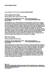
prevention has lagged far behind. Recent studies on CHD still cite that approximately 80-90% of the cases encountered at birth have a multifactorial origin.2 This implies that for the majority no single causative mechanism or agent is known. Rather, a general interaction model of genetic susceptibility and intra-uterine exposures is proposed. A genetic component of CHD was initially implicated by CHD recurrences in families.19 The pattern of genetic inheritance was often not clear, however, as apparently different CHD phenotypes arise within one family and mild defects are sometimes discounted or undiagnosed. Chromosomal anomalies such as Trisomy 21, 18, 13 and the 22q11 deletion are the strongest known contributors to CHD. Moreover, genetic studies in humans and knock-out embryos have identified numerous genes, such as TBX5, NKX2-5, GATA4, CX43, NOTCH1 and VEGF that are responsible for both inherited and sporadic cases of CHD.20 Epidemiological data also point to environmental influences. Currently, identified environmental risk factors for CHD are maternal illnesses such as diabetes mellitus, phenyl ketonuria, obesity and lifestyle factors such as nutrition, exposure to toxins, i.e. nicotine and alcohol, and medication use.21 Research on the role of nutrition in the aetiology of CHD has mainly been focused on maternal use of multivitamin supplements containing folic acid and several epidemiological studies reported a protective effect.22-24 A low dietary intake of vitamin B12 may also be an independent risk factor for CHD.25 The B-vitamins folate and vitamin B12 are important substrates in the homocysteine pathway (Figure 1). Animal studies have demonstrated the involvement of the homocysteine pathway in heart development. In chick embryos, homocysteine was shown to stimulate the proliferation and migration of neural crest cells and exposure to mild hyperhomocysteinaemia resulted in CHD in most of these embryos.18,26 Also in humans, maternal hyperhomocysteinaemia has been associated with a three-to-ten fold increased risk of CHD.16,17,27 Plasma total homocysteine concentrations are maintained within narrow ranges by efficient functioning of several genes encoding enzymes that participate in the homocysteine pathway, such as methylene tetrahydrofolate reductase (MTHFR), methionine synthase reductase (MTRR) and cystathionine β-synthase. Moreover, besides folate and vitamin B12, also vitamin B2 (riboflavin) and B6 (pyridoxine) determine efficient remethylation of homocysteine into methionine because these micronutrients are cofactors or substrates in this pathway (Figure 1). Compromised concentrations of these vitamins result in mild to moderate hyperhomocysteinaemia.
10
General introduction
Chapter
Folate
1
methionine
THF
SAM 5,10-methylene THF
Methyl group
Vitamin B12
NNMT gene SAH
Vitamin B2 5-methyl THF
Vitamin B3
Trans-methylation pathway
Medicine detoxification
Homocysteine cystathionine
Trans-sulfuration pathway
Vitamin B6 cysteine
VEGF gene
Vitamin B6 gluthatione Other anti-oxidants Vitamin E/retinol NADPH
Lipid peroxidation ROOH
NADP+H+
ROH
Vitamin B2
Oxidative pathway
Vitamin B3
Oxidized gluthatione
Figure 1. Homocysteine pathway and oxidative balance. THF, tetrahydrofolate. SAM, s-adenosylmethionine. SAH, s-adenosylhomocysteine. NNMT, nicotinamide N-methyl-transferase. VEGF, vascular endothelial growth factor. NADPH, nicotinamide adenine dinuc-cleotide phosphate-oxidase. ROOH, lipid hydroperoxides.
The relationship between maternal nutrition and fetal development is complex, involving numerous factors that contribute to or confound the effects of nutrition on the immediate-, intermediate- and long-term health of the human fetus. It is now widely accepted that the incidence of cardiovascular disease in adulthood can have its roots in the early stages of fetal development.28 Recently, several studies have suggested that the atherogenic process begins during fetal life, possibly as a result of maternal and fetal hypercholesterolemia, which contributes to the development and progression of atherosclerosis during the perinatal period and increases long-term susceptibility of offspring for adult coronary heart disease.29 It is intriguing to hypothesize that the same
11
Chapter 1
maternal nutritional and metabolic derangements predispose to cardiovascular disease in adult life30 and also affect the vascular structures of both embryonic and placental tissues, resulting in pregnancy complications such as CHD. Hyperhomocysteinaemia, hyperglycaemia and obesity are nice examples of metabolic derangements influencing both adult cardiovascular health and the development of CHD. In addition, also maternal hypercholesterolemia might result in both fetal and adult atherosclerosis and CHD in the offspring. During early embryogenesis, fatty acids play an important role in formation of membrane lipids and are involved as transcription factors in the regulation of gene expression. Oxidation of fatty acids is dependent of vitamin B2 (riboflavin), also cofactor in the homocysteine pathway, and B3 (nicotinamide). Interestingly, maternal use of the western dietary pattern, i.e., high saturated fats and low B-vitamins including riboflavin, increases the risk of orofacial clefts almost two-fold.31 Furthermore, low dietary intakes of riboflavin and nicotinamide have been associated with an increased risk of orofacial clefts and spina bifida. 32, 33 These malformations share similarities in the pathogenesis of CHD, as all three originate from disturbances in neural crest cell behavior by hyperhomocysteinaemia and low folate. Therefore, it is conceivable that these nutritional factors play a role in the aetiology of CHD as well. Hyperhomocysteinaemia is associated with alterations in the transsulfuration pathway resulting in lower levels of the endogenous anti-oxidant glutathione.34 The latter is substantiated by the finding that oxidized glutathione, a marker of oxidative stress, was increased in mothers with CHD affected pregnancies.35 Besides endogenous antioxidants, also exogenous antioxidants such as vitamin E and retinol are necessary to counteract damage from reactive oxygen species. Vitamin E and retinol are essential lipid soluble vitamins and play a role in immune mechanism, growth and cellular differentiation36, while lipids are the building blocks of cell membranes and are involved in gene regulation. Insufficient intake of vitamin E and to a lesser extent retinol might lead to oxidative stress due to a reduced anti-oxidant defense. However, high doses can also act as pro-oxidant. Therefore, low dietary intake of antioxidants may be harmful to the developing embryo, but also excessive intakes may be associated with potential risks. In addition, also an unfavorable lipid surplus due to a high cholesterol diet disturbs the precise oxidative balance by altering lipid levels and lipid peroxidation as well as glutathione levels and antioxidant enzyme activities. Elevated homocysteine concentrations result from inadequate intake of B-vitamins as well as from subtle variations in genes, such as methylene tetrahydrofolate reductase (MTHFR).37 Recently, nicotinamide N-methyltransferase (NNMT) was identified as a new candidate gene for hyperhomocysteinaemia.38 The NNMT enzyme is involved
12
General introduction
in the conversion of S-adenosylmethionine (SAM) to S-adenosylhomocysteine (SAH), intermediates in the homocysteine pathway, and catalyses the N-methylation of nicotinamide and other pyridines. The NNMT enzyme is also important for the detoxification of medicines that undergo methylation via methyltransferases. Since the thalidomide disaster, several other drugs have been recognized as a cardiac teratogen. Maternal intake of isotretinoin, a retinoid, has been shown to cause CHDs in addition to other malformations.39 Also ACE-inhibitors, anticonvulsants, trimethoprim, non-steroid anti-inflammatory drugs and antihistamines are among the drugs that have been suggested to increase the risk of heart anomalies.21 Antihistamines are the first choice medicines for nausea and vomiting during pregnancy, symptoms that affect 60-80% of pregnant women and that primarily occur during the sensitive period of cardiogenesis. Antihistamine medication is widely prescribed to pregnant women although its safety has not been established explicitly and studies reported conflicting results.40,41 Homocysteine is also involved in the regulation of several genes, of which vascular endothelial growth factor-A (VEGF) is one example. Recent data suggest a critical role for VEGF during heart development. VEGF heterozygous knockout mice showed impaired blood vessel formation and died at mid-gestation.42 A key process in the development of heart septa and valves is the formation of the endocardial cushions in the atrioventricular canal and outflow tract. Numerous lines of research have implicated a strict spatio-temporal expression pattern of VEGF in the control of endocardial cushion development.43 In human, VEGF expression shows a marked interindividual variability that is partly determined by single nucleotide polymorphisms (SNPs). First studies suggest associations between the VEGF SNPs -2578 C/A, -1154 G/A and -634 G/C and several CHD phenotypes, i.e., Tetralogy of Fallot, perimembranous ventricular septal defects and valvuloseptal defects.44-46 Besides homocysteine, several other maternal exposures regulate VEGF expression during cardiogenesis such as hyperglycaemia and hypoxia. Despite potential differences with human embryology, chick embryos are a good model to study the influence of environmental exposures on VEGF expression and the development of CHDs and unravel new mechanisms in their aetiology. The stages of heart development in human and chick are largely comparable and well described and the chicken embryo is easy accessible for manipulations without the need to sacrifice the mother animal.
13
Chapter
1
Chapter 1
Objectives of the thesis The main goal of the work presented in this thesis was to unravel the impact of periconceptional maternal lifestyle factors and genetic determinants, partly related to the homocysteine pathway, in the aetiology of CHD offspring. New information may in future contribute to improvement of the periconception and intrauterine environment thereby providing the optimal condition for reproductive performance and prevention of CHD. Specific objectives were: 1.
To assess associations between the maternal lipid status, comprising lipid concentrations in blood, dietary intake of fatty acids, related vitamins, and the risk of CHD in the offspring (Part I).
2.
To study both maternal exposure to medicines, particularly antihistamines for nausea and vomiting, in the first 10 weeks of pregnancy and the interaction between medication use and a new polymorphism in the NNMT gene and the risk of CHD (Part II).
3.
To investigate whether three functional polymorphisms in the VEGF gene are associated with human cardiac outflow tract defects and to study the expression of VEGF and its receptor VEGFR2 in the chicken embryonic heart (Part III).
Chapter Outline This thesis displays the results of the HAVEN study, which is an ongoing case-control family study in the Western part of the Netherlands. This study was designed to investigate environmental and genetic determinants in the pathogenesis and prevention of CHD and was carried out from June 2003 onwards at the Department of Obstetrics and Gynaecology / Division of Obstetrics and Prenatal Medicine of the Erasmus MC, University Medical Center in Rotterdam, The Netherlands. For the recruitment of cases and controls, we collaborated with the Departments of Paediatric Cardiology of the same hospital, Leiden University Medical Centre in Leiden, VU University Medical Centre and Academic Medical Center in Amsterdam and with the child health centres of ‘Careyn’ in the surroundings of Rotterdam. The first part of this thesis describes the maternal nutritional and biochemical lipid status and related vitamins in association with the risk of having a child with
14
General introduction
CHD. Chapter 2 focuses on concentrations in the maternal blood of lipids and total homocysteine because these biomarkers partly reflect the effects of the nutritional intake and lipid metabolism. Fatty acids play a central role in embryonic development, and the B-vitamins riboflavin and nicotinamide are co-enzymes in lipid metabolism. Therefore, in Chapter 3, we explored the association between the maternal dietary intake of fats, riboflavin and nicotinamide, and CHD risk. In Chapter 4, we assessed the role of dietary and supplement intake of the lipid soluble antioxidants vitamin E and retinol as a risk factor for CHD in the offspring. Part II of this thesis addresses the maternal use of medication in the periconception period as a risk factor for CHD. In Chapter 5, we present data on the interactions between a new polymorphism in the nicotinamide N-methyltransferase (NNMT) gene, low maternal dietary nicotinamide intake, and medication use in the periconception period. Chapter 6 shows an analysis of the maternal intake of antihistamines, indicated for nausea and vomiting, during the first trimester of pregnancy. Part III concerns the vascular endothelial growth factor (VEGF) gene, which is essential for endocardial cushion formation. In Chapter 7, we investigated whether three functional polymorphisms in the VEGF gene are associated with cardiac outflow tract defects. In Chapter 8, we present results on the expression of VEGF and VEGFR2 in the chick embryonic heart by using the new Optical Projection Tomography (OPT) technique to produce high-resolution 3-dimensional images. Chapter 9 provides a general discussion on the combined results of the studies in a broader perspective. We recommend future research and elaborate on the implications regarding preconception care.
15
Chapter
1
Chapter 1
References 1.
Department of Medical genetics, University Medical Centre Groningen, Groningen, the Netherlands. European Registration Of Congenital Anomalies and Twins (EUROCAT) 1981 - 2004; [www.eurocatnederland.nl] Accessed February 25 2007.
2.
Botto LD, Correa A. Decreasing the burden of congenital heart anomalies: an epidemiologic evaluation of risk factors and survival. Prog Pediatr Cardiol 2003;18:111-21.
3.
Hoffman JI. Incidence of congenital heart disease: II. Prenatal incidence. Pediatr Cardiol 1995;16:155-65.
4.
Anderson RN, Smith BL. Deaths: leading causes for 2002. Natl Vital Stat Rep 2005;53:1-89.
5.
Rosano A, Botto LD, Botting B, Mastroiacovo P. Infant mortality and congenital anomalies from 1950 to 1994: an international perspective. J Epidemiol Community Health 2000;54:660-6.
6.
Lawoko S, Soares JJ. Quality of life among parents of children with congenital heart disease, parents of children with other diseases and parents of healthy children. Qual Life Res 2003;12:65566.
7.
Economic costs of birth defects and cerebral palsy--United States, 1992. MMWR Morb Mortal Wkly Rep 1995;44:694-9.
8.
Boneva RS, Botto LD, Moore CA, Yang Q, Correa A, Erickson JD. Mortality associated with congenital heart defects in the United States: trends and racial disparities, 1979-1997. Circulation 2001;103:2376-81.
9.
Larsen W. Development of the heart. In: Schmitt WR, Otway M, Bowman-Schulman E, editors. Human embryology. Churchill Livingstone, New York, USA , 1993:131-165.
10.
Markwald RR, Fitzharris TP, Manasek FJ. Structural development of endocardial cushions. Am J Anat 1977;148:85-119.
11.
Markwald R, Eisenberg C, Eisenberg L, Trusk T, Sugi Y. Epithelial-mesenchymal transformations in early avian heart development. Acta Anat (Basel) 1996;156:173-86.
12.
Anderson RH, Webb S, Brown NA, Lamers W, Moorman A. Development of the heart: (3) formation of the ventricular outflow tracts, arterial valves, and intrapericardial arterial trunks. Heart 2003;89:1110-8.
13.
Kirby ML, Gale TF, Stewart DE. Neural crest cells contribute to normal aorticopulmonary septation. Science 1983;220:1059-61.
14.
Bartelings MM, Wenink AC, Gittenberger-De Groot AC, Oppenheimer-Dekker A. Contribution of the aortopulmonary septum to the muscular outlet septum in the human heart. Acta Morphol Neerl Scand 1986;24:181-92.
15.
Gittenberger-de Groot AC, Bartelings MM, Deruiter MC, Poelmann RE. Basics of cardiac development for the understanding of congenital heart malformations. Pediatr Res 2005;57:16976.
16.
Hobbs CA, Cleves MA, Melnyk S, Zhao W, James SJ. Congenital heart defects and abnormal maternal biomarkers of methionine and homocysteine metabolism. Am J Clin Nutr 2005;81:14753.
17.
Botto LD, Mulinare J, Erickson JD. Do multivitamin or folic acid supplements reduce the risk for congenital heart defects? Evidence and gaps. Am J Med Genet A 2003;121:95-101.
18.
Boot MJ, Steegers-Theunissen RP, Poelmann RE, van Iperen L, Gittenberger-de Groot AC. Cardiac outflow tract malformations in chick embryos exposed to homocysteine. Cardiovasc Res 2004;64:365-73.
19.
Nora JJ. Causes of congenital heart diseases: old and new modes, mechanisms, and models. Am Heart J 1993;125:1409-19.
20.
Bruneau BG. The developmental genetics of congenital heart disease. Nature 2008;451:943-8.
16
General introduction
21.
Jenkins KJ, Correa A, Feinstein JA, et al. Noninherited risk factors and congenital cardiovascular defects: current knowledge: a scientific statement from the American Heart Association Council on Cardiovascular Disease in the Young: endorsed by the American Academy of Pediatrics. Circulation 2007;115:2995-3014.
22.
Shaw GM, O’Malley CD, Wasserman CR, Tolarova MM, Lammer EJ. Maternal periconceptional use of multivitamins and reduced risk for conotruncal heart defects and limb deficiencies among offspring. Am J Med Genet 1995;59:536-45.
23.
Botto LD, Khoury MJ, Mulinare J, Erickson JD. Periconceptional multivitamin use and the occurrence of conotruncal heart defects: results from a population-based, case-control study. Pediatrics 1996;98:911-7.
24.
Czeizel AE. Periconceptional folic acid containing multivitamin supplementation. Eur J Obstet Gynecol Reprod Biol 1998;78:151-61.
25.
Verkleij-Hagoort AC, de Vries JH, Ursem NT, de Jonge R, Hop WC, Steegers-Theunissen RP. Dietary intake of B-vitamins in mothers born a child with a congenital heart defect. Eur J Nutr 2006;45:478-86.
26.
Boot MJ, Steegers-Theunissen RP, Poelmann RE, Van Iperen L, Lindemans J, Gittenberger-de Groot AC. Folic acid and homocysteine affect neural crest and neuroepithelial cell outgrowth and differentiation in vitro. Dev Dyn 2003;227:301-8.
27.
Verkleij-Hagoort AC, Verlinde M, Ursem NT, et al. Maternal hyperhomocysteinaemia is a risk factor for congenital heart disease. BJOG 2006;113:1412-8.
28.
Tappia PS, Gabriel CA. Role of nutrition in the development of the fetal cardiovascular system. Expert Rev Cardiovasc Ther 2006;4:211-25.
29.
Al MD, van Houwelingen AC, Hornstra G. Long-chain polyunsaturated fatty acids, pregnancy, and pregnancy outcome. Am J Clin Nutr 2000;71:285S-91S.
30.
Steegers-Theunissen RP, Steegers EA. Nutrient-gene interactions in early pregnancy: a vascular hypothesis. Eur J Obstet Gynecol Reprod Biol 2003;106:115-7.
31.
Vujkovic M, Ocke MC, van der Spek PJ, Yazdanpanah N, Steegers EA, Steegers-Theunissen RP. Maternal Western dietary patterns and the risk of developing a cleft lip with or without a cleft palate. Obstet Gynecol 2007;110:378-84.
32.
Groenen PM, van Rooij IA, Peer PG, Ocke MC, Zielhuis GA, Steegers-Theunissen RP. Low maternal dietary intakes of iron, magnesium, and niacin are associated with spina bifida in the offspring. J Nutr 2004;134:1516-22.
33.
Krapels IP, van Rooij IA, Ocke MC, van Cleef BA, Kuijpers-Jagtman AM, Steegers-Theunissen RP. Maternal dietary B vitamin intake, other than folate, and the association with orofacial cleft in the offspring. Eur J Nutr 2004;43:7-14.
34.
Huang RF, Hsu YC, Lin HL, Yang FL. Folate depletion and elevated plasma homocysteine promote oxidative stress in rat livers. J Nutr 2001;131:33-8.
35.
Hobbs CA, Cleves MA, Zhao W, Melnyk S, James SJ. Congenital heart defects and maternal biomarkers of oxidative stress. Am J Clin Nutr 2005;82:598-604.
36.
Debier C, Larondelle Y. Vitamins A and E: metabolism, roles and transfer to offspring. Br J Nutr 2005;93:153-74.
37.
Hobbs CA, James SJ, Parsian A, et al. Congenital heart defects and genetic variants in the methylenetetrahydroflate reductase gene. J Med Genet 2006;43:162-6.
38.
Souto JC, Blanco-Vaca F, Soria JM, et al. A genomewide exploration suggests a new candidate gene at chromosome 11q23 as the major determinant of plasma homocysteine levels: results from the GAIT project. Am J Hum Genet 2005;76:925-33.
39.
Rothman KJ, Moore LL, Singer MR, Nguyen US, Mannino S, Milunsky A. Teratogenicity of high vitamin A intake. N Engl J Med 1995;333:1369-73.
17
Chapter
1
Chapter 1
40.
Queisser-Luft A, Eggers I, Stolz G, Kieninger-Baum D, Schlaefer K. Serial examination of 20,248 newborn fetuses and infants: correlations between drug exposure and major malformations. Am J Med Genet 1996;63:268-76.
41.
Seto A, Einarson T, Koren G. Pregnancy outcome following first trimester exposure to antihistamines: meta-analysis. Am J Perinatol 1997;14:119-24.
42.
Carmeliet P, Ferreira V, Breier G, et al. Abnormal blood vessel development and lethality in embryos lacking a single VEGF allele. Nature 1996;380:435-9.
43.
Armstrong EJ, Bischoff J. Heart valve development: endothelial cell signaling and differentiation. Circ Res 2004;95:459-70.
44.
Lambrechts D, Devriendt K, Driscoll DA, et al. Low expression VEGF haplotype increases the risk for tetralogy of Fallot: a family based association study. J Med Genet 2005;42:519-22.
45.
Xie J, Yi L, Xu ZF, et al. VEGF C-634G polymorphism is associated with protection from isolated ventricular septal defect: case-control and TDT studies. Eur J Hum Genet 2007;15:1246-51.
46.
Vannay A, Vasarhelyi B, Kornyei M, et al. Single-nucleotide polymorphisms of VEGF gene are associated with risk of congenital valvuloseptal heart defects. Am Heart J 2006;151:878-81.
18
Chapter Part2I A derangement of the maternal lipid profile Maternal lipidsrisk and is associated with an elevated of related vitamins congenital heart disease in the offspring
H.P.M. Smedts E.M. van Uitert O.Valkenburg J.S.E. Laven M.J.C. Eijkemans J.Lindemans E.A.P. Steegers R.P.M. Steegers-Theunissen Nutr Metab Cardiovasc Dis; in press
Chapter 2 A derangement of the maternal lipid profile is associated with an elevated risk of congenital heart disease in the offspring
H.P.M. Smedts E.M. van Uitert O. Valkenburg J.S.E. Laven M.J.C. Eijkemans J. Lindemans E.A.P. Steegers R.P.M. Steegers-Theunissen Nutr Metab Cardiovasc Dis; in press
Chapter 2
Abstract Background and aims: Maternal hyperglycaemia and hyperhomocysteinaemia are risk factors for congenital heart disease (CHD). Metabolic derangements and deranged lipid levels are associated with adult cardiovascular disease. We examined whether maternal lipid levels are associated with the risk of CHD offspring. Methods and Results: From 2003 onwards, a case-control study was conducted. Participants were mothers of children with (n=261) and without (n=325) CHD. At around 16 months after the index-pregnancy maternal lipid levels were determined. Maternal characteristics and lipid levels were compared by Student’s T-test. In a multivariable logistic regression model, risk estimates were calculated for associations between CHD and lipid levels. Adjustments were made for maternal age, diabetes, ethnicity, BMI, parity, periconception folic acid use and total homocysteine levels. Outcome measures are presented in (geometric) means (P5-P95) and odds ratios (OR) with 95% confidence intervals (CI). Case mothers showed higher cholesterol (4.9 vs. 4.7 mmol/L, P5.0
Cut-off value
1.6 (1.02-2.4)
1.6 (1.2-2.3)
1.8 (1.3-2.5)
0.9 (0.7-1.3)
1.8 (1.2-2.5)
1.5 (1.05-2.1)
1.0 (0.7-1.4)
0.8 (0.5-1.1)
1.5 (1.02-2.1)
OR (95%CI)
1Adjusted
1.6 (1.1-2.5)
1.7 (1.2-2.5)
1.9 (1.3-2.7)
1.0 (0.7-1.4)
1.9 (1.3-2.7)
1.6 (1.1-2.3)
0.9 (0.6-1.3)
0.8 (0.5-1.1)
1.5 (1.05-2.2)
OR (95%CI)1
-
1.7 (1.1-2.5)
1.8 (1.2-2.6)
0.9 (0.6-1.3)
1.8 (1.2-2.7)
1.5 (1.02-2.2)
0.9 (0.6-1.3)
0.7 (0.5-1.1)
1.4 (0.9-2.0)
OR (95%CI)2
-
0.01
0.004
0.43
0.003
0.04
0.47
0.13
0.11
P-value2
OR, odds ratio. CI, confidence interval. Cut-off values are upper or lower tertiles based on control mothers. for maternal age, parity, BMI, ethnicity, family history of CHD, periconception smoking, folic acid and/or (multi)vitamin use and diabetes. 2Adjusted for all before mentioned covariates and total homocysteine. 3natural log transformed values. 4Upper quintile based on control mothers.
59 / 57
n=210/284
122 / 108 117 / 108
Apolipoprotein B, mg/dL
Homocysteine, μmol/L3,4
83 / 108
n=258/322
104 / 107
Apolipoprotein A-1, mg/dL
Total / HDL
LDL-cholesterol, mmol/L3 cholesterol3
74 / 107 97 / 107
HDL-cholesterol, mmol/L
102 / 107
n=229/320 64 / 107
mmol/L3
Cases/controls
Triglycerides, mmol/L3
Total cholesterol,
Biomarkers
Table 3. Associations between CHD risk and maternal lipid and total homocysteine concentrations with and without adjustment for potential confounders
Maternal lipid profile
Chapter
2
29
Chapter 2
Table 4. Maternal lipid profiles in association with adjusted risks of CHD stratified for periconception smoking Cases/
Cut-off
Periconception
Periconception
controls
value
smoking yes
smoking no
n=43/72
n=186/248
Total cholesterol, mmol/L1
>5.0
1.0 (0.4-2.4)
1.5 (0.96-2.3)
Triglycerides, mmol/L1
>1.14
0.7 (0.3-1.7)
0.8 (0.5-1.2)
HDL-cholesterol, mmol/L
3.3
1.7 (0.7-4.0)
1.5 (0.96-2.3)
>3.5
2.8 (1.1-6.9)
1.7 (1.1-2.6)
n=49/72
n=209/250
Apolipoprotein A-1, mg/dL
85.0
2.4 (1.0-5.9)
1.6 (1.1-2.5)
>0.6
3.4 (1.4-8.4)
1.4 (0.9-2.2)
n=42/62
n=168/222
0.7 (0.2-2.6)
1.6 (0.8-3.1)
Biomarkers
n=229/320
LDL-cholesterol,
mmol/L1
Total / HDL cholesterol1 n=258/322
Apolipoprotein B/ A-1 n=210/284 Homocysteine,
μmol/L1,2
>12.7
OR, odds ratio. CI, confidence interval. Cut-off values are upper or lower tertiles based on control mothers. 1natural log transformed values. 2Upper quintile based on control mothers. Odds ratios are adjusted for maternal age, parity, BMI, ethnicity, family history of CHD, periconception folic acid and/or (multi)vitamin use and diabetes.
30
n=258/322
1.2 (1.01-1.5)
1.3 (1.1-1.6)
1.3 (1.1-1.5)
0.9 (0.8-1.1)
1.4 (1.02-1.9)
1.3 (1.1-1.5)
1.0 (0.8-1.2)
1.0 (0.8-1.2)
1.2 (1.03-1.5)
OR(95%CI)1
-
1.3 (1.04-1.6)
1.3 (1.1-1.6)
1.0 (0.8-1.2)
1.3 (0.95-1.8)
1.3 (1.1-1.6)
1.0 (0.8-1.2)
1.0 (0.8-1.2)
1.2 (1.03-1.5)
OR(95%CI)2
-
0.02
0.01
0.73
0.12
0.01
0.92
0.63
0.02
P-value 2
Odds ratio (OR) with confidence interval (CI) of CHD risk per standard deviation (SD) increase in lipids and homocysteine. One SD total cholesterol = 0.88, triglycerides = 0.47, HDL-cholesterol = 0.36, LDL-cholesterol = 0.82, total cholesterol / HDL-cholesterol = 1.64, apolipoprotein A = 21.7, apolipoprotein B = 17.5, apolipoprotein B / A-1 = 0.14, total homocysteine = 4.2. 1Adjusted for maternal age, parity, BMI, ethnicity, family history of CHD, periconception smoking, folic acid and/or (multi)vitamin use and diabetes. 2Adjusted for all before mentioned covariates and total homocysteine.
1.2 (1.01-1.)
1.3 (1.1-1.5)
Apolipoprotein B/ A-1 n=210/284
1.3 (1.1-1.5)
Apolipoprotein B
Total homocysteine
0.9 (0.8-1.1)
1.3 (1.01-1.8)
Total/ HDL cholesterol
Apolipoprotein A-1
1.0 (0.8-1.1) 1.2 (1.05-1.5)
LDL-cholesterol
1.0 (0.8-1.2)
HDL-cholesterol
1.2 (1.01-1.4)
Triglycerides
n=229/320
OR (95%CI)
Total cholesterol
Biomarkers
Cases/controls
Table 5. Associations between maternal lipids and total homocysteine as continuous variables and CHD risk, adjusted for potential confounders
Maternal lipid profile
Chapter
2
31
32
Odds ratio (95% Confidence Interval)
>5.0 >3.3 >85.0 mmol/L mmol/L mg/dL
-
ApoA-I >3.3 mmol/L mmol/L
>85.0 mg/dL
25 ≤ BMI ≤ 30
ApoA-I
>85.0 mg/dL
BMI > 30 UB= UB= UB= 8.9 8.9 8.8
>3.3 mmol/L mmol/L
UB= UB= 5.1 5.1
-
UB= 5.3
ApoA-I
UB= 9.1
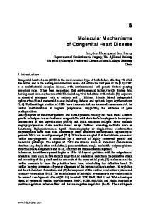
CHD, periconception smoking, folic acid and/or (multi)vitamin use and diabetes.
were based on the upper tertiles based on the control mothers. Odds ratios adjusted for maternal age, parity, BMI, ethnicity, family history of
C, total cholesterol. CI, confidence interval. HDL-C, HDL-cholesterol. LDL-C, LDL-cholesterol. OR, odds ratio. UB, upper bound. Cut-off values
Figure 1. Maternal lipid profiles in association with adjusted risks of CHD stratified for BMI. ApoA-I, apolipoprotein A-I. ApoB, apolipoprotein B.
0
0,5
1
1,5
2
2,5
3
3,5
4
BMI 120 mg/dL, 3.4% of the case and 1.5% of the control mothers are at risk for cardiovascular disease. Only 4% of case mothers and 3% of control mothers have total cholesterol levels above 6.5 mmol/L. LDL-cholesterol levels are >4.2 mmol/L in 9% of case mothers and 12% of control mothers. Data on embryonic exposure to maternal lipids are not available. It is known, however, that fetal aortas contain more and larger fatty streak lesions from mothers with total cholesterol >5.0 mmol/l compared with normocholesterolaemic mothers.29 If the same situation is present during cardiogenesis this may suggest that only a minimal increase in lipoprotein levels might disturb normal heart development. The reference values for adult cardiovascular disease risk or congenital heart disease may therefore be different. Furthermore, genetic variations contribute to variation in lipoprotein levels and may increase the susceptibility for cardiovascular disease, even when levels are slightly increased.30 A direct effect of an unfavorable maternal lipid profile on embryonic heart development is biologically plausible. Dependent on the genetic background of the embryo, exposure to a slightly deranged maternal lipid profile may have differential effects on genes and tissues, i.e., neural crest, involved in cardiogenesis. Although the embryo can synthesize cholesterol, the majority of cholesterol is derived from the mother via the yolk sac and embryonic coelomic and amniotic fluids. This is substantiated by studies in mice.31 As apolipoprotein B is abundantly expressed in the yolk sac of rodents32
34
Maternal lipid profile
and humans33, it is likely that apolipoprotein B – containing lipoproteins participate in lipid transport to the embryo. In mice, a maternal diet high in saturated fats resulted in dyslipidemia in the offspring, programmed by suppression of hepatic expression of LDLreceptor mRNA.34 Also in a mouse model, a maternal high fat diet primed offspring fatty liver disease, mediated through impaired mitochondrial metabolism and up-regulated hepatic lipogenesis, inflammatory pathways and oxidative stress.35 It is possible that these changes are already present during cardiogenesis. In our study, oxidative stress may be increased in mothers of a child with CHD as total homocysteine, sometimes used as biomarker of oxidative stress35, was higher in this group. This is supported by Hobbs et al. demonstrating that biomarkers of oxidative stress involved in the transsulfuration pathway were higher in mothers of a child with CHD than in controls.37 Cell machinery crucial for normal cardiogenesis can be disturbed via complex mechanisms, including oxidation sensitive signalling pathways regulating gene expression and translation. For example, oxidized LDL-cholesterol modulates the expression of genes involved in cell differentiation and proliferation regulated by nuclear factor kappa B and influences the expression of apoptotic factors activated through Fas and TNF receptors and c-Mycdependent transcription factors.38,39 Moreover, oxidized LDL regulates the expression of vascular endothelial growth factor, a crucial regulator of endocardial cushion formation.40 Thus, a window of vulnerability may exist during which slightly elevated maternal lipid levels in combination with excessive oxidative stress in the mother and embryo may result in disturbed cardiogenesis. Here, we show that mildly elevated maternal cholesterol, LDL-cholesterol and apolipoprotein B are significantly associated with an almost two-fold increased risk of CHD offspring. Studies on subtle genetic variations in lipid pathways and prospective studies are needed to show the causality of these associations, and in specific CHD phenotypes. Moreover, relationships between mildly elevated maternal lipid levels in the reproductive period and the development of cardiovascular disease in later life warrant further investigation.
35
Chapter
2
Chapter 2
References 1.
March of Dimes Birth Defects Foundation. Global report on birth defects. The hidden toll of dying and disabled children. New York, USA: White Plains; 2006:28.
2.
Botto LD, Correa A, Erickson JD. Racial and temporal variations in the prevalence of heart defects. Pediatrics 2001;107:E32.
3.
Ray JG, O’Brien TE, Chan WS. Preconception care and the risk of congenital anomalies in the offspring of women with diabetes mellitus: a meta-analysis. QJM 2001;94:435-44.
4.
Gilboa SM, Correa A, Botto LD, et al. National Birth Defects Prevention Study. Association between prepregnancy body mass index and congenital heart defects. AJOG 2010;202:51.e1-51.e10.
5.
Verkleij-Hagoort AC, Verlinde M, Ursem NT, et al. Maternal hyperhomocysteinaemia is a risk factor for congenital heart disease. BJOG 2006;113:1412-8.
6.
Hobbs CA, Malik S, Zhao W, James SJ, Melnyk S, Cleves MA. Maternal homocysteine and congenital heart defects. JACC 2006;47:683-5.
7.
Huxley R, Barzi F, Woodward M. Excess risk of fatal coronary heart disease associated with diabetes in men and women: meta-analysis of 37 prospective cohort studies. BMJ (Clinical research ed.) 2006;332:73-8.
8.
Boushey CJ, Beresford SA, Omenn GS, Motulsky AG. A quantitative assessment of plasma homocysteine as a risk factor for vascular disease. Probable benefits of increasing folic acid intakes. JAMA 1995;274:1049-57.
9.
Expert Panel on Detection Evaluation and Treatment of High Blood Cholesterol in Adults. Executive Summary of The Third Report of The National Cholesterol Education Program (NCEP) Expert Panel on Detection, Evaluation, And Treatment of High Blood Cholesterol In Adults (Adult Treatment Panel III). JAMA 2001;285:2486-97.
10.
Steegers-Theunissen RP, Steegers EA. Nutrient-gene interactions in early pregnancy: a vascular hypothesis. Eur J Obstet Gynecol Reprod Biol 2003;106:115-7.
11.
Khan IY, Dekou V, Douglas G, et al. A high-fat diet during rat-pregnancy of suckling induces cardiovascular dysfunction in adult offspring. Am J Physiol Regul Integr Comp Physiol 2005;288:R127-33.
12.
Leeson CP, Kattenhorn M, Morley R, Lucas A, Deanfield JE. Impact of low birth weight and cardiovascular risk factors on endothelial function in early adult life. Circulation 2001;103:1264-8.
13.
Innis SM. Fatty acids and early human development. Early Hum Dev 2007;83:761-6.
14.
Devine CM, Bove CF, Olson CM. Continuity and change in women’s weight orientations and lifestyle practices through pregnancy and the postpartum period: the influence of life course trajectories and transitional events. Soc Sci Med 2000;50:567-82.
15.
Willet W. Nature of variation in Diet. In: Willet W, ed. Nutritional Epidemiology. New York, NY: Oxford University Press;1998:33-50.
16.
Van Driel LMJW, Zwolle LJH, de Vries JHM, et al. The maternal nutritional status at one year after delivery is comparable with the preconception period. Reproductive Sciences 2010; 16 (supplement): 239A. Abstract.
17.
Potter JM, Nestel PJ. The hyperlipidemia of pregnancy in normal and complicated pregnancies. Am J Obstet Gynecol 1979;133:165-70.
18.
Lewis CE, Funkhouser E, Raczynski JM, Sidney S, Bild DE, Howard BV. Adverse effect of Pregnancy on High Density Lipoprotein (HDL) Cholesterol in Young Adult Women. Am J Epid 1996;144:24457.
19.
Larsson A, Palm M, Hansson LO, Axelsson O. Reference values for clinical chemistry tests during normal pregnancy. BJOG 2008;115:874-81.
36
Maternal lipid profile
20.
Smedts HP, Rakhshandehroo M, Verkleij-Hagoort AC, et al.. Maternal intake of fat, riboflavin and nicotinamide and the risk of having offspring with congenital heart defects. Eur J Nutr 2008;47:35765.
21.
Rosenquist TH, Ratashak SA, Selhub J. Homocysteine induces congenital defects of the heart and neural tube: effect of folic acid. Proc Natl Acad Sci U S A 1996; 93:15227-32.
22.
Botto LD, Mulinare J, Erickson JD. Do multivitamin or folic acid supplements reduce the risk for congenital heart defects? Evidence and gaps. Am J Med Genet 2003;121:95–101.
23.
Boot MJ, Steegers-Theunissen RP, Poelmann RE, van Iperen L, Gittenberger-de Groot AC. Cardiac outflow tract malformations in chick embryos exposed to homocysteine. Cardiovasc Res 2004;64:365–73.
24.
Valkenburg O, Steegers-Theunissen RP, Smedts HP, et al. A more atherogenic serum lipoprotein profile is present in women with polycystic ovary syndrome: a case-control study. J Clin Endocrinol Metab 2008;93:470-6.
25.
Statistics Netherlands. Classification of educational level and ethnicity. Voorburg/Heerlen, the Netherlands. 2005. http://www.cbs.nl/en-GB/meny/methoden/methoden-per-thema/default.thm. Accessed on July 10, 2007.
26.
Walldius G, Jungner I. Apolipoprotein B and apolipoprotein A-I: risk indicators of coronary heart disease and targets for lipid-modifying therapy. J Intern Med 2004;255:188-205.
27.
Sattar N, Greer IA. Pregnancy complications and maternal cardiovascular risk: opportunities for intervention and screening? BMJ 2002;325:157-60.
28.
Germain AM, Romanik MC, Guerra I, et al. Endothelial dysfunction: a link among preeclampsia, recurrent pregnancy loss, and future cardiovascular events? Hypertension 2007;49:90-5.
29.
Napoli C, D’Armiento FP, Mancini FP, et al. Fatty streak formation occurs in human fetal aortas and is greatly enhanced by maternal hypercholesterolemia. Intimal accumulation of low density lipoprotein and its oxidation precede monocyte recruitment into early atherosclerotic lesions. J Clin Invest 1997;100:2680-90.
30.
Benn M. Apolipoprotein B levels, APOB alleles, and risk of ischemic cardiovascular disease in the general population, a review. Atherosclerosis. 2009;206:17-30.
31.
Yoshida S, Wada Y. Transfer of maternal cholesterol to embryo and fetus in pregnant mice. J Lipid Res 2005;46:2168-74.
32.
Demmer LA, Levin MS, Elovson J, Reuben MA, Lusis AJ, Gordon JI. Tissue-specific expression and developmental regulation of the rat apolipoprotein B gene. Proc Natl Acad Sci U S A 1986;83:8102-6.
33.
Hopkins B, Sharpe CR, Baralle FE, Graham CF. Organ distribution of apolipoprotein gene transcripts in 6-12 week postfertilization human embryos. J Embryol Exp Morphol 1986;97:177-87.
34.
Chechi K, McGuire JJ, Cheema SK. Developmental programming of lipid metabolism and aortic vascular function in C57BL/6 mice: a novel study suggesting an involvement of LDL-receptor. Am J Physiol Regul Integr Comp Physiol 2009;296:1029-40.
35.
Bruce KD, Cagampang FR, Argenton M, et al. Maternal high-fat feeding primes steatohepatitis in adult mice offspring, involving mitochondrial dysfunction and altered lipogenesis gene expression. Hepatology 2009;50:1796-808.
36.
Sibrian-Vazquez M, Escobedo JO, Lim S, Samoei GK, Strongin RM. Homocystamides promote free-radical and oxidative damage to proteins. Proc Natl Acad Sci U S A 2010;107:551-4.
37.
Hobbs CA, Cleves MA, Zhao W, Melnyk S, James SJ. Congenital heart defects and maternal biomarkers of oxidative stress. Am J Clin Nutr 2005;82:598-604.
38.
Napoli C, Quehenberger O, De Nigris F, Abete P, Glass CK, Palinski W. Mildly oxidized low density lipoprotein activates multiple apoptotic signaling pathways in human coronary cells. Faseb J 2000;14:1996-2007.
37
Chapter
2
Chapter 2
39.
de Nigris F, Youssef T, Ciafré S, et al. Evidence for oxidative activation of c-Myc-dependent nuclear signaling in human coronary smooth muscle cells and in early lesions of Watanabe heritable hyperlipidemic rabbits: protective effects of vitamin E. Circulation 2000;102:2111-7.
40.
Ramos MA, Kuzuya M, Esaki T, et al. Induction of macrophage VEGF in response to oxidized LDL and VEGF accumulation in human atherosclerotic lesions. Arterioscler Thromb Vasc Biol 1998;18:1188-96.
38
Chapter 3 Maternal intake of fat, riboflavin and nicotinamide and the risk of having offspring with congenital heart defects
H.P.M. Smedts M. Rakhshandehroo A.C. Verkleij-Hagoort J.H.M. de Vries J. Ottenkamp E.A.P. Steegers R.P.M. Steegers-Theunissen Eur J Nutr 2008;47:357-65
Chapter 3
Abstract Background: With the exception of studies on folic acid, little evidence is available concerning other nutrients in the pathogenesis of congenital heart defects (CHDs). Fatty acids play a central role in embryonic development, and the B-vitamins riboflavin and nicotinamide are co-enzymes in lipid metabolism. Aim of the study: To investigate associations between the maternal dietary intake of fats, riboflavin and nicotinamide, and CHD risk in the offspring. Methods: A case-control family study was conducted in 276 mothers of a child with a CHD comprising of 190 outflow tract defects (OTD) and 86 non-outflow tract defects (non-OTD) and 324 control mothers of a non-malformed child. Mothers filled out general and food frequency questionnaires at 16 months after the index-pregnancy, as a proxy of the habitual food intake in the preconception period. Nutrient intakes (medians) were compared between cases and controls by Mann-Whitney U-test. Odds ratios (OR) for the association between CHDs and nutrient intakes were estimated in a logistic regression model. Results: Case mothers, in particular mothers of a child with OTD, had higher dietary intakes of saturated fat, 30.9 vs 29.8 g/d; PA (r2=0.52); between VEGF -2578 C>A and -634 G>C (r2=0.49); and between VEGF
119
Chapter
7
Chapter 7
-1154 G>A and -634 G>C (r2=0.26). The distributions of the VEGF alleles and genotypes are presented in Table 2. None of the genotype frequencies in the control population deviated from Hardy-Weinberg equilibrium. The genotype distributions of VEGF -2578 C>A, -1154 G>A and -634 G>C were not significantly different between the children with ECD and controls. The VEGF -2578 C and -1154 G alleles were more frequently present, albeit not significantly, in children with ECD than in controls. The distribution of the genotypes stratified per ECD phenotype is summarized in Table 3. In children with pulmonary valve stenosis the distributions of the -2578 C>A variants deviated from Hardy-Weinberg equilibrium and in these children the C-allele tended to be more frequently present than in controls (P=0.07). We inferred haplotypes from the three VEGF polymorphisms and compared the frequencies. Four of the eight possible haplotypes occurred at an appreciable frequency in both children with ECD and controls, i.e., AAG, CGC, CGG and AGG whereby each letter refers to the allele of the -2578, -1154, and -634 SNP, respectively (Table 4). Consistent with the genotype distributions, the haplotype analyses revealed that the CGG haplotype was present in 17% of children with ECD and in 13% of control children (P=0.08). The AAG haplotype was associated with a reduced risk of the ECD phenotype (OR 0.8, 95% CI 0.6-1.0). The results of the FBAT analyses are depicted in Table 5 and point out that the -2578C and -1154G alleles were transmitted to children with an ECD more frequently than expected by Mendelian inheritance (59% of all transmissions). FBAT-o, which also incorporates information from the control triads, revealed even stronger evidence for over transmission of the -2578 C allele (P=0.003) and the -1154 G allele (P=0.002) to offspring with the ECD phenotype. These P-values survived the rather conservative Bonferroni correction for multiple testing of 5x3 independent test with α=0.0033. Stratification for five separate ECD phenotypes revealed that the -2578 C allele (P=0.012) and the -1154 G allele (P=0.006) were significantly overtransmitted to children with pVSD. Consistently, the HBAT-o results suggested that the CGC haplotype was overtransmitted to children with ECD (P=0.057) and the AAG haplotype was significantly less frequently transmitted than expected to children with ECD (41% of transmissions, which was significant at an Bonferroni α-level of 0.0025) (Table 6), in particular to children with pVSD (P=0.022). Empirical P-values obtained by permutation analyses (HBAT-p) substantiated these associations. The global P-value with four degrees of freedom for differences in the transmission of the four haplotypes to children with ECD was P=0.03. We did not find any evidence for a parent-of-origin effect of risk alleles in the VEGF candidate loci (data not shown).
120
VEGF gene polymorphisms
Table 1. General characteristics of the case children with ECD and control children ECD
Controls
n=190
n=317
16.2 (15.1-19.2)
16.1 (15.1-18.0)
96 (51)
170 (54)
18 (10)
17(5)
177 (93)
289 (91)
13 (7)
28 (9)
Child Age at the study moment in months, median (interquartile range) Boys, n (%) Family history for Ethnicity2,
CHD1,
n (%)
n (%)
Dutch Natives European Others 1Any
ECD, outflow tract defect. CHD, congenital heart disease. congenital heart disease of family members in the first, second or third degree. 2Children were classified as Dutch natives when both parents and grandparents are born in the Netherlands or one of the parents is born in another country, but both grandparents are born in the Netherlands. If one of the parents or grandparents is born in a European country, or is from European origin and living in the USA, Australia or Indonesia we classified the child in the category European Others.
Table 2. Distribution of the VEGF genotypes in case children with ECD and control children VEGF SNPs
ECD
Controls
n=185
n=312
Χ2
Fisher’s P
OR (95% CI)
2.52
0.28
0.8 (0.6-1.1)
-2578, AA
44 (24)
88 (28)
CA
88 (48)
153 (49)
0.8 (0.5-1.2)
CC
53 (29)
71 (23)
1.0 (Reference)
-
0.77
194 (52)
295 (47)
n=187
n=307
-1154, AA
18 (10)
43 (14)
GA
79 (42)
130 (42)
0.9 (0.6-1.3)
GG
90 (48)
134 (44)
1.0 (Reference)
HWE p-value C-allele
HWE p-value
2.38
0.12 Chapter
2.32
0.31
-
0.20
G-allele
259 (69)
398 (65)
n=184
n=303
-634, CC
22 (12)
39 (13)
GC
85 (46)
133 (44)
1.1 (0.7-1.6)
GG
77 (42)
131 (43)
1.0 (Reference)
HWE p-value G-allele
-
0.57
239 (65)
395 (65)
1.55
0.21
0.26
0.88
0.00
7
0.8 (0.6-1.1)
1.0 (0.7-1.3)
1.00
Values are numbers (percentage). OR, odds ratio. CI, confidence interval. ECD, outflow tract defect. HWE, Hardy-Weinberg Equilibrium.
121
Chapter 7
Table 3. Distribution of the VEGF genotypes in case children with ECD, stratified for ECD phenotypes, and control children VEGF SNPs
ECD pVSD
PS
TOF
Controls AVSD
AoS
n=79
n=42
n=29
n=29
n=6
n=312
-2578, AA
15 (19)
12 (29)
6 (21)
9 (31)
2 (33)
88 (28)
CA
44 (56)
14 (33)
16 (55)
11 (38)
3 (50)
153 (49)
CC
20 (25)
16 (38)
7 (24)
9 (31)
1 (17)
71 (23)
0.25
0.07
0.68
0.47
0.93
Χ2; P-value1 HWE p-value C-allele Χ2; P-value2
-
-
-
-
-
0.77
84 (53)
46 (55)
30 (52)
29 (50)
5 (42)
295 (47)
0.16
0.18
0.49
0.65
0.72
n=79
n=43
n=29
n=30
n=6
n=307
-1154, AA
6 (8)
6 (14)
2 (7)
3 (10)
1 (17)
43 (14)
GA
36 (46)
13 (30)
11 (38)
15 (50)
4 (67)
130 (42)
GG
37 (47)
24 (56)
16 (55)
12 (40)
1 (17)
134 (44)
0.31
0.27
0.39
0.68
0.40
-
-
-
-
-
0.20
110 (70)
61 (71)
43 (74)
39 (65)
6 (50)
398 (65)
0.24
0.28
0.16
1.00
0.36
n=79
n=43
n=29
n=27
n=6
Χ 2;
P-value1
HWE p-value G-allele Χ2; P-value2 -634, CC
n=303
9 (11)
6 (14)
2 (7)
4 (15)
1 (17)
39 (13)
GC
43 (54)
16 (37)
14 (48)
10 (37)
2 (33)
133 (44)
GG
27 (34)
21 (49)
13 (45)
13 (48)
3 (50)
131 (43)
Χ2; P-value1
0.24
0.71
0.64
0.79
0.87
HWE p-value G-allele Χ2; P-value2
-
-
-
-
-
0.57
97 (61)
58 (67)
40 (69)
36 (67)
8 (67)
395 (65)
0.35
0.75
0.54
0.81
1.00
Values are numbers (percentages), tested between case-children with ECD and controls by Chi-square tests (Χ2). 1Comparison of genotypes between cases and controls. 2Comparison of alleles between cases and controls. ECD, outflow tract defect; pVSD, perimembranous ventricular septal defect; PS, pulmonary valve stenosis; TOF, Tetralogy of Fallot; AVSD, atrioventricular septal defect; AoS, aortic valve stenosis.
122
VEGF gene polymorphisms
Table 4. Distribution of VEGF haplotypes in case children with ECD and control children ECD
Controls
Χ2
Fisher’s P
OR (95% CI)
AAG
114.4 (30)
225.1 (36)
3.92
0.05
0.76 (0.58-1.00)
CGC
133.4 (35)
220.2 (35)
0
1
-
CGG
65.0 (17)
82.4 (13)
3.07
0.08
1.37 (0.96-1.95)
AGG
65.1 (17)
104.8 (17)
0
1
-
VEGF Haplotypes 2578/-1154/-634
Values are numbers (percentage). OR, odds ratio. CI, confidence interval. ECD, outflow tract defects.
Table 5. Family based association analysis of VEGF alleles transmitted from heterozygous parents to case children with the separate ECD phenotype FBAT Allele
Frequency
VEGF ECD
pVSD
PS
TOF
AVSD
AoS
Informative
FBAT-o P-value
Trios
Informative
P-value
Trios
-2578 C
0.490
136
0.015
366
0.0031
-1154 G
0.663
131
0.017
331
0.0021
-634 G
0.648
132
0.125
345
0.066
-2578 C
0.490
64
0.052
294
0.012
-1154 G
0.664
59
0.039
259
0.006
-634 G
0.639
63
0.185
276
0.099
-2578 C
0.494
32
0.170
262
0.060
-1154 G
0.668
32
0.217
232
0.063
-634 G
0.647
28
0.398
241
0.256
-2578 C
0.489
18
0.239
248
0.109
-1154 G
0.670
19
0.221
219
0.084
-634 G
0.646
19
0.127
232
0.081
-2578 C
0.490
19
0.564
249
0.300
-1154 G
0.663
18
0.835
218
0.456
-634 G
0.642
19
0.852
232
0.892
Chapter
7
NT
FBAT-o incorporates information from control trios. NT, sample size is too low. ECD, outflow tract defect. pVSD, perimembranous ventricular septal defect. PS, pulmonary valve stenosis. TOF, Tetralogy of Fallot. AVSD, atrioventricular septal defect. AoS, aortic valve stenosis. 1P-values surviving Bonferroni correction for 5 x 3 independent tests, with Χ= 0.0033.
123
Chapter 7
Table 6. Family based association analysis of VEGF haplotypes transmitted from parents to case children with the separate ECD phenotype VEGF Haplotypes
HBAT
-2578-1154-634
Frequency
CGC
0.344
HBAT-o
Trios ECD
pVSD
PS
TOF
AVSD
AoS
HBAT-p
Informative P-value Informative P-value 113.9
P-value
Trios 0.063
294.8
0.057
0.064 0.007
AAG
0.330
109.9
0.008
288.8
0.0021
AGG
0.173
76.0
0.612
202.9
0.492
0.611
CGG
0.148
78.0
0.538
184.0
0.299
0.489
CGC
0.354
56
0.056
239
0.045
0.060
AAG
0.327
55
0.028
231
0.006
0.022
AGG
0.172
41
0.764
169
0.629
0.699
CGG
0.142
35
0.873
144
0.573
0.828
CGC
0.352
20
0.117
191
0.090
0.098
AAG
0.322
26
0.366
192
0.129
0.400
AGG
0.174
15
0.819
142
0.946
0.743
CGG
0.151
20
0.853
129
0.840
0.741
CGC
0.350
18
0.276
189
0.220
0.371
AAG
0.320
15
0.221
181
0.090
0.322
AGG
0.180
13
0.816
140
0.944
0.717
CGG
0.149
14
0.590
123
0.435
0.462
CGC
0.359
13
0.513
184
0.650
0.614
AAG
0.326
12
0.405
178
0.227
0.449
AGG
0.174
10
0.808
137
0.709
0.654
CGG
0.140
11
0.166
120
0.113
0.205
NT
-
-
-
-
-
-
HBAT-o incorporates information from control trios. HBAT-p = 10,000 permutations. NT, sample size is too low. ECD, outflow tract defect. pVSD, perimembranous ventricular septal defect. PS, pulmonary valve stenosis. TOF, Tetralogy of Fallot. AVSD, atrioventricular septal defect. AoS, aortic valve stenosis. 1P-value surviving Bonferroni correction for 5 x 4 independent tests, with α=0.0025.
Discussion This study provides evidence for an association between the VEGF gene, in particular the VEGF -2578 C and -1154 G alleles and AAG haplotype, and ECD in human. Recently, Vannay et al. demonstrated a higher frequency of the VEGF -634 C allele in Hungarian children with congenital heart disease.14 They included several congenital
124
VEGF gene polymorphisms
heart disease phenotypes with valvular and/or septal defects, which may explain the different results. In a Chinese study the VEGF -634 C allele was shown to reduce the risk of a child with the pVSD phenotype.15 Our sample size of 79 children with pVSD and 317 controls was too limited to detect the association with this phenotype (power of 41%, OR 0.7, risk allele frequency 35%, type I error of 0.05, congenital heart disease population risk of 0.008). Lambrechts et al. reported an increased risk of the VEGF -2578/-1154/-634 AAG haplotype for both nonsyndromic and DiGeorge syndrome related TOF.13 We show, however, that the same AAG haplotype was associated with a reduced risk for in particularly ECD. In our study the VEGF -2578 A and -1154 A alleles were less frequent in children with TOF, although not significantly different from controls. The genetic differences that may exist between our Dutch population and their mixed population of Caucasians, Afro-Americans, Hispanics and subjects from unknown origin, might explain the differences in results. Furthermore, it cannot be excluded that selective survival is confounding their results as no information was provided on the age of the children with congenital heart disease at the moment of investigation. Moreover, maternal exposures in early pregnancy that seem to influence VEGF expression may also have led to the different results. The effect of the studied VEGF SNPs on the expression in human, particularly the VEGF isoforms, is not fully understood. In vegf120/120 mouse embryos, lacking the 164 and 188 isoforms, a spatiotemporal increase of VEGF coincides with hyperplasia of the outflow tract cushions and abnormally high levels of apoptosis. This manifests in later stages than TOF, pulmonary stenosis and ventricular septal defects.10 In humans, higher VEGF production was observed in cells from individuals with the -2578 C and -1154 G alleles compared with -2578 A and -1154 A individuals in stimulated peripheral blood mononuclear cells from healthy volunteers.26 Individuals homozygous for the haplotypes containing the -1154 AA and -2578 AA genotypes also showed lower circulating VEGF than heterozygous individuals, whereas the VEGF AAG haplotype resulted in lower plasma VEGF
concentrations.27 We
speculate that the AAG haplotype is associated with
a reduced risk for ECD by modifying the spatiotemporal VEGF expression during heart development. Our findings suggest that the polymorphic promoter region of the VEGF gene, in particular the -2578 C and -1154 G alleles, contribute to a genetic predisposition to ECD. The genetic variants might cause a spatiotemporal increase in VEGF expression during endocardial cushion formation, superimposed on the developmental regulated program of expression. Hypoxia, hyperglycaemia and hyperhomocysteinemia are suggested to modify VEGF expression during cardiogenesis.5,6,8 The interaction between VEGF and
125
Chapter
7
Chapter 7
homocysteine is of particular interest because both epidemiological and experimental studies showed that maternal hyperhomocysteinemia increases the risk of offspring with in particular ECD.16-18 Folate shortage results in hyperhomocysteinemia and both folic acid and homocysteine affect the behaviour of neural crest cells that are implicated in endocardial cushion formation.28 Periconception use of folic acid supplements was shown to reduce the risk of in particular neural crest related congenital heart disease.19 VEGF functions as an endothelial-cell specific growth factor and affects epithelial, mesenchymal and neural cells. It is unknown whether VEGF also influences neural crest cells. However, in future studies with large sample sizes these interactions should be further studied. Some limitations and strengths of the study have to be addressed. Our dataset consisted of a relatively large set of ECD, however sample sizes of the individual ECD phenotypes were relatively small, especially that of aortic valve stenosis. Therefore, with an a priori hypothesis and to minimize type I errors possibly occurring when testing the individual phenotypes, at first we pooled the five ECD phenotypes as they share similarities in pathogenetic background. However, as there may be differential effects between the phenotypes, we also presented the FBAT and HBAT results per ECD phenotype. We acknowledge the selection of only surviving children of 16 months of age with and without a congenital heart defect which may have resulted in an overor underestimation of the risk estimates. Only a few data have been published on the mortality rates of congenital heart defects in the first year of life. To provide some estimation of the impact on the risk estimates we calculated that the overall mortality rate of ECD in our hospital over the previous years was on average 2%. The European Association of Cardio-Thoracic Surgeons (EACTS) reported mortality rates varying between 0.76% for aortic valve stenosis to 6.98% for complete atrioventricular septal defects.29 The overall infant mortality rate in the first year of life in the developed world is 0.8%. Therefore, it is not very likely that the selection of only surviving case and control children had a great impact on the conclusions of this study. Strength of our study is the detailed description of the ECD phenotypes and the ethnic homogeneity of case and control families. Moreover, the VEGF genotypes are reliable as the distributions in the controls are largely comparable with healthy control subjects in other European study populations.27 Bonferroni correction is rather conservative when considering SNPs in linkage disequilibrium and may increase the risk of false negative results. However, to minimize the risk for type I errors, Bonferroni adjusted probability values were calculated for 5x3 and 5x4 independent tests for alleles and haplotypes, respectively. It is of note that the associations between the VEGF -2578 C (P=0.003) and -1154 G (P=0.002) allele
126
VEGF gene polymorphisms
as well as the AAG haplotype (P=0.002) with ECD, as revealed from the FBAT-o and HBAT-o analyses, were of such magnitude that they would remain significant even after Bonferroni correction. The family based association test (FBAT) looks for distortions in the transmission frequencies of a given allele, compared to the assumption of random transmission. A TDT-like design such as FBAT is an attractive method because it is robust against population admixture or stratification.30 Another advantage of FBAT over standard TDT is the FBAT-o option, which also incorporates information on the control triads. The observed associations between the VEGF polymorphisms at position -2578 and -1154, the AAG haplotype and ECD phenotypes require replication in an independent study group. The rapid expansion of knowledge regarding genetic profiling will in future lead to a more detailed characterization of congenital heart disease. Large studies addressing interactions between VEGF and periconception environmental exposures as well as those investigating the signalling pathway upstream and downstream of VEGF during cardiac cushion formation should be encouraged to modify ECD risk in future.
Chapter
7
127
Chapter 7
References 1.
March of Dimes Birth Defects Foundation 2006 Global report on birth defects. The hidden toll of dying and disables children. White Plains, New York, USA, p 28.
2.
Yoon PW, Olney RS, Khoury MJ, Chavez GF, Taylor D. Contribution of birth defects and genetic diseases to pediatric hospitalizations. A population-based study. Arch Pediatr Adolesc Med 1997;151:1096-103.
3.
Botto LD, Correa A. Decreasing the burden of congenital heart anomalies: an epidemiologic evaluation of risk factors and survival. Progr Paediatr Cardiol 2003;18:111-21.
4.
Steegers-Theunissen RP, Steegers EA. Nutrient-gene interactions in early pregnancy: a vascular hypothesis. Eur J Obstet Gynecol Reprod Biol 2003;106:115-7.
5.
Armstrong EJ, Bischoff J. Heart valve development: endothelial cell signalling and differentiation. Circ Res 2004;95:459-70.
6.
Dor Y, Camenisch TD, Itin A, et al. A novel role for VEGF in endocardial cushion formation and its potential contribution to congenital heart defects. Development 2001;128:1531-8.
7.
Enciso JM, Gratzinger D, Camenisch TD, Canosa S, Pinter E, Madri JA. Elevated glucose inhibits VEGF-A-mediated endocardial cushion formation: modulation by PECAM-1 and MMP-2. J Cell Biol 2003;160:605-15.
8.
Roybal CN, Yang S, Sun CW, et al. Homocysteine increases the expression of vascular endothelial growth factor by a mechanism involving endoplasmic reticulum stress and transcription factor ATF4. J Biol Chem 2004;279:14844-52.
9.
Van den Akker NM, Molin DG, Peters PP, et al. Tetralogy of fallot and alterations in vascular endothelial growth factor-A signaling and notch signaling in mouse embryos solely expressing the VEGF120 isoform. Circ Res 2007;100:842-9.
10.
Van den Akker NM, Caolo V, Wisse LJ, et al. Developmental coronary maturation is disturbed by aberrant cardiac vascular endothelial growth factor expression and Notch signalling. Cardiovasc Res 2008;78:366-75.
11.
Brogan IJ, Khan N, Isaac K, Hutchinson JA, Pravica V, Hutchinson IV. Novel polymorphisms in the promoter and 5’ UTR regions of the human vascular endothelial growth factor gene. Hum Immunol 1999;60:1245-9.
12.
Watson CJ, Webb NJ, Bottomley MJ, Brenchley PE. Identification of polymorphisms within the vascular endothelial growth factor (VEGF) gene: correlation with variation in VEGF protein production. Cytokine 2000;12:1232-5.
13.
Lambrechts D, Devriendt K, Driscoll DA, et al. Low expression VEGF haplotype increases the risk for tetralogy of Fallot: a family based association study. J Med Genet 2005;42:519-22.
14.
Vannay A, Vasarhelyi B, Kornyei M. Single-nucleotide polymorphisms of VEGF gene are associated with risk of congenital valvuloseptal heart defects. Am Heart J 2006;151:878-81.
15.
Xie J, Yi L, Xu ZF, et al. VEGF C-634G polymorphism is associated with protection from isolated ventricular septal defect: case-control and TDT studies. Eur J Hum Genet 2007;15:1246-51.
16.
Verkleij-Hagoort AC, Verlinde M, Ursem NT, et al. Maternal hyperhomocysteinaemia is a risk factor for congenital heart disease. BJOG 2006;113:1412-8.
17.
Hobbs CA, Cleves MA, Melnyk S, Zhao W, James SJ. Congenital heart defects and abnormal maternal biomarkers of methionine and homocysteine metabolism. Am J Clin Nutr 2005;81:14753.
18.
Boot MJ, Steegers-Theunissen RP, Poelmann RE, van Iperen L, Gittenberger-de Groot AC. Cardiac outflow tract malformations in chick embryos exposed to homocysteine. Cardiovasc Res 2004;64:365-73.
128
VEGF gene polymorphisms
19.
Botto LD, Mulinare J, Erickson JD. Do multivitamin or folic acid supplements reduce the risk for congenital heart defects? Evidence and gaps. Am J Med Genet A 2003;121:95-101.
20.
Lao O, van Duijn K, Kersbergen P, de Knijff P, Kayser M. Proportioning whole-genome singlenucleotide-polymorphism diversity for the identification of geographic population structure and genetic ancestry. Am J Hum Genet 2006;78:680-90.
21.
International Pediatric and Congenital Cardiac Code. http://www.ipccc.net/. Retrieved at July 25, 2009.
22.
Dudbridge F. Pedigree disequilibrium tests for multilocus haplotypes. Genet Epidemiol 2003;25:115-21.
23.
Barrett JC, Fry B, Maller J, Daly MJ. Haploview: analysis and visualization of LD and haplotype maps. Bioinformatics 2005;21:263-5.
24.
Horvath S, Xu X, Laird NM. The family based association test method: Strategies for studying general genotype–phenotype associations. Eur J Hum Genet 2001;9:301-6.
25.
Hu YQ, Zhou JY, Sun F, Fung WK. The transmission disequilibrium test and imprinting effects test based on case-parent pairs. Genet Epidemiol 2007;31:273-87.
26.
Shahbazi M, Fryer AA, Pravica V. Vascular endothelial growth factor gene polymorphisms are associated with acute renal allograft rejection. J Am Soc Nephrol 2002;13:260-4.
27.
Lambrechts D, Storkebaum E, Morimoto M, et al. VEGF is a modifier of amyotrophic lateral sclerosis in mice and humans and protects motoneurons against ischemic death. Nat Genet 2003;34:38394.
28.
Boot MJ, Steegers-Theunissen RP, Poelmann RE, Van Iperen L, Lindemans J, Gittenberger-de Groot AC. Folic acid and homocysteine affect neural crest and neuroepithelial cell outgrowth and differentiation in vitro. Dev Dyn 2003;227:301-8.
29.
EACTS Congenital Database. http://www.eactscongenitaldb.org/db/public-reports.py?fnc=r42&d bname=database. Retrieved at July 25, 2009.
30.
Ewens WJ, Spielman RS. The transmission/disequilibrium test: history, subdivision, and admixture. Am J Hum Genet 1995;57:455-64.
Chapter
7
129
Chapter 8 Vascular Endothelial Growth Factor expression in the developing chicken embryonic heart using Optical Projection Tomography
H.P.M. Smedts R.P.M. Steegers-Theunissen B. Dumee E. de Graaff A.H.J. Koning L. van Iperen A.C. Gittenberger-de Groot P.J. van der Spek A.J.M.H. Verkerk Submitted for publication
Chapter 9 General discussion
Chapter 9
The aim of this thesis was to investigate periconceptional maternal nutritional and lifestyle factors and genetic determinants, partly related to the homocysteine pathway, in the aetiology of congenital heart disease (CHD). The studies described in this thesis are performed in the HAVEN-study, which is an ongoing case-control family study in the Western part of the Netherlands. We focused on three topics: 1.
The maternal lipid status, comprising of lipid concentrations in blood and dietary intake of fatty acids and related vitamins.
2.
The maternal use of medicines, particularly antihistamines for nausea and vomiting, in the first 10 weeks of pregnancy and the interaction with a new polymorphism in the NNMT gene.
3.
Three functional polymorphisms in the VEGF gene in the family study and the expression of VEGF and its receptor VEGFR2 in the chicken embryonic heart.
A mildly deranged maternal lipid profile comprising total cholesterol, LDL-cholesterol and total cholesterol / HDL-cholesterol ratio, and in particular apolipoprotein B and apolipoprotein B / apolipoprotein A-I ratio, was associated with an almost two-fold higher risk of CHD. Consistently, mothers of a child with CHD had higher intakes of saturated fats and lower intakes of B-vitamins riboflavin and nicotinamide than control mothers. On the other hand, high maternal intake of vitamin E increased CHD risk. CHD risk was almost nine-fold increased for children carrying the NNMT AG/AA genotype who were exposed to both periconception medication and low nicotinamide. Early pregnancy use of antihistamines was associated with a more than 3-fold increased CHD risk, independent of its indication nausea and vomiting. Further, our study provides evidence for an association between the VEGF gene, in particular the VEGF 2578 C and 1154 G alleles and AAG haplotype, and endocardial cushion defects. In a whole mount chicken embryo model, we demonstrated expression of VEGF in the ventricular epicardium and myocardium and of its receptor VEGFR2 in the ventricular endocardium. Also, a broad saddle shaped band of VEGFR2 expression was found in the distal outflow tract. Epidemiological research is an important tool to identify risk factors for CHD with prevention as the ultimate goal. Especially widespread exposure to ‘low-level’ risk factors, which must have a pathogenetic basis and thus may be considered teratogens, can be expected to have a major impact on the occurrence of congenital malformations. A small increase in risk of affected progeny may lead to a significant increase of cases if the exposure-rate is high. However, studies to identify cardiac teratogens face many challenges. Therefore, before we can make inferences on our findings, we first consider several methodological issues of the studies described in this thesis and place
152
General discussion
our findings in the context of the literature. We end this chapter with a discussion on potential implications for future research and public health.
Methodological issues Study design A prospective preconception study would be the first choice to investigate associations between risk factors and CHD. The major drawback of this design, however, is the enormous number of participants needed due to the relatively low birth prevalence rate of 6-8/1000 of CHD, and thereby the high financial costs. As second best we therefore conducted a large case-control study with a standardized study moment. This is a relatively inexpensive, rapid and reliable design to investigate associations between exposures and a rare outcome. The investigation was done around 16 months of age, which is relatively short after pregnancy, to minimize recall bias regarding periconception lifestyle behaviors and to increase compliance of the participants. Furthermore, most CHD are diagnosed during the first year of life and therefore misclassification of cases and controls is avoided. At least one year after delivery, dietary intake has been shown to be largely comparable with the preconception intake when illnesses, dieting, pregnancy and lactation are taken into account.1,2 Moreover, the standardized investigation at 16 months after the indexpregnancy is 2 years after conception and equals the season of the periconception period. The seasonal influences on food intake are thus comparable between the groups. Also maternal metabolism, such as cholesterol and total homocysteine levels change during pregnancy and lactation and are returned to preconceptional values within 1 year after delivery.3-5 Thus, we assume that the maternal and metabolic status determined at 16 months after pregnancy is the best estimate for those variables in the periconceptional period. A drawback of this retrospective study design is that investigation of the exact timing and dose of the exposure is very difficult, which increases the chance of type I errors.
Study population We recruited the case families, consisting of a mother, father and child, in collaboration with the Departments of Paediatric Cardiology of the Erasmus MC in Rotterdam, Leiden University Medical Centre in Leiden, VU University Medical Centre and Academic Medical Centre in Amsterdam, The Netherlands. Case children were recruited by two paediatric
153
Chapter
9
Chapter 9
cardiologists who were trained in the same hospital. This enhances the uniformity of the CHD phenotypes diagnoses. All diagnoses were confirmed by ultrasound and/or cardiac catheterization and/or surgery. The selection of the CHD phenotypes was based on experimental and epidemiological studies showing that hyperhomocysteinaemia and folic acid use are associated with CHD.6-8 Included CHD phenotypes comprised of Tetralogy of Fallot, atrioventricular septal defects, perimembranous ventricular septal defect, aortic valve stenosis, pulmonary valve stenosis, coarctation of the aorta, transposition of the great vessels, hypoplastic left heart syndrome and miscellaneous. CHDs can be classified anatomically, clinically, epidemiologically and developmentally, but the knowledge of these mechanisms, while advancing, is far from complete. In some of the studies described in this thesis, we classified CHDs as outflow tract defects or endocardial cushion defects and non-outflow tract defects to increase homogeneity and thereby increase the chance to detect risk factors. We invited case children at 16 months of age, which minimized misclassification of cases and controls as nearly all CHD are diagnosed during the first postnatal year. We acknowledge that the selection of only surviving children may have resulted in an over- or underestimation of the risk estimates. The European Association of CardioThoracic Surgeons (EACTS) reported mortality rates varying between 0.76% for AoS and 6.98% for complete AVSD9. The overall infant mortality rate in the first year of life in the developed world is 0.8%. Therefore, it is not very likely that the selection of only surviving case and control children had a great impact on the conclusions of this study. Compared with controls, the rate of close-related family members with a CHD is higher in the case group (8% and 5%, respectively, P=0.07). These data further substantiate the validity of our case group. At the start of the HAVEN-study, we aimed to recruit the control families via the Departments of Otorhinolaryngology at the participating hospitals through which cases and controls would be from exact the same domain population. However, the Medical Ethics Committees of these hospitals did not give permission because of the burden for these children. Therefore, control children and their parents were enrolled in collaboration with the child health centres of “Thuiszorg Nieuwe Waterweg Noord” in the surroundings of Rotterdam, at which each child is regularly checked on growth and development. This increased the compliance rates of controls. The Rotterdam area is located in the Western part of the Netherlands from where the cases are derived. Also, the main group of cases is derived from the Rotterdam region. The exposures studied in cases are not expected to be different in controls. Control children without major congenital malformations according to the medical records and from the regular health
154
General discussion
checks by the physician at the child health centre were randomly selected. Controls were not eligible to participate if there was a familial relationship with one of the cases.
Comparability of information Response rates were higher in cases than in controls, 79 versus 53% respectively. The differences in response rated may be explained by different motivations to participate. Parents of a child affected with CHD often seek new information on the aetiology of CHD and the possibilities to prevent CHD in a new pregnancy. On the other hand, parents of control children are willing to participate because they know someone with an affected child, they seek a health check up, or because of altruism. The explanation not to participate was rather similar between case and control families, i.e. practical considerations, such as travel distance or the need for taking a day off from work, and both medical and emotional reasons such as chronic disease of a family member, a new pregnancy or a divorce. Furthermore, differential recall bias of cases and controls can not be excluded. Parents of case children actively seek an explanation for the CHD, and may thereby have a better recall. This might be the case for some exposures, but only when the exposure is well known to be associated with the disease or is socially undesirable.10 Even for alcohol or cigarette use with a negative image regarding pregnancy, differential misclassification had only a minor effect.11 In this thesis we studied mainly new associations between diet, lifestyle, biomarkers, single nucleotide polymorphisms (SNPs) and CHD risk and participants were not aware of the specific hypotheses of this study. We used a study moment relatively short after pregnancy compared with other studies. Furthermore, periconceptional exposures, e.g., use of vitamin supplements, alcohol and cigarettes, are equally distributed between cases and control mothers. Thus it is not likely that differential recall bias will have distorted our findings. With regard to our dietary analyses, investigating current dietary intake, reporting bias might exist. Energy adjustment minimized possible underreporting bias. Furthermore, the ratio of energy intake and basal metabolic rate (BMR) was comparable between cases and controls representative of long-term habitual dietary intake.12 Selection bias seems not an important issue in our study, since all general characteristics are comparable between cases and controls, except for maternal age and periconceptional use of medication. Mothers of a child with CHD are significantly but only slightly older then control mothers (33.1 and 32.7 years, respectively). Therefore, we adjusted all analyses for maternal age. The distributions of the ethnic groups were comparable between cases and controls. Furthermore, these distributions corresponded to the numbers for the Western part of the
Netherlands.13
All single nucleotide
155
Chapter
9
Chapter 9
polymorphisms (SNP) in the control population were in Hardy Weinberg equilibrium and the frequencies of the variant alleles were all comparable with those reported in other studies, supporting the absence of population selection.
Accuracy of the data We measured four categories of potential risk factors in relation to CHD, i.e. dietary intake, medication use, biomarkers and genetic polymorphisms. The questionnaires have been filled out at home and were checked for completeness and consistency by the researcher during the hospital visit. We used a semi quantitative food frequency questionnaire (FFQ), validated and adjusted for the estimation of B-vitamin intake14,15, to measure the current intake reflecting the preconception status. Median nutrient intakes were similar to those of the Dutch National food consumption survey (FCS),16 indicating that dietary intakes of case and control mothers generally reflect the intakes of Dutch non-pregnant women aged 22-–50 years. Although random measurement errors can not be excluded, the estimated dietary intakes are adjusted for nutrient loss due to processing of the food and also energy adjusted. Medication use during the first 10 weeks of pregnancy was obtained from the general questionnaire and coded according to the internationally accepted Anatomical Therapeutic Chemical (ATC) classification that was controlled by the World Health Organization Collaborating Centre for Drug Statistics Methodology. Medication was coded based on their pharmacological and chemical properties. The retrieval of prescribed antihistamine medication could not be checked at the pharmacies due to ethical constraints. Another limitation is the lack of detailed information on the timing, duration and dose. A child was considered to be exposed to the medicine during the first 10 weeks of pregnancy if the mother had taken the medicine at least one day during this period. All blood samples were analysed anonymously in a standardized way in the same clinical chemistry laboratory at the Erasmus MC, University Medical Centre, Rotterdam, The Netherlands. Immediately after blood sampling, an EDTA tube was put on ice and a serum separator tube was kept at room temperature. Both tubes were centrifuged at 4,000 x g for ten minutes at 4°C and separated within one hour. All samples were stored at -80°C. This process is very important for accurate measurement of the biomarkers.17 Possible measurement errors were random as the samples were analysed in batches. In addition, all concentrations were comparable with reference values established at the clinical chemistry laboratory (chapter 4).
156
General discussion
Measurement errors in the genotyping are not likely to play a role in our study. We used TaqMan techniques according to the protocol provided by the manufacturer (TaqMan, Applied Biosystems, Foster City, CA, USA). Each genotype plate contained no DNA template (water) controls. To check consistency, we randomly chose 5% of samples and repeated the measurements. Reproducibility was 100%. The success rate for genotyping was 96% and the sparse inconsistent triads were excluded from analysis.
Power We have used a population CHD risk of 0.008 and a type I error of 0.05 in the power calculations. Calculations revealed that we have identified low dietary intake of riboflavin and nicotinamide as a risk factor for CHD with a power of 71% (control prevalence of 25%, cases n=276, OR 1.6). We detected an increased CHD risk for increased apolipoprotein B with a power of 90% (control prevalence of 33%, cases n=258, OR 1.8). The sample size of 283 case families in chapter 5 might have been too small to detect a 1.5-fold increased CHD risk in children carrying the NNMT AG/AA genotype (risk allele frequency 0.20, power 65%). Although we recruited a sufficient number of case children, the power to detect risk alleles and haplotypes of the VEGF gene for the specific CHD phenotypes was still limited, e.g. perimembranous ventricular septal defects n=79, controls n=312, power of 41%, OR 0.7, risk allele frequency 35%.
Inferences on our findings Maternal lipids and related vitamins The window and environment in which the embryo develops is crucially influenced by nutrition, maternal lifestyles, metabolism, and physical exposures, all against the genetic background of both mother and embryo. Thus, a particular nutritional deficiency or excessive exposure may occur, even if the dietary intake of a particular nutrient is within the normal range. We demonstrate that a mildly deranged maternal lipid profile comprising total cholesterol, LDL-cholesterol and total cholesterol / HDL-cholesterol ratio, and in particular apolipoprotein B and apolipoprotein B / apolipoprotein A-I ratio, is associated with an almost two-fold higher risk of CHD, independent of the homocysteine level. Apolipoprotein B is the strongest predictor of CHD risk. Data on embryonic exposure to maternal lipids in relation to CHD are not available. It is known however, that fetal aortas contain more and larger fatty streak lesions from mothers with total cholesterol >5.0 mmol/l compared with normocholesterolaemic
157
Chapter
9
Chapter 9
mothers.18 If the same situation is present during cardiogenesis this may suggest that only a minimal increase in lipoprotein levels might disturb normal heart development. Complex mechanisms including endothelial cell dysfunction, up regulated lipogenesis, inflammatory pathways and oxidative stress are thought to be involved. Hyperlipidemia is a well established risk factor for adult cardiovascular incidents19 and animal studies have suggested that in utero exposure to a maternal diet rich in fat may lead to an increased risk of adult cardiovascular disease in the offspring.20 The rising evidence that maternal hyperlipidaemia during pregnancy affects both the fetal and adult cardiovascular system is fascinating.21 It can not be excluded that the observed deranged lipid profiles in case mothers are a proxy for the maternal risk of developing cardiovascular diseases in later life. Maternal nutrition plays an important role in the development of congenital malformations including CHD, and the preventive effect of folic acid containing multivitamin use in the periconception period is probably best known.7 A positive association has been demonstrated between CHD risk and obesity,22,23 which is a phenotype of unhealthy life style factors such as excessive intake of energy dense food. We demonstrate that mothers of a child with CHD have higher intakes of saturated fats than control mothers, which is in line with our findings on the maternal lipid concentrations in blood. Also, a low dietary intake of cholesterol was associated with a reduced risk of having a child with a non-outflow tract defect. To our knowledge, other research on the association between dietary fats and CHD risk is lacking. CHDs share many similarities in their pathogenesis with orofacial clefts and neural tube defects as these birth defects originate from disturbances in neural crest cell behavior by for example hyperhomocysteinemia and low folate.24-26 Though, our findings are in line with the two-fold increased risk for orofacial clefts in mothers who used a western dietary pattern, i.e. high intake of saturated fats and insufficient intake of B-vitamins such as folate, riboflavin and nicotinamide.27 We also demonstrate that a dietary intake of riboflavin and nicotinamide below 1.20 and 13.5 mg/d, respectively, more than 2-fold increased the risk of CHD, especially in mothers who did not use vitamin supplements in the periconceptional period. CHD risk decreased with increasing intakes of nicotinamide, which seemed to be independent of dietary folate and riboflavin intake. Low dietary intake of B-vitamins riboflavin and nicotinamide has been associated with spina bifida28 and orofacial clefts29, but other studies on CHD risk are lacking. Riboflavin and nicotinamide are coenzymes in fat metabolism31,31 and are important in molecular biological processes crucial for normal embryonic heart development, such as reduction of lipid peroxides, cholesterol and steroid synthesis, glycolysis, regulation
158
General discussion
of numerous oxidoreductases to reduce oxidative stress and DNA repair mechanisms.32 Riboflavin is also a co-factor in the folate pathway, and might exert its effect via both folate and fat metabolism. In our study, oxidative stress may be increased in mothers of a child with CHD as lipids and total homocysteine, sometimes used as biomarker of oxidative stress33, were higher in this group. Hobbs et al. also found that several biomarkers of oxidative stress were elevated in mothers of a child with CHD.34 We demonstrate that high maternal intake through diet or supplements of vitamin E, an essential lipid soluble vitamin with strong anti-oxidant but also pro-oxidant capacity was associated with a 1.7 fold increased CHD risk. This effect was independent of folic acid supplement use. Interestingly, periconception users of vitamin E containing supplements demonstrated a five- to nine-fold higher CHD risk together with dietary vitamin E intakes above 14.8 mg/day. We could not examine a dose–response relationship and determine a safe threshold for vitamin E intake as the total vitamin E intake from diet and supplements could not be added up due to the absence of brand names of the supplements used. Evidence on the safety of high intakes of vitamin E during pregnancy is lacking but an adverse effect is biologically plausible. Vitamin E functions as a signaling molecule and regulator of gene expression and may, thereby, modify specific genes involved in embryonic heart development.35 Vitamin E may also inhibit human cytosolic glutathione S-transferases that contribute to the detoxification of drugs and endogenous toxins.36 Several studies using a ferrous oxidation xylenol assay suggested a pro-oxidant effect of high concentrations of vitamin E in vivo.37,38
Medication use In this thesis we demonstrate a positive association between CHD risk and nicotinamide, which is a cofactor for the NNMT-gene and plays a role in the detoxification of certain medicines and toxins.39 Several medicines have been shown to be teratogens for heart development.40 We demonstrate an almost nine fold increased CHD risk for children carrying the NNMT AG/AA genotype who were exposed to both periconception medication and low nicotinamide (chapter 5). Thus, children are at increased CHD risk when the detoxification of medicine exposure via the mother is compromised due to polymorphisms in the NNMT gene and diminished availability of nicotinamide. Unfortunately, we were not able to make a distinction between the different types of medication due to low numbers. It is suggested that the demonstrated additive risk may affect the control of the expression of specific embryonic cardiac genes. However, this hypothesis needs to be tested in experimental studies.
159
Chapter
9
Chapter 9
During pregnancy, antihistamines for the treatment of nausea and vomiting are more often prescribed than any other medication except for vitamin and iron preparations.41 We found that the maternal use of antihistamines in early pregnancy was associated with a more than 3-fold increased overall CHD risk and even a 6- to 12-fold increased risk of perimembranous and atrioventricular septal defects. Our results suggest that not pregnancy related nausea and vomiting, but rather early pregnancy exposure to antihistamines seems to increase the risk of CHD. Severe nausea and vomiting is associated with a reduced risk of CHD, which is consistent with a body of literature on pregnancy related nausea and several congenital malformations.42,43 Our findings are in line with the previous studies reporting that antihistamine use was associated with a 7- to 9- fold increased risk of CHD and musculoskeletal anomalies.44 Recent data from the large National Birth Defect Prevention Study also revealed positive associations between doxylamine, the major compound of Bendectin®, and spina bifida, cleft lip, left ventricular outflow tract obstruction defects and hypoplastic left heart syndrome.45 On the other hand, in two prospective cohort studies, meclozine and cetirizine were not identified as risk factor for birth defects.46-48 However, one study was hampered by a limited number of CHD cases, whereas in the other study all kind of congenital malformation were pooled which may have diluted a potential effect. Our results suggest that antihistamine medication might be a cardiac teratogen. However, an alternative hypothesis is that it is not the antihistamine medication itself, but rather the resulting reduction in nausea and vomiting that increases CHD risk. A reduction in nausea and vomiting during early pregnancy might result in increased fetal exposure to harmful agents and unknown teratogens. Our ability to distinguish between the exposure to antihistamines from nausea and vomiting is as a major strength of the study and significantly minimizes the potential for confounding by indication. When investigating medication use in a case control study, recall bias cannot be excluded. The retrieval of prescribed antihistamine medication could not be checked at the pharmacies due to ethical constraints. Another limitation is the lack of detailed information on the timing, duration and dose of medicine use. These studies show that early pregnancy use of medication or combined with other unfavorable genetic or lifestyle factors, play a role in the aetiology of CHD. Further studies should be stimulated to confirm our original data and to shed light on underlying mechanisms.
160
General discussion
Vascular endothelial growth factor Previous studies have implicated a strict spatiotemporal expression of the VEGF gene in the control of endocardial cushion development49 and derangements in VEGF expression can result in CHD.50,51 VEGF gene expression shows a marked individual variability, which is partly determined by single nucleotide polymorphisms (SNPs).52,53 Our study provides evidence for an association between the VEGF gene, in particular the VEGF 2578 C and 1154 G alleles and AAG haplotype, and endocardial cushion defects. For this analysis, we selected from the total case group of the Haven study, only the endocardial cushion related defects, i.e. perimembranous ventricular septal defect, pulmonary valve stenosis, Tetralogy of Fallot, aortic valve stenosis and atrioventricular septal defect. To date, there are four other studies on the association between VEGF SNPs and CHD risk, with conflicting results.54-57 Griffin et al. recently published new data on the association of VEGF alleles with all types of CHD and specifically Tetralogy of Fallot (including the related conditions pulmonary stenosis/VSD and ‘‘Fallot-type’’ double outlet right ventricle).57 Their study did not support the hypothesis that either common or rare genetic variation in VEGF alleles significantly predisposes to the risk of CHD. However, they do not describe which CHD phenotypes were included and both CHD and Tetralogy of Fallot group were heterogeneous, thereby significantly reducing the chance to detect an association. The genetic variants might cause a spatiotemporal increase in VEGF expression during endocardial cushion formation, superimposed by environmental exposures, such as hypoxia, hyperglycaemia, and hyperhomocysteinemia.50,58,59 These interactions are of particular interest because of their independent associations with CHD risk.60-63 Large human studies addressing interactions between VEGF and periconception environmental exposures as well as animal studies investigating the signaling pathway upstream and downstream of VEGF during cardiogenesis may contribute to modify CHD disease risk in future. We have investigated the expression patterns of VEGF-A and VEGFR2 by means of mRNA in situ hybridization in whole mount chicken embryonic hearts at Hamburger and Hamilton (HH) stages 26-28.64 These stages are crucial for endocardial cushion development and outflow tract septation. VEGF expression was detected abundantly in the ventricular walls, including epicardium and myocardium. Its receptor, VEGFR2, was also expressed in the ventricular myocardium as well as endocardium. A remarkable broad band of intense VEGFR2 signal was shown stage 26 in both the endothelium and mesenchymal compartment of the distal outflow tract and the signal gradually decreased in the ventricles and outflow tract but was still present in HH stage 28. One of the major characteristics of this study is the use of the sophisticated high resolution
161
Chapter
9
Chapter 9
OPT microscopy65 which enables the possibility of a new phase of 3D descriptive embryology. From the OPT-scanned images holograms were made with V-scope software on the floor, left, right and front wall of our virtual reality system (Barco I-space, Barco N.V., Kortrijk, Belgium), which allowed us to perceive depth and to interact with the whole mount labeled hearts. With a joystick, we could visualize all possible cross-cuts in every direction resulting in detailed expression data of all segments of the heart.
Future perspectives Our data are original but as in other research it also raises new questions. We are curious about the mechanisms by which imbalances in maternal lipid and nutritional status affect fetal cardiac development. What is the extent of the differences between CHD phenotypes in response to maternal dietary changes? Both human and animal studies are needed to solve these issues. The chicken embryo is a good model to study cardiogenesis and is particularly suitable to study the impact of differences in timing and character of insults on fetal cardiac development. It also poses a useful model to broaden the horizon in investigating etiological pathways and new gene-environment interactions. For example, it would be interesting to study the occurrence of specific CHD in chicken embryos exposed to high concentrations of homocysteine, glucose or apolipoprotein B. Further investigations have to be performed on the critical balance between antioxidant activity and pro-oxidant exposures that are modifiable by genetic and/or environmental factors. Also studies focusing on epigenetic control of gene expression, particularly regarding DNA methylation, should be encouraged. For example, the link between CHD and epigenetic modification of hyperhomocysteinemia and low folate, but conceivable also low riboflavin and nicotinamide concentrations as co-factors in the methylation pathway, is an interesting field to study. As it is clear that the effect of an unhealthy diet is not just through a single nutrient, further studies should not only unravel the specific nutrients involved, but also determine dietary patterns associated with an increased CHD risk. Interesting examples are the adherence to the Mediterranean diet in association with a reduced risk of spina bifida in the offspring, the adherence to the Western diet in association with an increased risk of cleft lip and/or palate and the use of a diet rich in fish and seafood that reduced CHD risk.23,66,69 The prospective cohort study built-in in a clinical setting, e.g., Predict Study of the department of Obstetrics and Gynaecology of Erasmus MC Rotterdam, is the best design to investigate the effects of the periconception nutritional and biomarker status on reproductive outcome. In this
162
General discussion
design the actual periconceptional dietary intake and levels of e.g. vitamins, lipids and markers of oxidative stress can be measured. Furthermore, recall bias does not occur and multiple neonatal as well as maternal outcomes can be studied. This requires a very large cohort study over many years due to the low prevalence of CHD and it is highly time-consuming and expensive. Genome wide association studies may reveal new genetic pathways involved in the pathogenesis of CHD. Epidemiologic studies with complete data on large series of early abortions, including karyotyping and / or genotyping, extensive post-mortem examination, clinical data on periconception exposures, i.e. nutrition, lifestyle, biomarkers, and clinical data on relatives are another potential source of information currently neglected but with great potential. A concerted action of epidemiologists, obstetricians, pediatric cardiologists, embryologists, geneticists and pharmacologists should be encouraged and may also add to the current 15% of CHD explained by a specific factor rather than by the repository of multifactorial causes. One of the most important but difficult questions to answer is how to spread the knowledge effectively to the target population, i.e. couples who are planning to become pregnant, and how to actually change their unfavorable lifestyle habits? In 2007, the “Gezond Zwanger Spreekuur”, a separate consulting-hour for preconception advice on dietary habits and lifestyle, started at the outpatient clinic of the department of Obstetrics and Gynaecology of the Erasmus MC in Rotterdam.67 The results from this outpatient clinic will provide more knowledge about targeting preconception harmful lifestyles.
Implications for public health practice We should not only study the relationship between intrauterine exposures and the risk of CHD and publish the results in international scientific journals. The scientific community should also take responsibility in informing both health care providers, such as general practitioners and midwives, and especially the general population about new findings. Last but not least, the target population, i.e. couples planning to become pregnant, should be educated about teratogenic risks. Ideally, couples contact their general practitioner or midwife when they want to finish contraception to become pregnant. At that time, the couple can be informed about teratogenic risks and benefits. Although recent years have seen increasing attention focused on preconception care, no widespread and consequent preconception care is available yet. Since August 2006, Rotterdam was the first city in the Netherlands providing preconception care
163
Chapter
9
Chapter 9
to future parents. Programs developed for those at highest risk are implemented and will be gradually expanded to all future parents. In 2007 The Health Council of the Netherlands advised to implement preconception care.68 Recently, the Dutch minister of Health recognized the importance of implementing preconception care. He called up the professional groups to define preconceptional guidelines and agreed to include preconception care in the health insurance packages starting from January 1st 2011 onwards. We were the first to demonstrate an association between mildly elevated maternal concentrations of total cholesterol, LDL-cholesterol and Apolipoprotein B and the risk of CHD in the offspring. If these findings are confirmed, standardized screening for hypercholesterolemia in preconception care programs should be considered. A cholesterol lowering diet or in some instances medication tested for safety during pregnancy might be advised to optimize the maternal periconception lipid status, thereby potentially reducing CHD risk. A maternal diet high in saturated fats, but low in riboflavin and nicotinamide seems to contribute to CHD risk. Increasing dietary nicotinamide intake was associated with a reduced CHD risk, independent of dietary folate. It may be argued whether a folic acid containing multivitamin supplement rather than a supplement containing folic acid only should be advised. A randomized controlled trial would be the exquisite method to give the answer, but is unrealistic. However, it should be stressed that multivitamin supplements cannot replace a healthy diet and also require a high compliance. Importantly, as was demonstrated by our results on vitamin E intake, vitamin supplements may also induce excessive intakes thereby posing potential risks to the developing fetus. The number of women using multivitamin supplements specifically adjusted for pregnancy requirements in our studies was too low to recommend their use. Furthermore, vitamin supplements are widely available in all different types and doses and evidence on the safety and benefit threshold for most micronutrients is lacking. Therefore, we consider it too early to advice all women who want to become pregnant to use specific nutritional supplements other than folic acid. On the contrary, these women should be educated on the importance of a healthy dietary pattern low in saturated fat and vitamin E but high in B-vitamins. CHD risk was increased by maternal periconception medication use, particularly in combination with low nicotinamide intake and the child having the A-allele of the NNMT polymorphism. Furthermore, maternal use of antihistamines was suggested to increase CHD risk, independent of the main indication during pregnancy, which is nausea and vomiting. On the basis of our results, also considering the previous literature, it is too early to advice against use of any antihistamines in the periconceptional period,
164
General discussion
since detailed information on the teratogenic mechanisms, dose and timing of exposure is not available. However, a more cautious attitude should be taken by health care professionals and mothers-to-be towards the prescription and intake of medicines, particularly antihistamines, during the critical period of organogenesis. The results on the role of VEGF alleles 2578-C and 1154 G in the pathogenesis of CHD are mainly of scientific interest and my give more insight into the genetic basis of CHDs. Once the genetic basis modifying CHD risk has been elucidated, e.g., the genes involved have been recognized, it may become possible to develop diagnostic tests to identify individuals at risk. A next step may be to offer treatment to those individuals by means of gene-therapy or advice changes in lifestyle factors that interact with the genes involved. In conclusion, studies to identify new cardiac teratogens are challenging and a fascinating research topic. Derangements in the maternal lipid status, unfavorable dietary intake, use of medication and single nucleotide polymorphisms in the VEGF gene appear as new risk factors for the development of CHD. The obtained knowledge adds to a better understanding of the aetiology of CHD and will contribute to develop strategies for primary prevention.
Chapter
9 165
Chapter 9
References 1.
Devine CM, Bove CF, Olson CM. Continuity and change in women’s weight orientations and lifestyle practices through pregnancy and the postpartum period: the influence of life course trajectories and transitional events. Soc Sci Med 2000;50:567-82.
2.
Willett W. Nature of variation in Diet; in: Willet W (ed). Nutritional Epidemiology. New York, NY, Oxford University Press, 1998, pp 33-50.
3.
Potter JM, Nestel PJ. The hyperlipidemia of pregnancy in normal and complicated pregnancies. Am J Obstet Gynecol 1979;133:165-70.
4.
Lewis CE, Funkhouser E, Raczynski JM, Sidney S, Bild DE, Howard BV. Adverse effect of pregnancy on high density lipoprotein (HDL) cholesterol in young adult women. The CARDIA Study. Coronary Artery Risk Development in Young Adults. Am J Epidemiol 1996;144:247-54.
5.
Cikot RJ, Steegers-Theunissen RP, Thomas CM, de Boo TM, Merkus HM, Steegers EA. Longitudinal vitamin and homocysteine levels in normal pregnancy. Br J Nutr 2001;85:49-58.
6.
Rosenquist TH, Ratashak SA, Selhub J. Homocysteine induces congenital defects of the heart and neural tube: effect of folic acid. Proc Natl Acad Sci U S A 1996;93:15227-32.
7.
Botto LD, Mulinare J, Erickson JD. Do multivitamin or folic acid supplements reduce the risk for congenital heart defects? Evidence and gaps. Am J Med Genet A 2003;121:95-101.
8.
Boot MJ, Steegers-Theunissen RP, Poelmann RE, van Iperen L, Gittenberger-de Groot AC. Cardiac outflow tract malformations in chick embryos exposed to homocysteine. Cardiovasc Res 2004;64:365-73.
9.
International Pediatric and Congenital Cardiac Code. http://www.ipccc.net/. Retrieved at July 25, 2009.
10.
Infante-Rivard C, Jacques L. Empirical study of parental recall bias. Am J Epidemiol 2000;152: 480-86.
11.
Verkerk PH, Buitendijk SE, Verloove-Vanhorick SP. Differential misclassification of alcohol and cigarette consumption by pregnancy outcome. Int J Epidemiol 1994;23:1218-25.
12.
Goldberg GR, Black AE, Jebb SA, et al. Critical evaluation of energy intake data using fundamental principles of energy physiology: 1. Derivation of cut-off limits to identify under-recording. Eur J Clin Nutr 1991;45:569-81.
13.
Statistics Netherlands. Classification of educational level and ethnicity. Voorburg / Heerlen, the Netherlands, 2006. http:/www.cbs.nl/en-GB/menu/methoden-per-thema/default.htm. Accessed on January 22,2007.
14.
Feunekes GI, Van Staveren WA, De Vries JH, Burema J, Hautvast JG. Relative and biomarker-based validity of a food-frequency questionnaire estimating intake of fats and cholesterol. Am J Clin Nutr 1993;58:489-96.
15.
Verkleij-Hagoort AC, de Vries JH, Stegers MP, Lindemans J, Ursem NT, Steegers-Theunissen RP. Validation of the assessment of folate and vitamin B12 intake in women of reproductive age: the method of triads. Eur J Clin Nutr 2007;61:610-5.
16.
Dutch National Food Consumption Survey Netherlands Nutrition Centre 1998. The Hague, The Netherlands, Netherlands Nutrition Centre, 1998.
17.
Refsum H, Smith AD, Ueland PM, et al. Facts and recommendations about total homocysteine determinations: an expert opinion. Clin Chem 2004;50:3-32.
18.
Napoli C, D’Armiento FP, Mancini FP, et al: Fatty streak formation occurs in human fetal aortas and is greatly enhanced by maternal hypercholesterolemia. Intimal accumulation of low density lipoprotein and its oxidation precede monocyte recruitment into early atherosclerotic lesions. J Clin Invest 1997;100:2680-90.
166
General discussion
19.
Expert Panel on Detection EaToHBCiA. Executive Summary of The Third Report of The National Cholesterol Education Program (NCEP) Expert Panel on Detection, Evaluation, And Treatment of High Blood Cholesterol In Adults (Adult Treatment Panel III). Jama 2001;285:2486-97.
20.
Khan IY, Dekou V, Douglas G, et al. A high-fat diet during rat pregnancy or suckling induces cardiovascular dysfunction in adult offspring. Am J Physiol Regul Integr Comp Physiol 2005;288:R127-33.
21.
Steegers-Theunissen RP, Steegers EA. Nutrient-gene interactions in early pregnancy: a vascular hypothesis. Eur J Obstet Gynecol Reprod Biol 2003;106:115-7.
22.
Watkins ML, Rasmussen SA, Honein MA, Botto LD, Moore CA: Maternal obesity and risk for birth defects. Pediatrics 2003;111:1152-8.
23.
Waller DK, Shaw GM, Rasmussen SA, et al. Prepregnancy obesity as a risk factor for structural birth defects. Arch Pediatr Adolesc Med 2007;161:745-50.
24.
Boot MJ, Steegers-Theunissen RP, Poelmann RE, Van Iperen L, Lindemans J, Gittenberger-de Groot AC. Folic acid and homocysteine affect neural crest and neuroepithelial cell outgrowth and differentiation in vitro. Dev Dyn 2003;227:301-8.
25.
Brauer PR, Rosenquist TH. Effect of elevated homocysteine on cardiac neural crest migration in vitro. Dev Dyn 2002;224:222-30.
26.
Rosenquist TH, Bennett GD, Brauer PR, Stewart ML, Chaudoin TR, Finnell RH. Microarray analysis of homocysteine-responsive genes in cardiac neural crest cells in vitro. Dev Dyn 2007;236:104454.
27.
Vujkovic M, Ocke MC, van der Spek PJ, Yazdanpanah N, Steegers EA, Steegers-Theunissen RP. Maternal Western dietary patterns and the risk of developing a cleft lip with or without a cleft palate. Obstet Gynecol 2007;110:378-84.
28.
Groenen PM, van Rooij IA, Peer PG, Ocke MC, Zielhuis GA, Steegers-Theunissen RP. Low maternal dietary intakes of iron, magnesium, and niacin are associated with spina bifida in the offspring. J Nutr 2004;134:1516-22.
29.
Krapels IP, van Rooij IA, Ocke MC, van Cleef BA, Kuijpers-Jagtman AM, Steegers-Theunissen RP. Maternal dietary B vitamin intake, other than folate, and the association with orofacial cleft in the offspring. Eur J Nutr 2004;43:7-14.
30.
Powers HJ: Riboflavin (vitamin B-2) and health. Am J Clin Nutr 2003;77:1352-60.
31.
Rivlin RS, Pinto JT. Riboflavin (Vitamin B2) in Rucker RB, Suttie JW, McCormick DB, Machlin LJ (eds). Handbook of vitamins, third edition. New York, Marcel Dekker, Inc, 2001, pp 255-273.
32.
Kirkland JB, Rawling JM. Niacin; in Rucker RB, Suttie JW, McCormick DB, Machlin LJ (eds). Handbook of Vitamins, Third Edition. New York, Marcel Dekker, Inc, 2001, pp 213-254.
33.
Sibrian-Vazquez M, Escobedo JO, Lim S, Samoei GK, Strongin RM. Homocystamides promote free-radical and oxidative damage to proteins. Proc Natl Acad Sci U S A;107:551-4.
34.
Hobbs CA, Cleves MA, Zhao W, Melnyk S, James SJ. Congenital heart defects and maternal biomarkers of oxidative stress. Am J Clin Nutr 2005;82:598-604.
35.
Schneider C: Chemistry and biology of vitamin E. Mol Nutr Food Res 2005;49:7-30.
36.
van Haaften RI, Haenen GR, van Bladeren PJ, Bogaards JJ, Evelo CT, Bast A. Inhibition of various glutathione S-transferase isoenzymes by RRR-alpha-tocopherol. Toxicol In Vitro 2003;17:245-51.
37.
Bowry VW, Ingold KU, Stocker R. Vitamin E in human low-density lipoprotein. When and how this antioxidant becomes a pro-oxidant. Biochem J 1992;288:341-4.
38.
Pearson P, Lewis SA, Britton J, Young IS, Fogarty A. The pro-oxidant activity of high-dose vitamin E supplements in vivo. BioDrugs 2006;20:271-3.
39.
Kurpius MP, Alexander B. Rates of in vivo methylation of desipramine and nortriptyline. Pharmacotherapy 2006;26:505-10.
167
Chapter
9
Chapter 9
40.
Jenkins KJ, Correa A, Feinstein JA, et al. Noninherited risk factors and congenital cardiovascular defects: current knowledge: a scientific statement from the American Heart Association Council on Cardiovascular Disease in the Young: endorsed by the American Academy of Pediatrics. Circulation 2007;115:2995-3014.
41.
Miller F. Nausea and vomiting in pregnancy: the problem of perception--is it really a disease? Am J Obstet Gynecol 2002;186:S182-183.
42.
Flaxman SM, Sherman PW. Morning sickness: a mechanism for protecting mother and embryo. Q Rev Biol 2000;75:113-148.
43.
Huxley RR. Nausea and vomiting in early pregnancy: its role in placental development. Obstet Gynecol 2000;95:779-82.
44.
Queisser-Luft A, Eggers I, Stolz G, Kieninger-Baum D, Schlaefer K. Serial examination of 20,248 newborn fetuses and infants: correlations between drug exposure and major malformations. Am J Med Genet 1996 63:268-76.
45.
Gilboa SM, Strickland MJ, Olshan AF, Werler MM, Correa A. Use of antihistamine medications during early pregnancy and isolated major malformations. Birth Defects Res A Clin Mol Teratol 2009;85:137-50.
46.
Seto A, Einarson T, Koren G. Pregnancy outcome following first trimester exposure to antihistamines: meta-analysis. Am J Perinatol 1997;14:119-24.
47.
Kallen B, Mottet I. Delivery outcome after the use of meclozine in early pregnancy. Eur J Epidemiol 2003;18:665-9.
48.
Weber-Schoendorfer C, Schaefer C. The safety of cetirizine during pregnancy. A prospective observational cohort study. Reprod Toxicol 2008;26:19-23.
49.
Armstrong EJ, Bischoff J. Heart valve development: endothelial cell signaling and differentiation. Circ Res 2004;95:459-70.
50.
Dor Y, Camenisch TD, Itin A, et al. A novel role for VEGF in endocardial cushion formation and its potential contribution to congenital heart defects. Development 2001;128:1531-8.
51.
van den Akker NM, Molin DG, Peters PP, et al. Tetralogy of fallot and alterations in vascular endothelial growth factor-A signaling and notch signaling in mouse embryos solely expressing the VEGF120 isoform. Circ Res 2007;100:842-9.
52.
Brogan IJ, Khan N, Isaac K, Hutchinson JA, Pravica V, Hutchinson IV. Novel polymorphisms in the promoter and 5’ UTR regions of the human vascular endothelial growth factor gene. Hum Immunol 1999;60:1245-9.
53.
Watson CJ, Webb NJ, Bottomley MJ, Brenchley PE. Identification of polymorphisms within the vascular endothelial growth factor (VEGF) gene: correlation with variation in VEGF protein production. Cytokine 2000;12:1232-5.
54.
Lambrechts D, Devriendt K, Driscoll DA, et al. Low expression VEGF haplotype increases the risk for tetralogy of Fallot: a family based association study. J Med Genet 2005;42:519-22.
55.
Vannay A, Vasarhelyi B, Kornyei M, et al. Single-nucleotide polymorphisms of VEGF gene are associated with risk of congenital valvuloseptal heart defects. Am Heart J 2006;151:878-81.
56.
Xie J, Yi L, Xu ZF, et al. VEGF C-634G polymorphism is associated with protection from isolated ventricular septal defect: case-control and TDT studies. Eur J Hum Genet 2007;15:1246-51.
57.
Griffin HR, Hall DH, Topf A, et al. Genetic variation in VEGF does not contribute significantly to the risk of congenital cardiovascular malformation. PLoS One 2009;4:e4978.
58.
Enciso JM, Gratzinger D, Camenisch TD, Canosa S, Pinter E, Madri JA. Elevated glucose inhibits VEGF-A-mediated endocardial cushion formation: modulation by PECAM-1 and MMP-2. J Cell Biol 2003;160:605-15.
59.
Roybal CN, Yang S, Sun CW, et al. Homocysteine increases the expression of vascular endothelial growth factor by a mechanism involving endoplasmic reticulum stress and transcription factor ATF4. J Biol Chem 2004;279:14844-52.
168
General discussion
60.
Ray JG, O’Brien TE, Chan WS. Preconception care and the risk of congenital anomalies in the offspring of women with diabetes mellitus: a meta-analysis. Qjm 2001;94:435-44.
61.
Huxley R, Barzi F, Woodward M. Excess risk of fatal coronary heart disease associated with diabetes in men and women: meta-analysis of 37 prospective cohort studies. Bmj 2006;332:73-8.
62.
Hobbs CA, Cleves MA, Melnyk S, Zhao W, James SJ. Congenital heart defects and abnormal maternal biomarkers of methionine and homocysteine metabolism. Am J Clin Nutr 2005;81:14753.
63.
Verkleij-Hagoort AC, Verlinde M, Ursem NT, et al. Maternal hyperhomocysteinaemia is a risk factor for congenital heart disease. BJOG 2006;113:1412-8.
64.
Hamburger V, Hamilton HL. A series of normal stages in the development of the chick embryo. 1951. Dev Dyn 1992;195:231-72.
65.
Sharpe J, Ahlgren U, Perry P, et al. Optical projection tomography as a tool for 3D microscopy and gene expression studies. Science 2002;296:541-5.
66.
Vujkovic M, Steegers EA, Looman CW, Ocke MC, van der Spek PJ, Steegers-Theunissen RP. The maternal Mediterranean dietary pattern is associated with a reduced risk of spina bifida in the offspring. Bjog 2009;116:408-15.
67.
Hammiche F, Temel S, Laven JSE, Verhagen-van den Graaf IMJ, Steegers EAP, Steegers-Theunissen RPM. Fertile advices. In Dutch. Medisch Contact 2008;41:1672-4.
68.
Health Council of The Netherlands. Preconception care: a good beginning. The Hague, Health Council of the Netherlands, 2007, p 19.
69.
Vujkovic M, Obermann-Borst SA, de Vries JH, Wildhagen MF, de Jonge R, Steegers EAP, SteegersTheunissen RPM. A Periconceptional Maternal Methyl-Rich Dietary Pattern Is Associated with a 70% Reduced Risk of Congenital Heart Defects. Reprod Sciences (supplement) 2010;17 :160 (abstract).
Chapter
9 169
Summary
Chapter 2
Samenvatting
A derangement of the maternal lipid profile List of abbreviations is associated with an elevated risk of About the author congenital heart disease in the offspring Publications and related awards PhD Portfolio Dankwoord
H.P.M. Smedts E.M. van Uitert O.Valkenburg J.S.E. Laven M.J.C. Eijkemans J.Lindemans E.A.P. Steegers R.P.M. Steegers-Theunissen Nutr Metab Cardiovasc Dis; in press
Sumario
Summary Congenital heart disease (CHD) is among the most common congenital abnormalities, arises in the first trimester of pregnancy and occurs in many forms. The reported CHD birth prevalence rate ranges from 6 per 1,000 newborns in the Netherlands to 9 per 1,000 newborns in the United States of America. CHD is the leading cause of infant mortality and contributes to 30-40% of all deaths during infancy and early childhood. Quality of life of these patients and their families is often compromised due to severe physical and psychological problems. Only approximately 15% of CHD can be attributed to a known cause. For the majority, a general interaction model of genetic susceptibility and intra-uterine exposures is proposed. Therefore, when we gain more insight into the embryogenesis of the heart and the role of genes and environmental factors in the aetiology, primary prevention of CHD comes within reach. The aim of this thesis was to investigate maternal nutritional and lifestyle factors and genetic determinants, partly related to the homocysteine pathway, in the aetiology of CHD. This thesis displays the results from the HAVEN study, a case-control family study coordinated from June 2003 onwards at the Department of Obstetrics and Gynaecology, Erasmus MC, Rotterdam, The Netherlands. In the last chapter we present a chicken embryo model to assess the expression pattern of the vascular endothelial growth factor (VEGF) gene and its receptor during heart development. A general introduction is presented in Chapter 1.
Part I The first part of this thesis focuses on associations between the maternal lipid status, comprising of the dietary intake of fatty acids, related vitamins, and concentrations in blood, and the risk of CHD in the offspring. In Chapter 2, we examined whether maternal lipid levels are associated with CHD risk. Mildly deranged maternal lipid levels were associated with an increased CHD risk. Case mothers showed higher cholesterol, LDL-cholesterol, apolipoprotein B and homocysteine concentrations than controls. LDL-cholesterol above 3.3 mmol/L and especially apolipoprotein B above 85.0 mg/dL were associated with an almost two-fold increased CHD risk. In Chapter 3, we assessed whether maternal dietary intake of fats, riboflavin and nicotinamide are
172
Summary
associated with CHD risk. Case mothers had higher mean dietary intakes of saturated fat. We demonstrated a dose response relationship towards a reduced risk of outflow tract defects with increasing intakes of riboflavin. Low dietary intakes of both riboflavin (