Psychiatria Danubina, 2013; Vol. 25, No. 1, pp 62-67 © Medicinska naklada - Zagreb, Croatia
Original paper
COMPARISON OF HEART RATE VARIABILITY IN PATIENTS WITH PANIC DISORDER DURING COGNITIVE BEHAVIORAL THERAPY PROGRAM Tomas Diveky1, Jan Prasko2, Dana Kamaradova2, Ales Grambal2, Klara Latalova2, Petr Silhan3, Radko Obereigneru4, Jiri Salinger5, Jaroslav Opavsky6 & Ingrid Tonhajzerova7 1
Department of Psychiatry, University Hospital Trencin, Slovak Republic Department of Psychiatry, University Hospital Olomouc, Czech Republic 3 Department of Psychiatry, University Hospital Ostrava, Czech Republic 4 Department of Psychology, University Hospital Olomouc, Faculty of Medicine and Dentistry, Palacky University Olomouc, Czech Republic 5 Department of Biomechanics and Technical Cyberentics, Faculty of Physical Culture, Palacky University Olomouc 6 Department of Physiotherapy, Faculty of Physical Culture, Palacky University Olomouc 7 Department of Physiology, Jessenius Medical Faculty, Comenius University Martin 2
received: 30.11.2011;
revised: 28.8.2012;
accepted: 17.12.2012
SUMMARY Background: Many authors suggest that there is low reactivity of autonomic nervous system and reduced heart rate variability in patients with panic disorder. The patients are therefore exposed to increased cardiac mortality. Power spectral analysis is a successful tool in detecting autonomic instabilities in many disorders. Subjects and methods: The aim of our study is to monitor the activity of the autonomic nervous system through heart rate variability measured in the beginning and end of a therapeutic cognitive behavioral therapy (CBT) program in patients with panic disorder. We measured 31 patients with panic disorder in the beginning (1st measurement) and end of a therapeutic CBT program (2nd measurement). The autonomic nervous system (ANS) has been evaluated in three positions (supine – standing – supine). The evaluated parameters of the HRV linear analysis were: RR interval, HF, LF, VLF band and VLF + LF / HF ratio. Results: Spectral activity in the very low frequency band was significantly higher in the 2nd measurement compared to the 1st measurement in the standing position. The ratio of the spectral activity at lower frequencies (VLF+LF) to high frequency (HF) was significantly lower in the supine position. Conclusion: This study demonstrated an improvement of neurocardiac control regulation after a therapeutic CBT program in patients suffering from panic disorder.
Key words: autonomic nervous system - sympathicus - parasympathicus - heart rate variability - power spectral analysis cognitive behavioral therapy
* * * * * INTRODUCTION The prevalence of panic disorder is 4.1% to 8.8% (Bandelow 2003). Panic disorder is a serious disorder that can influence common daily wellbeing in the patients suffering from this disorder. It is characterized by unexpected panic attacks, which are not exactly bounded to a specific situation. There are specific physiological and psychological symptoms characterrizing the panic disorder and the physiological symptoms are often associated with autonomic activation controlled by autonomic activation - hyperventilation, palpitations, dizziness, tremor, chest discomfort, sweating, and hot and cold flashes and gastrointestinal problems. Patients with panic disorder were found to be unique in their familial aggregation, development of agoraphobia and panic-induction responsiveness to sodium lactate infusion. The most useful treatment approach in treating panic disorder is combination of pharmacotherapy and psychotherapy – especially cognitive behavioral psychotherapy – CBT (Prasko et al. 2004).
Many authors suggest that there is an autonomic dysfunction and reduced heart rate variability in patients with panic disorder (Klein et al. 1995, Ito et al. 1999, Carney et al. 2005, Blechert et al. 2007, Latalova et al. 2010, Prasko et al. 2011). Patients with panic disorder have a higher baseline heart rate and periods of tachycardia which coincide with panic symptoms (Freedman et al. 1985, Liebowitz et al. 1985). The autonomic imbalance could be an important pathophysiological mechanism leading to increased cardiac mortality and morbidity in panic disorder (Katerndahl 2008). Respiration has a strong influence on HR changes and is commonly included as a covariate in statistical analysis of the relationship between stress and HRV changes (Berntson et al. 1997). Respiratory sinus arrhythmia (RSA) is one of the basic mechanisms participating in heart rate variability origin. RSA is known as an index of cardiac parasympathetic activity and usually decreases under acute psychological stress (Houtveen et al. 2002). Heart rate variability has been found to be the outcome of rapidly reacting cardiovascular control
62
Tomas Diveky, Jan Prasko, Dana Kamaradova, Ales Grambal, Klara Latalova, Petr Silhan, Radko Obereigneru, Jiri Salinger, Jaroslav Opavsky & Ingrid Tonhajzerova: COMPARISON OF HEART RATE VARIABILITY IN PATIENTS WITH PANIC DISORDER DURING COGNITIVE BEHAVIORAL THERAPY PROGRAM Psychiatria Danubina, 2013; Vol. 25, No. 1, pp 62–67
systems, namely, the sympathetic and parasympathetic branches of the autonomic nervous system (Pagani et al 1997). Continuous changes in sympathetic and parasympathetic neural impulses on the sinoatrial node exhibit alterations in HR and cause oscillations of the R–R interval around its mean value (HRV). Increasingly refined calculations have been developed to measure HRV. The most used method to quantify heart rate variability oscillations is linear - spectral analysis. This conventional method is known to be a particularly successful tool in the detection of autonomic instabilities in various clinical disorders (Berntson et al. 1997). The spectral analysis can provide information about physiological mechanisms influencing three frequency bands - high frequency (HF), low frequency (LF) and very low frequency (VLF). The most important problem is how to interpret these frequency bands according to the function of branches of the autonomic nervous system – sympathicus and parasympathicus. There are clear suggestions that the HF represents parasympathethic activity (Task Force, 1996), but we must be cautious in the interpretation of the LF and VLF frequencies. Some authors (Malliani et al. 1994, Pagani et al. 2009) suggested that the LF represents the activity of sympathicus, but actual research shows, that this is not so clear. Other authors suggest, that the LF band represents both sympathicus and parasympathicus activity (Moak et al. 2009, Goldstein et al. 2011), especially because of the baroreflex activity, but there are other factors that influence LF – central oscillator, vasomotor noise etc. The physiological interpretation of the VLF frequency is also ambiguous. It is hypothesized to be under the influence of thermoregulation, peripheral vasomotoric tone and the rennin – angiotenzine – aldosterone system. There are many studies that reflect heart rate variability in patients with psychiatric disorders (Yeragani et al. 1994, Klein et al. 1995, Slaap et al. 2002, Tonhajzerova et al. 2009, Prasko et al. 2011, Latalova et al. 2011). These studies indicated the reduced heart rate variability in patients with mental disorders, especially with panic disorders. The aim was to study the effect of 6-weeks of cognitive behavioral therapy on cardiac autonomic control using short-term heart rate variability spectral analysis in adult patients suffering from panic disorder.
SUBJECTS AND METHODS Evaluation of psychopathological symptoms After study enrolment, patients were assessed during the first two days of hospitalization. Inclusion criteria were: (a) precisely defined diagnosis of panic disorder according to ICD-10; (b) Non-responders on SSRIs (at
least 6 weeks treatment before the screening into the study; (c) Age 18-60 years; Excluding criteria were: (a) Comorbid psychiatric diagnoses (e.g. major depression); (b) High risk of suicidality; (c) Organic psychiatric disorder; (d) Psychotic disorder in history; (e) Abuse of alcohol or other drugs; (f) Serious somatic disease; (g) Pregnancy or lactation. Inclusion and exclusion criteria were confirmed by 2 independent raters. The diagnosis of panic disorder was confirmed according to the clinical interview by two experienced clinicians. Diagnosis was confirmed using M.I.N.I. (MINI-international neuropsychiatric interview; Lecrubier et al. 1997). The severity of the disorder was assessed using the CGI (Clinical Global Impression, Guy 1976), BAI (Beck Anxiety Inventory (Beck & Emery 1985), BDI (Beck Depression Inventory, Beck et al. 1961). Psychological dissociative symptoms were examined using the Dissociative Experiences Scale (DES, Carlson et al. 1991, 1993). The measurements were done in three changing positions (supine – standing – supine) in an interval of 5 min. change. Investigation was carried out in accordance with the latest version of the Declaration of Helsinki and the written informed consent was obtained from all subjects after the nature of the procedures had been fully explained. The local ethic Committee of University Hospital Olomouc approved this project.
Data recording and analysis The HRV measurement was done with microcomputer HRV system VarCor PF 7 which enables radio transmission of the ECG signal to the receiver connected by an USB cable to the PC. This system evaluates HRV by spectral (frequency) analysis, using the fast Fourier transformation algorithm. Because the distribution of variance of the frequency bands (HF, LF, VLF) exhibited skewness, we used natural logarithmic transformation to adjust this skewness. This was also recommended for appropriate statistical analysis using parametric tests (Kuo et al. 1999).
Statistical analysis Demographic and baseline clinical characteristics were analyzed using column statistics. Normal distribution of the demographic and clinical variables was determined by the Shapiro-Wilk W test. Group differences between patients with panic disorder and healthy controls were analyzed using unpaired t-tests. The chi2 test or Fisher´s exact test were used for the analyses of categorical data. The relationships between variables with normal distribution were calculated using Pearson correlation analysis, while Spearman rank correlation was used for variables with non-normal distribution. GraphPad PRISM version 5.0 was used and the level of significance was set at 5% (http://www.graphpad.com/prism/prism.htm).
63
Tomas Diveky, Jan Prasko, Dana Kamaradova, Ales Grambal, Klara Latalova, Petr Silhan, Radko Obereigneru, Jiri Salinger, Jaroslav Opavsky & Ingrid Tonhajzerova: COMPARISON OF HEART RATE VARIABILITY IN PATIENTS WITH PANIC DISORDER DURING COGNITIVE BEHAVIORAL THERAPY PROGRAM Psychiatria Danubina, 2013; Vol. 25, No. 1, pp 62–67
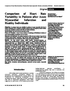
We have also found statistically significant differences in HRV frequency ratios in the third position in the case of a decrease in VLF + LF / HF ratio. We can also see decreasing activity in VLF and LF band in the standing position in the 1st measurement and an increase in VLF and LF band in the standing position in the 2nd measurement (Figure 2).
RESULTS Demographic data 31 subjects (74.2% females) were recruited from the inpatients department of the Psychiatry Department University Hospital Olomouc. All patients had been hospitalized for panic disorder. The diagnosis was confirmed after detailed examination by a specialist. The mean age of the patients was 39.45+10.32 years. The age at the disorder onset was 32.71+11.48 years; the duration of the disorder was 6.74+7.14 years. All patients used psychotropic medication. This included antidepressants (n=26; mean defined daily dosage of antidepressant was 23.27+8.825 mg of daily paroxetine equivalent); and some patients also used antipsychotics (n=10, mean defined daily dosage of antipsychotics was 1.87+1.984 mg of daily haloperidol equivalent) and benzodiazepines (n=20; mean defined daily dosage of benzodiazepines was 11.5+6.708 mg of daily diazepam equivalent) (Table 1). Doses of drugs were converted to defined daily doses using data provided by the Czech State Institute of Drug Control (SÚKL 2010).
Table 1. Demographic and clinical characteristics of the patients Patients (n=31) Age 39.45+10.32 Sex: Males 9 Females 22 Age of the disorder onset 32.71+11.48 Length of the disorder 6.74+7.14 Antidepressants index 23.27+8.825 (n=26) Benzodiazepine index 11.5+6.708 (n=20) Antipsychotics index 1.87+1.984 (n=10) CGI 4.13+1.23 BAI 27.41+13.38 BDI 20.48+11.38 DES 8.75+10.06
HRV measures There were no correlations between age of the patient and the parameters of the autonomic nervous system. We have found statistically significant differences in HRV frequencies before and after the CBT program in VLF in the standing position (Figure 1).
CGI = Clinical Global Impression-Severity of disorder; DES=Dissociative Experience Scale; BAI=Beck Anxiety Inventory; BDI=Beck Depression Inventory
Comparison of HRV Frequencies 9
8
*
7
Frequency
6
5 BT AT
4
3
2
1
0 VLF I1
VLF I2
VLF I3
LF I1
LF I2
LF I3
HF I1
HF I2
HF I3
Band
BT = Before Treatment; AT = After Treatment; VLF= very low frequency band (0.0033 – 0.04 Hz); LF= low frequency band (0.04 – 0.15 Hz); HF= high frequency band (0.15 – 0.40 Hz); I1= supine; I2= standing; I3= supine; *unpair t-test: p