286
J. VIDYA et al.: L-Asparaginase II from E. coli MTCC 739, Food Technol. Biotechnol. 49 (3) 286–290 (2011)
original scientific paper
ISSN 1330-9862 (FTB-2748)
Cloning, Functional Expression and Characterization of L-Asparaginase II from E. coli MTCC 739 Jalaja Vidya1, Ushasree Mrudula Vasudevan1, Carlos Ricardo Soccol2 and Ashok Pandey1* 1
Biotechnology Division, National Institute for Interdisciplinary Science and Technology (NIIST), 695 019 Trivandrum, India 2
Bioprocess Engineering and Biotechnology Division, Federal University of Paraná (UFPR), P.O. Box 19011, CEP 81531-970 Curitiba, PR, Brazil Received: September 13, 2010 Accepted: January 11, 2011
Summary L-Asparaginase is an antineoplastic agent that selectively decreases the level of L-asparagine in blood and diminishes the proliferation of the cancerous cells. L-Asparaginases from Escherichia coli are widely used for clinical application because of their high substrate specificity and limited glutaminase activity. L-Asparaginase II-encoding gene ansB was isolated by excluding the native signal from E. coli MTCC 739, cloned in frame with pelB leader sequence of prokaryotic expression vector pET20b and expressed in E. coli DE3 cells. Overexpression of recombinant protein was achieved with an optimized final concentration of 10 mM of isopropyl b-D-1-thiogalactopyranoside (IPTG). The protein was expressed as soluble protein. The recombinant protein contained hexahistidine tag at C-terminus and was purified using nickel-nitrilotriacetic acid chromatography. Enzymatic properties such as optimum temperature, pH and the effect of temperature on the stability of L-asparaginase II from E. coli MTCC 739 were determined and the purified protein showed an optimum activity at 37 °C and pH=6.
Key words: functional expression, L-asparaginase II, enzymatic property
Introduction Bacterial L-asparaginases have obtained considerable attention as a prerequisite for the treatment of acute lymphoblastic leukaemia and lymphosarcoma for the past 30 years following the first report in 1967 (1). Among the bacterial asparaginases, type II asparaginase (L-asparagine aminohydrolase, EC 3.5.1.1) displays high specific activity against L-asparagine and hence is used for clinical applications. After the catalysis, the enzyme converts L-asparagine to L-aspartate and ammonia via an acyl-enzyme intermediate. The chemotherapeutic nature of the enzyme relies on its ability to maintain low L-asparagine level in blood and thus eliminate the source of L-asparagine for tumour cells, which require a huge amount of amino acids for their protein synthetic machinery.
To date numerous microbial genera have been screened for L-asparaginase activity, and asparaginase with tumour inhibitory activity, encompassing reports from both prokaryotic as well as eukaryotic sources. In spite of a number of reports of L-asparaginase production from Pseudomonas flourescens (2), Mycobacterium (3) Staphylococcus (4) and Serratia marcescens (5), their intrinsic glutaminase activity, which could result in serious side effects like neurotoxicity, hepatitis and other dysfunctions, restricts their clinical applications. However, enzymes from Gram-negative bacteria such as Escherichia coli (6) and Erwinia carotovora (7) were found as most effective, owing to less severe immunorelated side effects as they possess strong preference to asparagine over glutamine. In order to improve the half life of L-asparaginase in blood during clinical trials, efforts have been made to modify the protein
*Corresponding author; Phone: ++91 471 251 5279; Fax: ++91 471 249 1712; E-mail:
[email protected],
[email protected]
J. VIDYA et al.: L-Asparaginase II from E. coli MTCC 739, Food Technol. Biotechnol. 49 (3) 286–290 (2011)
by replacement of certain amino acid residues by site-directed mutagenesis (8), attachment of some chemical moieties to the purified protein (9) or immobilization of the asparaginase in nanoparticles (10) or microparticles of natural silk sericin protein (11). The aim of the current study is to construct an expression cassette of L-asparaginase II gene from E. coli MTCC 739 bearing a C-terminal His6 tag, in frame with pelB leader sequence of pET20b under the control of a T7 inducible promoter. The ansB gene from E. coli MTCC 739 was cloned and expressed in E. coli DE3 cells. The expressed protein was purified and studied for operational properties.
Materials and Methods Bacterial strains and plasmids
287
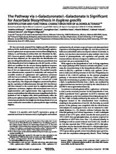
Periplasmic and cytosolic fractions were collected and analysed to establish the location of expressed recombinant protein in the cell compartments. For the preparation of periplasmic fractions, 50-mL culture was pelleted and resuspended in Tris-sucrose buffer (30 mM Tris, pH=8, 20 % sucrose and 1 mM EDTA) followed by shaking for 10 min and pelleting at 8000×g for 20 min. The pellet was resuspended again in 10 mL of 5 mM MgSO4, kept in an ice bath and shaken for 10 min, and after centrifugation at 8000×g for 20 min the supernatant was collected as periplasmic fraction. The cytosolic fraction was prepared by sonicating the pellet collected from 50 mL of culture in lysis buffer (50 mM NaH2PO4, 300 mM NaCl and 10 mM imidazole). For recombinant protein expression analysis, total cell lysate made from all the samples including induced and uninduced cultures was analysed on 12 % SDS-PAGE and then stained with Coomasie brilliant blue R-250 (Fig. 1).
E. coli MTCC 739 was used as a source of the L-asparaginase II gene. E. coli strains BL21(DE3) and DH5a, and pET-20b plasmid as an expression vector for ansB gene were obtained from Novagen (Milan, Italy).
Construction of recombinant plasmid and cloning of ansB gene Genomic DNA of E. coli MTCC 739 (NII08131) was used as template for polymerase chain reaction (PCR) amplification of L-asparaginase II (ansB) gene by excluding the native signal sequence using AnsBF (forward, 5’-GCGGAATTCGTTACCCAATATCACCA-3’ and AnsBR (reverse, 5’-GGCGAAGCTTGTACTGATTGAAGA-3’) with EcoRI and HindIII restriction sites underlined respectively. The PCR conditions were as follows: initial denaturation at 94 °C for 4 min, denaturation at 95 °C for 40 s, annealing at 57.4 °C for 30 s, extension at 72 °C for 90 s for 30 cycles, and a final extension at 72 °C for 10 min. The 981-bp amplicons, digested by EcoRI and HindIII, were cloned in pET20b vector having the same restriction termini. The recombinant construct thus obtained (pET20b-M2His) bearing an N-terminal pelB leader and a C-terminal His6 tag was then transformed into chemically competent E. coli DH5a cells. The clones were analyzed by a restriction digestion for an insert release of the recombinant plasmid isolated from the confirmed clones. The plasmid DNA was isolated from the confirmed clone of E. coli DH5a cells and used for transformation of E. coli DE3 competent cells for protein expression.
Soluble expression and purification of the recombinant enzyme To optimize the expression of L-asparaginase gene in E. coli, the effect of various isopropyl b-D-1-thiogalactopyranoside (IPTG) concentrations was studied. E. coli DE3 cells harbouring the expression plasmid pET20b-M2His was grown in Luria-Bertani broth containing 0.5 % glucose and 50 mg/mL of ampicillin and induced with different IPTG concentrations starting from 10 to 400 mM at the A600 nm of 0.6. The culture was then allowed to grow for a postinduction period of 3 h at 37 °C and 200 rpm. After the harvest, absorbance (at 600 nm) was measured and all the samples were diluted to an absorbance of 1 for a normalised SDS-PAGE and enzymatic assays.
Fig. 1. SDS-PAGE profile of total cell lysate from samples after expression. Lane 1: molecular mass marker (from top 200, 150, 120, 100, 85, 70, 60, 50, 40, 30, 25, 20, 15 and 10 kDa), lane 2: total cell lysate of host cell DE3, lane 3: total cell lysate of uninduced sample, lanes 4–7: total cell lysate from induced samples with 10, 50, 100 and 400 mM IPTG
The His6-tagged protein was purified by affinity chromatography using nickel-nitrilotriacetic acid (Ni-NTA) spin column (Quiagen, Hilden, Germany) and the bound protein was eluted in elution buffer (50 mM NaH2PO4, 300 mM NaCl and 250 mM imidazole). The purified enzyme was dialysed over night against 50 mM Tris buffer, pH=7, and used for further characterization.
Enzyme assay Activity analysis of L-asparaginase II was performed according to Imada et al. (12) comprising the following steps. The samples were mixed with 0.04 M L-asparagine in 0.05 M Tris buffer, pH=7, and 200 mL of assay mixture were incubated for 10 min at 37 °C for enzymatic reaction. The reaction was interrupted with 50 mL of 1.5 M trichloroacetic acid and the samples were centrifuged before the addition of Nessler’s reagent to measure the released ammonia after L-asparagine hydrolysis. All the measurements were done spectrophotometrically
288
J. VIDYA et al.: L-Asparaginase II from E. coli MTCC 739, Food Technol. Biotechnol. 49 (3) 286–290 (2011)
at 450 nm. Experimental values were calculated as an average of three independent measurements. The enzyme activity of recombinant protein was determined using an ammonium sulphate calibration curve. One unit of enzyme activity was defined as the amount of enzyme required to release 1 mM of ammonia per minute.
Functional features of purified enzyme Optimum conditions of temperature and pH The analysis of optimum temperature and the effect of pH on the activity of purified enzyme was carried out in the range of 40–70 °C and different pH from 4–10 using appropriate buffer systems respectively. Optimum conditions were determined and maintained stable for further assays to determine the thermostability. Thermostability The enzyme was incubated at 50 °C and the activities were measured at various time intervals of 15, 30, 45 and 60 min. Percentage of residual activities was calculated based on the untreated control activity, which is taken as 100 %.
currently with the increase of IPTG concentration above 50 mM. Hence, an inducer concentration of 10 mM was selected for further expression and purification studies. Moreover, extracellular expression of L-asparaginase by E. coli K12 was reported with optimal IPTG concentration of 100 mM from pET22b vector (14). SDS-PAGE profiles distinguished the His6-tagged asparaginase in the induced samples as 37 kDa band, which corresponds to the expected molecular mass of E. coli L-asparaginase II.
Purification and functional features of the enzyme Expression vector contained the gene for asparaginase fused to a C-terminal His6 tag, enabling the purification in a one-step procedure using affinity chromatography. The bound recombinant protein was eluted from the nickel affinity column in 250 mM imidazole and the eluted fractions were analysed on native PAGE gel to reveal oligomeric molecular mass of 150 kDa (Fig. 3) and a simultaneous SDS-PAGE gel of 12 % showed a subunit molecular mass of 37 kDa (Fig. 4a).
Results and Discussion Cloning and overexpression of L-asparaginase L-Asparaginase gene encoding asnB region was cloned downstream to T7 promoter of pET20b vector allowing the insertion of pelB sequence at the N-terminus and His6 tag at the C-terminus and overexpressed in E. coli DE3 cells. The enzyme activity of the recombinant ansB in cytosolic and periplasmic fractions of E. coli DE3 was determined and found to be more in cytosol compared to the collected periplasmic fraction. Previous reports have already revealed an inhibitory effect of the C-terminal His6 tag in targeting the recombinant protein to the E. coli periplasm (13), which may account for the limited activity of the recombinant protein in periplasm compared to the intracellular fraction. With an objective to determine an optimized expression conditions for the recombinant construct, the effect of various inducer concentrations on recombinant protein expression was studied. Of the different inducer concentration used (10, 50, 100 and 400 mM) for expression studies, 10 mM IPTG was optimum for the expression of L-asparaginase gene as determined by activity assay (Fig. 2). Furthermore, native protein in the soluble fraction was decreased con-
Fig. 2. L-Asparaginase activity in the soluble fractions
Fig. 3. Molecular mass determination of the oligomeric L-asparaginase through native PAGE. Lane 1: purified L-asparaginase in tetrameric form having a molecular mass of around 150 kDa, lane 2: gene protein molecular mass marker for native PAGE
Effect of temperature and pH on enzyme The effect of temperature on the activity of purified enzyme was checked from 37 to 70 °C. As shown in Fig. 4b, the purified L-asparaginase showed a maximum activity at 37 °C. However, the activity of the enzyme sharply decreased above 37 °C. Optimum temperature of L-asparaginase II from E. coli W3110 was at 40 °C for free enzyme, and a 10-degree rise in temperature was observed for immobilized enzyme preparation (15). The enzyme activity profile was analyzed at various pH from 4 to 9. The enzyme was optimally active at pH=6 and there was no significant reduction in the enzyme activity in the alkaline range beyond the pH optima (Fig. 4c). Enzyme with similar activity, EC-2, was reported from E. coli B, which showed a very little change of activity over the pH range from 6.0 to 8.4, with an
J. VIDYA et al.: L-Asparaginase II from E. coli MTCC 739, Food Technol. Biotechnol. 49 (3) 286–290 (2011)
289
Fig. 4. Purification and functional features of His6-tagged L-asparaginase: a) lane 1: purified L-asparaginase through Ni-NTA spin column, lane 2: protein molecular mass marker; b) temperature optimum for purified L-asparaginase; c) pH optimum for L-asparaginase (buffer systems used were acetate buffer, pH=4–6, Tris-HCl, pH=7–8, and glycine NaOH, pH=9; d) thermal stability of the enzyme at 50 °C for various time intervals of 15, 30, 45 and 60 min, and residual activity compared to control
optimum at pH=7 (16). According to Ehrman (17), the release of aspartate was high at pH=5.6 when b-aspartohydroxamate was used as substrate. The pH activity profile of E. coli W3110 was reported at pH=7 and both the free and immobilized enzymes were active at broad range of alkaline pH, up to 10.
Thermostability assays The thermal stability of the purified enzyme at 50 °C was studied to find out the extent of temperature resistance of the enzyme. Around 26 % of the initial activity was retained by the purified enzyme after 30 min of incubation at 50 °C. The activity was significantly reduced after 45 min and decreased to less than 10 % in 1 h of incubation at the same temperature (Fig. 4d). The earlier reports on the thermostability of different L-asparaginase preparations indicate that the modified enzyme was more stable at elevated temperatures for a greater extent than the native enzymes (8,15) and the native enzyme from E. coli W3110 retained about 22 % of its activity at 60 °C after an incubation of 30 min.
Conclusions Aaparaginases are widely distributed in the microbial kingdom, and L-asparaginase from E. coli is well known in clinical application. This study comprises clon-
ing and effective expression of asnB gene from E. coli MTCC 739 and determination of its functional properties. Investigations to find out the potential of amino acid residues for modification of this enzyme using computational methods are currently underway and this enzyme could be used to improve the properties like thermostability and substrate specificity in order to obtain the best characteristics for therapeutic application.
Acknowledgements Jalaja Vidya gratefully acknowledges the award of Junior Research Fellowship by the Council of Scientific and Industrial Research (CSIR), New Delhi, India, to carry out this study.
References 1. J.M. Hill, J. Roberts, E. Loeb, A. Khan, A. Maclellan, R.W. Hill, L-asparaginase therapy for leukemia and other malignant neoplasms. Remission in human leukemia, J. Am. Med. Assoc. 202 (1967) 882–888. 2. A. Nilolaev, N.N. Sokolov, E.A. Kozlov, M.E. Kutsman, Isolation and properties of a homogeneous L-asparaginase preparation from Pseudomonas fluorescens AG, Biokhimiia, 40 (1975) 984–989. 3. I. Pastuszak, M. Szymona, Purification and properties of L-asparaginase from Mycobacterium phlei, Acta Biochim. Pol. 23 (1976) 37–44.
290
J. VIDYA et al.: L-Asparaginase II from E. coli MTCC 739, Food Technol. Biotechnol. 49 (3) 286–290 (2011)
4. J. Mikucki, J. Szarapin b ska-Kwaszewska, Z. Krzemin b ski, Factors influencing L-asparaginase production by staphylococci, Zentralbl. Bakteriol. Parasitenkd. Infektionskr. Hyg. 132 (1977) 135–142. 5. J.W. Boyd, A.W. Phillips, Purification and properties of L-asparaginase from Serratia marcescens, J. Bacteriol. 106 (1971) 578–587. 6. Y. Wang, S. Qian, G. Meng, S. Zhang, Cloning and expression of L-asparaginase gene in E. coli, Appl. Biochem. Biotechnol. 95 (2001) 93–101. 7. G.A. Kotzia, N.E. Labrou, Cloning, expression and characterization of Erwinia carotovora L-asparaginases, J. Biotechnol. 119 (2005) 309–323. 8. L.Z. Li, T.H. Xie, H.J. Li, C. Qing, G.M. Zhang, M.S. Sun, Enhancing the thermostability of Escherichia coli L-asparaginase II by substitution with Pro in predicted hydrogen-bonded turn structures, Enzyme Microb. Technol. 41 (2007) 523–527. 9. J.F. Zhang, L.Y. Shi, D.Z. Wei, Chemical modification of L-asparaginase from Escherichia coli with a modified polyethyleneglycol under substrate protection conditions, Biotechnol. Lett. 26 (2004) 753–756. 10. E. Teodor, S.C. Litescu, V. Lazar, R. Somoghi, Hydrogel-magnetic nanoparticle with immobilizes L-asparaginase for biomedical applications, J. Mater. Sci: Mater. Med. 20 (2009) 1307–1314.
11. Y.Q. Zhang, M.L. Tao, W.D. Shen, Y.Z. Zhou, Y. Ding, Y. Ma, W.L. Zhou, Immobilization of L-asparaginase on the microparticles of the natural silk sericin protein and its characters, Biomaterials, 25 (2004) 3751–3759. 12. A. Imada, S. Igarasi, K. Nakahama, M. Isono, L-Asparaginase and glutaminase activities of micro-organisms, J. Gen. Microbiol. 76 (1973) 85–99. 13. A. Khushoo, Y. Pal, K.J. Mukherjee, Optimization of extracellular production of recombinant asparaginase in Escherichia coli in shake-flask and bioreactor, Appl. Microbiol. Biotechnol. 68 (2005) 189–197. 14. A. Khushoo, Y. Pal, B.N. Singh, K.J. Mukherjee, Extracellular expression and single step purification of recombinant Escherichia coli L-asparaginase II, Protein Expr. Purif. 38 (2004) 29–36. 15. M.M. Youssef, M.A. Al-Omair, Cloning, purfication, characterization and immobilization of L-asparaginase II from E. coli W3110, Asian J. Biochem. 3 (2008) 337–350. 16. H.A. Campbell, L.T. Mashburn, E.A. Boyse, L.J. Old, Two L-asparaginases from Escherichia coli B. Their separation, purification, and antitumor activity, Biochemistry, 6 (1967) 721–730. 17. M. Ehrman, H. Cedar, J.H. Schwartz, L-Asparaginase II of Escherichia coli. Studies on the enzymatic mechanism of action, J. Biol. Chem. 246 (1971) 88–94.