RESIDENT & FELLOW SECTION Section Editor Mitchell S.V. Elkind, MD, MS
Jon Foss-Skiftesvik, MD Majken G. Hougaard, MD Vibeke A. Larsen, MD, PhD Klaus Hansen, MD, DMSc
Correspondence to Dr. Foss-Skiftesvik:
[email protected]
Clinical Reasoning: Partial Horner syndrome and upper right limb symptoms following chiropractic manipulation SECTION 1
A 60-year-old man with no medical history was admitted to the Department of Neurology with drooping of his right upper eyelid and an ipsilateral contracted pupil, combined with pain, weakness, and numbness in his upper right limb. The patient had experienced thoracic back pain of moderate intensity with radiating right-sided belt-like chest pain for 7 days when the discomfort suddenly intensified, causing him to seek chiropractic treatment. Following manipulations of the thoracic and
cervical spine, the pain intensity initially lessened. Approximately 1 hour after chiropractic treatment, the patient experienced onset of the eye and upper limb symptoms described above, for which he sought medical assistance 3 days later. No other symptoms were present at any time. Questions for consideration:
1. What is your differential diagnosis at this point? 2. What are the most common causes of Horner syndrome?
GO TO SECTION 2
From the Departments of Neurology (J.F.-S., M.G.H., K.H.) and Radiology (V.A.L.), Rigshospitalet, Copenhagen University Hospital, Denmark. Go to Neurology.org for full disclosures. Funding information and disclosures deemed relevant by the authors, if any, are provided at the end of the article. © 2015 American Academy of Neurology
e175
ª 2015 American Academy of Neurology. Unauthorized reproduction of this article is prohibited.
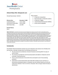
Figure 1
Photograph of the patient with right-sided blepharoptosis and moderate pupillary miosis
SECTION 2
A detailed neurologic examination revealed moderate right-sided ptosis and miosis (2 mm1/4 mm1 measured in light conditions) (figure 1), no facial anhidrosis, decreased strength of the intrinsic and opponens muscles of the right hand (Medical Research Council scale 4), and reduced cutaneous sensation corresponding to the T1 dermatome, with inability to discriminate pain and light touch. The remaining clinical examination, routine blood tests, and vital parameters were otherwise unremarkable. Questions for consideration:
1. Did the clinical examination alter your proposed diagnosis? 2. What potential lesion location may explain the described symptoms and clinical findings? 3. What are your suggested next investigative steps, and in what order of prioritization?
GO TO SECTION 3
e176
Neurology 84
May 26, 2015
ª 2015 American Academy of Neurology. Unauthorized reproduction of this article is prohibited.
Figure 2
MRI
SECTION 3
Brain CT scan and CT angiography including the aortic arch and neck vessels were performed and ruled out cerebral stroke and carotid artery dissection, respectively. As clinical signs of Horner syndrome and a concomitant radiating pain to the medial arm were considered suggestive of either a lower brachial plexopathy, i.e., due to a Pancoast tumor, or a radiculopathy, chest X-ray and electroneurography (ENG) were performed. No apical pulmonary pathology was detected. ENG of the right medial cutaneous antebrachial nerve demonstrated a normal sensory action potential (SAP), consistent with the lesion being located proximally to the dorsal spinal root ganglion, thus suggestive of a spinal nerve root lesion. A subsequent MRI of the thoracic spine showed a paramedian herniation of the T1-T2 intervertebral disc compressing the right T1 spinal nerve root (figure 2). The patient received no surgery, and follow-up examination 6 months later revealed near-complete recovery, with only mild paresthesia in the T1 segment of his right arm and a subtle ptosis remaining. Questions for consideration:
Sagittal and axial T2-weighted (upper row) and T1-weighted (lower row) sequences of the cervico-thoracic spine show osteochondrotic degenerative changes and a right-sided convex herniated intervertebral T1-T2 disc (arrow).
1. How can you explain the patient’s lack of anhidrosis? 2. What other lesion types and locations would you consider if the ENG had shown a decreased or absent SAP? 3. What topical pharmacological tests are used to (1) confirm the presence of Horner syndrome and (2) differentiate central and preganglionic from postganglionic Horner syndrome?
Neurology 84
May 26, 2015
e177
ª 2015 American Academy of Neurology. Unauthorized reproduction of this article is prohibited.
Figure 3
Illustration of the oculosympathetic pathway
Modified from Alstadhaug KB. Ervervet Horners syndrom. Tidsskr Nor Legeforen 2011;131:950–954. Printed with permission from © Kari C. Toverud.
The sympathetic innervation of the eye and the ocular adnexae is traditionally described as a 3-neuron pathway (figure 3). It consists of a central (first-order) neuron located in the posterolateral hypothalamus, which axon descends ipsilaterally and connects to a preganglionic (second-order) neuron in the intermediolateral columns of the spinal cord at level C8 through T2, and a postganglionic (third-order) neuron located in the superior cervical ganglion.1 Spanning from C8-T4, most fibers from the preganglionic neuron leave through the T1 ventral spinal nerve root, but sudomotor and vasomotor fibers to the ANATOMY
e178
Neurology 84
face primarily exit at the T2 and T3 level.2 The fibers rejoin in the sympathetic trunk, which ascends adjacent to spinal vertebrae, the apex of the lung, the first rib, and the common carotid artery (CCA) before synapsing with the postganglionic neuron in the superior cervical ganglion.1 The postganglionic fibers form a nerve plexus in the adventitia of the CCA. Most fibers follow the internal carotid artery (ICA) through the cavernous sinus, reaching the superior tarsal muscle by the ophthalmic artery, whereas fibers to the iris dilator muscle branch off and follow the nasociliar nerve. The sudomotor and vasomotor fibers, however, follow the external carotid artery to the face.1
May 26, 2015
ª 2015 American Academy of Neurology. Unauthorized reproduction of this article is prohibited.
Our patient’s ocular symptoms were consistent with Horner syndrome without anhidrosis, also referred to as partial Horner syndrome.3 Horner syndrome due to a herniated thoracic disc is considered very rare, and has to our knowledge only been reported 6 times in the English language literature,4 though never preceded by chiropractic manipulation. Symptomatic thoracic disc herniation is uncommon and has been estimated to less than 0.75% of all symptomatic spinal disc herniations.5 Furthermore, more than 75% of thoracic protrusions are located below T8, and only approximately 3% occur at the T1-T2 level,5 as in our patient. Only 40 cases with a T1-T2 disc herniation have been reported since Svien and Karavitz presented their first case in 1959.6,7 One of the most frequent causes of Horner syndrome is carotid artery dissection, which may occur spontaneously or due to local trauma to the neck region. Chiropractic manipulation as an independent risk factor for neck artery dissection and a consequent stroke is a controversial topic, though multiple cases of Horner syndrome due to ICA dissections subsequent to chiropractic manipulation have been reported.8 In our patient, an ICA dissection was considered unlikely due to the concomitant prominent radiating medial brachialgia and was furthermore ruled out by a CT angiogram of the neck vessels. Lack of anhidrosis is indicative of either a postganglionic lesion at the ICA level, since sudomotor fibers are separated from the carotid plexus at the level of the carotid bifurcation, or alternatively a preganglionic sympathetic lesion of the T1 root, as the sudomotor fibers exit the spinal cord at the T2-T3 level (figure 3). Thus, rare cases of partial Horner syndrome due to lesions at the T1 root level should be kept in mind, as illustrated by our patient. Horner syndrome combined with ipsilateral upper limb pain and weakness of the muscles of the hand also complies with the diagnostic criteria for Pancoast syndrome, suggestive of a malignant tumor in the apex of the lung affecting both the sympathetic trunk and the inferior trunks of the brachial plexus. Pulmonary pathology was excluded by chest X-ray. The subsequent ENG with a normal SAP in the medial cutaneous antebrachial nerve was compatible with a supraganglionic root lesion as later confirmed by MRI. In case of a Pancoast tumor with involvement of the lower brachial plexus, the ENG would be expected to reveal a decreased or absent SAP. Our patient experienced onset of Horner syndrome and ipsilateral upper limb symptoms shortly after chiropractic treatment, suggesting the cervico-thoracic manipulation as the cause of or at least worsening factor in the T1-T2 disc herniation. Approximately DISCUSSION
33%–50% of patients with a symptomatic thoracic disc herniation report a history of recent trauma,9 including torsion and athletic activity. Furthermore, several examples of disc herniations following chiropractic treatment have been reported.10 Pharmacologic pupillary testing can be utilized both to confirm the presence of Horner syndrome and to locate the lesion in the oculosympathetic pathway. Cocaine is a norepinephrine reuptake inhibitor and will fail to induce pupillary mydriasis if applied topically to a patient with disrupted sympathetic innervation of the iris dilator muscle, confirming Horner syndrome, irrespective of where the lesion is located. Topical hydroxyamphetamine, on the other hand, produces pupillary dilation by releasing norepinephrine from the presynaptic terminal. This effect will only occur if the postganglionic (third-order) neuron is intact, and may therefore be used to differentiate central and preganglionic from postganglionic lesions.1 Central Horner syndrome due to a brainstem stroke, i.e., Wallenberg syndrome, will rarely be confused with a peripheral cause due to concomitant and characteristic clinical findings consistent with brainstem pathology, including nystagmus, dissociated loss of sensibility, and ipsilateral cerebellar symptoms. Though considered extremely rare, Horner syndrome, and more specifically partial Horner syndrome, can occur due to an isolated herniated intervertebral disc at the T1-T2 level. The condition may result in diagnostic challenges, including considerations involving carotid artery lesions and malignant pulmonary pathologies. While the definite pathophysiologic mechanism in our patient remains unclear, it seems evident that manipulations as a minimum altered the configuration of an already existing disc protrusion. AUTHOR CONTRIBUTIONS Dr. Foss-Skiftesvik conceived the manuscript, drafted the initial version, and revised the final edition. Dr. Hougaard drafted the initial and revised the final manuscript. Dr. Larsen was responsible for the diagnostic imaging and revised the final manuscript. Dr. Hansen was responsible for the care of the patient, including diagnosis and treatment, and revised the initial and final manuscript.
STUDY FUNDING No targeted funding reported.
DISCLOSURE The authors report no disclosures relevant to the manuscript. Go to Neurology.org for full disclosures.
REFERENCES 1. Walton KA, Buono LM. Horner syndrome. Curr Opin Ophthalmol 2003;14:357–363. 2. Wasner G, Maag R, Ludwig J, et al. Harlequin syndrome: one face of many etiologies. Nat Clin Pract Neurol 2005; 1:54–59. Neurology 84
May 26, 2015
e179
ª 2015 American Academy of Neurology. Unauthorized reproduction of this article is prohibited.
3. 4.
5. 6.
e180
Flaherty PM, Flynn JM. Horner syndrome due to carotid dissection. J Emerg Med 2011;41:43–46. Morgan H, Abood C. Disc herniation at T1-2: report of four cases and literature review. J Neurosurg 1998;88: 148–150. Arce CA, Dohrmann GJ. Herniated thoracic disks. Neurol Clin 1985;3:383–392. Son ES, Lee SH, Park SY, Kim KT, Kang CH, Cho SW. Surgical treatment of t1-2 disc herniation with t1 radiculopathy: a case report with review of the literature. Asian Spine J 2012;6:199–202.
Neurology 84
7.
8.
9. 10.
Negovetic L, Cerina V, Sajko T, Glavic Z. Intradural disc herniation at the T1-T2 level. Croat Med J 2001;42: 193–195. Jeret JS, Bluth M. Stroke following chiropractic manipulation: report of 3 cases and review of the literature. Cerebrovasc Dis 2002;13:210–213. Kibler WB. Orthopaedic Knowledge Update, 7th ed. Rosemont, IL: American Academy of Orthopaedic Surgeons; 2002. Murphy DR. Herniated disc with radiculopathy following cervical manipulation: nonsurgical management. Spine J 2006;6:459–463.
May 26, 2015
ª 2015 American Academy of Neurology. Unauthorized reproduction of this article is prohibited.