Clinical Manifestations of Thoracic OPLL and OLF Morio Matsumoto1, Kazuhiro Chiba2, and Yoshiaki Toyama2
Introduction Ossification of the posterior longitudinal ligament (OPLL) is far less common in the thoracic spine than in the cervical spine; and the rate of occurrence, natural history, and optimal treatment of this condition are yet to be investigated. Because progressive deterioration of the neurological symptoms due to thoracic OPLL often severely impairs patients’ activities of daily living and quality of life, awareness of this clinical entity is important. Ossification of the ligamentum flavum (OLF) is mostly found in the thoracic spine and is a frequent cause of thoracic myelopathy. In this chapter, the clinical and neurological manifestations of thoracic OPLL and OLF are reviewed.
Clinical Characteristics of Thoracic OPLL and OLF OPLL Thoracic OPLL came to be recognized as a cause of thoracic myelopathy only during the 1970s following several reports of small case series. Although the occurrence rates of thoracic OPLL and OLF remain to be clearly determined, it appears that thoracic OPLL occurs far less frequently than cervical OPLL. Until now, several multicenter studies have been conducted nationwide by the Investigation Committee on the Ossification of the Spinal Ligaments of the Japanese Ministry of Public Health and Welfare to study the epidemiology of this condition [1–3]. In the study conducted in 1998 [3], a total of 207 patients (mean age 55.6 years) undergoing surgery for thoracic OPLL were registered. There were 62 men and 145 women, suggesting that thoracic OPLL appears preponderantly in female 1 Department of Musculoskeletal Reconstruction and Regeneration Surgery, Keio University, 35 Shinanomachi, Shinjuku-ku, Tokyo 160-8582, Japan 2 Department of Orthopaedic Surgery, Keio University, 35 Shinanomachi, Shinjuku-ku, Tokyo 160-8582, Japan
subjects. Thoracic OPLL extended over 4.8 intervertebral segments on average, and the apex of the ossification was located at T5 (range T1–T12). OLF was also found with OPLL in 113 of the 207 patients (54.5%). The results of this study suggest that thoracic OPLL often involves several segments, mainly of the mid-thoracic spine, and occurs more frequently in middle-aged to older women. Isolated thoracic OPLL is rather rare, accounting for only 10% of the patients. In the remaining 90%, thoracic OPLL is associated with OPLL in the cervical spine [1]. Because the motions of the thoracic spine are limited by the rib cage, dynamic factors may not play an important role in the development of myelopathic symptoms in cases of thoracic OPLL, unlike the case in patients with OPLL of the cervical or lumbar spine [4]. However, physiologic kyphosis of the thoracic spine renders the spinal cord vulnerable to pressure against the ventrally located OPLL. Moreover, thoracic OPLL occurs frequently in the mid-thoracic spine, where under physiological conditions the spinal cord receives scarce blood supply; this intramedullary hypocirculation may also render the spinal cord more vulnerable to compression by OPLL [4].
OLF Ossification of the ligamentum flavum develops in the thoracic spine, either alone or in combination with OPLL. The lower thoracic spine (T9–T12) is most often affected [5–9]. Unlike OPLL, OLF occurs more frequently in men than in women. Asymptomatic OLF may not be rare. In a cadaveric study, Hashizaki and Kaneko [10] found OLF bridging adjacent laminae in 21.7% and 30.4%, respectively, of men and women older than 30 years of age. Because the lower thoracic spine has more mobility than the upper or middle thoracic spine, it is thought that mechanical stress on the ligamentum flavum may contribute to the development and progression of the ossification. In most patients, OLF arises from the capsular portion of the ligamentum flavum and extends medially. There is often a difference in the thickness of the ossification between the right and left sides, causing asymmetry of the neurological
121
122
M. Matsumoto et al.
symptoms. Coexistence of posterior protrusion of degenerated thoracic intervertebral discs and posterior spurs may also contribute to the neurological symptoms in these cases.
Neurological Symptoms In patients with OPLL, OLF, or both, the development of neurological symptoms may be influenced by several factors, including the size of the ossified lesions, the segmental motions of the thoracic spine (especially of the lower thoracic spine), the blood supply of the spinal cord, and the inherent diameter of the spinal canal.
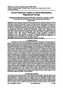
OPLL Patients with OPLL in the thoracic spine are asymptomatic unless the ossification has progressed sufficiently to compress the spinal cord. Otherwise, these patients may complain only of slight pain or discomfort in the back. Once myelopathy develops, it tends to deteriorate steadily [4,6]. Although the neurological deterioration is usually gradual in most patients, it is rapid in others, who are unable to walk within a short period of time. Thoracic OPLL has been classified radiographically into several types (Fig. 1). Among them, beak-type and continuous wave-type OPLL are notorious for causing severe thoracic myelopathy (Fig. 2). Miyasaka et al. [7] reported that the critical anteroposterior diameter of OPLL for the development of thoracic myelopathy was 7 mm. Before the development of thoracic myelopathy, some patients experience girdle pain in the chest at the level corresponding to compression of the spinal cord by the OPLL. Pain or numbness (or both) in the lower extremities are the initial clinical symptoms in some
patients. Patients who are myelopathic may complain of difficulty in walking as well as tightness and stiffness of the trunk and lower limbs. On neurological examination, they usually have hyperreflexia in the lower extremities. Pathological reflexes, such as Babinski’s reflex, are frequently positive. Gait disturbance may be observed with spasticity of the lower limbs; and with progression of the myelopathy, the patients become unable to walk. Sensory disturbances, including an impaired sense of light touch, pinprick, temperature, vibration, and position, are observed just below (sometimes far below) the dermatome corresponding to the level of the OPLL. Some patients have sensory disturbances beyond the dermatome corresponding to the level of the maximum OPLL, in which case the presence of concomitant cervical OPLL should be suspected. Urinary and bowel disturbances are not rare in severely myelopathic patients.
OLF Ossification of the ligamentum flavum develops in the thoracic spine, either alone or in combination with OPLL. The lower thoracic spine (T9–T12) is most commonly affected. The clinical manifestations of OLF differ depending on the level and magnitude of compression of the spinal cord [11]. Although thoracic OLF sometimes causes intercostal neuralgia [12], most symptomatic patients present with thoracic myelopathy. The OLF at the lower thoracic spine causes various neurological symptoms, sometimes mimicking those caused by lumbar spinal disease, motor neuron disease, or peripheral neuropathy because the epiconus, conus, and cauda equina are located at the lower thoracic and thoracolumbar levels and because their localization often varies among individuals (Fig. 3) [11]. For
Fig. 1. Classification of thoracic ossification of the posterior longitudinal ligament (OPLL) by the Investigation Committee on the Ossification of the Spinal Ligaments of the Japanese Ministry of Public Health and Welfare (1994)
Clinical Manifestations of Thoracic OPLL and OLF
123
Fig. 2. A 61-year-old male patient with beak-type OPLL at T3–T4. He had severe spastic paraparesis and could not walk without a walker. Note the severe compression of the spinal cord by the beak-type OPLL on the reconstruction computed tomography (CT) scan
example, OLF at the lower thoracic spine from T10 to T12 usually compresses the epiconus, which consists of spinal cord segments L4-S2, causing the “epiconus syndrome.” For example, patients with OLF at T11–T12 may have muscle weakness and sometimes atrophy of the quadriceps muscles, anterior tibial muscles, and gastrocnemius muscles (Fig. 4). Although the pattern of abnormalities of the deep tendon reflexes differs among patients, the patellar tendon reflex (PTR) is frequently normal or diminished, whereas the Achilles tendon reflex (ATR) is exaggerated. Babinski’s reflex may be present. Sensory disturbances are often observed below the level of the knees. Some patients complain of pain in the lower legs that resembles sciatic pain. Patients with the epiconus syndrome caused by OLF sometimes demonstrate only segmental signs, such as flaccid paralysis with muscle atrophy and stocking-type sensory disturbance, with no abnormality of the deep tendon reflexes. In such cases, compressive myelopathy by OLF must be differentiated from motor neuron disease or peripheral neuropathy not only by neurological examination but also by additional blood tests, electrophysiological examinations, biopsy of muscles or peripheral nerves, and intensive discussions with neurologists. OLF at T12-L1 usually compresses the L5S2 segments, resulting in a diminished Achilles tendon reflex. OLF at a more rostral level than T11–T12 usually presents with typical thoracic myelopathic symptoms,
Fig. 3. Lower thoracic and thoracolumbar spine and the spinal cord. The epiconus, consisting of L4 to S1 spinal cord segments, is located at the level of T10–T12. (adapted from ref. 14, with permission)
such as exaggerated PTR and ATR and sensory disturbances below the affected level, among others. Myelopathic intermittent claudication has been reported in patients with OLF at the lower thoracic spine [13]. This intermittent claudication is thought to be caused by ischemic changes of the spinal cord due to a diminished arterial blood supply or venous congestion. The patients cannot walk for more than a short distance, and they complain of fatigue or tightness in the lower extremities (or both) while walking. Their neurological abnormalities, such as abnormalities of the deep tendon reflexes and sensory disturbances are aggravated after walking. Myelopathic intermittent claudication must be differentiated from claudication caused by the cauda equina syndrome due to lumbar spinal diseases or arteriosclerosis of the lower extremities (Table 1). There have been reports of patients in whom thoracic OLF was first recognized because of the development of paraplegia following surgery on the
124
M. Matsumoto et al. Fig. 4. A 47-year-old woman with ossification of the ligamentum flavum (OLF) at T11–T12. She had muscle weakness in the right anterior tibial muscle and numbness in the right lower leg. She also had difficulty walking. a Magnetic resonance imaging (MRI). b CT myelography
Table 1. Differential diagnosis in patients with intermittent claudication Criteria Aggravation of symptoms by gait Symptom relief by bending posture Symptoms and signs Pain Dysesthesia Sensory disturbance Muscle weakness Deep tendon reflex Positive Babinski sign Bladder dysfunction Pulsation of distal artery Cyanosis in the foot
Myelopathic
Vascular
Cauda equina
+ ±
+ −
+ +
Sometimes Frequent Frequent Always Exaggerated Frequent Frequent Normal None
Frequent Rare None Rare Normal None None Absent Frequent
Frequent Frequent Frequent Sometimes Diminished None Frequent Normal None
lumbar spine or trauma to the thoracic spine (Fig. 5) [14].
Combined Lesions In patients with combined cervical and thoracic OPLL or those with combined OLF and OPLL at multiple
levels, the neurological abnormalities may be complex, making the correct diagnosis difficult. When the sensory disturbance spreads rostrally beyond the level of the thoracic OPLL, coexistence of cervical OPLL should be suspected. Usually, patients with symptomatic thoracic OPLL or OLF have a disproportionately greater sensory loss and muscle weakness with spasticity in the lower extremities compared to that in the upper extremities.
Clinical Manifestations of Thoracic OPLL and OLF
125
Fig. 5. A 61-year-old man with OLF at T11–T12. He fell down the stairs and became paraplegic. He obtained spontaneous neurological recovery without surgical intervention. MRI and CT myelography demonstrated severe compression of the spinal cord by OLF at T11–T12. a MRI. b CT myelography
References 7. 1. Tsuyama N, Kurokawa T (1977) Ossification of posterior longitudinal ligament in the thoracolumbar spine: analyses of nationwide investigation on OPLL. Rinshoseikeigeka 12:327–339 (in Japanese) 2. Investigation Committee on OPLL of the Japanese Ministry of Public Health and Welfare (1981) The ossification of the spine (OPLL). Nippon Seikeigeka Gakkai Zasshi (J Jpn Orthop Assoc) 55:425–440 (in Japanese) 3. Kaneda K, Abumi K, Hasegawa K, Harada S, Fujiwara N (1999) Postoperative outcomes and QOL of thoracic myelopathy due to ossification of the spinal ligaments: a review of patients with thoracic ossification of posterior longitudinal ligament treated surgically. In: Harada S (ed) Investigation committee report on the ossification of the spinal ligaments of the Japanese Ministry of Public Health and Welfare, Tokyo, pp 138–142 (in Japanese) 4. Fujimura Y, Nishi Y, Nakamura M, Watanabe M, Matsumoto M (1997) Myelopathy secondary to ossification of the posterior longitudinal ligament of the thoracic spine treated by anterior decompression and bony fusion. Spinal Cord 35:777–784 5. Miyasaka H, Tsuji H, Inoue S, Fujizuka M, Watanabe T, Nagase J (1977) Association between radiographic findings and neurological symptoms in patients with ossification of the thoracic spinal ligaments. Rinshoseikeigeka 12:381–386 (in Japanese) 6. Kaneda K, Sato S, Higuchi M, Nohara Y, Oguma T, Honma S, Mitsuzaki A, Fujiya M (1981) Thoracic spinal canal
8.
9.
10.
11.
12.
13.
14.
stenosis due to ossification of the spinal canal ligaments. Rinshoseikeigeka 16:63–74 (in Japanese) Miyasaka K, Kaneda K, Ito T, Takei H, Sugimoto S, Tsuru M (1982) Ossification of spinal ligaments causing thoracic radiculomyelopathy. Radiology 143:463–468 Epstein NE (1999) Ossification of the yellow ligament and spondylosis and/or ossification of the posterior longitudinal ligament of the thoracic and lumbar spine. Spinal Disord 12:250–256 Shiokawa K, Hanakita J, Suwa H, Saiki M, Oda M, Kajiwara M (2001) Clinical analysis and prognostic study of ossified ligamentum flavum of the thoracic spine. J Neurosurg 94(Suppl):221–226 Hashizaki T, Kaneko M (1979) Study of spinal canal stenosis with special reference to its bony factors. Sapporo Med J 48:143–156 (in Japanese) Yanagi T (1988) Myelopathy due to ossification of the ligaments of the thoracic spine. Seikeisaigaigeka (Orthop Surg Traumatol) 31:1397–1403 (in Japanese) Iihara K, Hanakita J, Suwa H, Nishihara K, Sakaida H (1991) Ossification of the thoracic ligamentum flavum presenting with intercostal neuralgia: case report. Neurol Med Chir (Tokyo) 31: 999–1002 Kikuchi S, Watanabe E, Hasue M (1996) Spinal intermittent claudication due to cervical and thoracic degenerative spine disease. Spine 21:313–318 Takeuchi A, Miyamoto K, Hosoe H, Shimizu K (2004) Thoracic paraplegia due to missed thoracic compressive lesions after lumbar spinal decompression surgery: report of three cases. J Neurosurg Spine 100(Suppl):71– 74