The Korean Journal of Internal Medicine: 21:230-235, 2006
Clinical Investigation of Cavitary Tuberculosis and Tuberculous Pneumonia Ki Man Lee, M.D., Kang Hyeon Choe, M.D. and Sung Jin Kim, M.D.2 Departments of Internal Medicine and Diagnostic Radiology2, Chungbuk National Universit yCollege of Medicine, Cheongju, Korea
Backgorund : The radiographic characteristics of tuberculous pneumonia in adults are similar to primary tuberculosis that occurs in childhood, and upper lobe cavitary tuberculosis is the hallmark of postprimary tuberculosis. The purpose of this study was to investigate the factors associated with tuberculous pneumonia by making comparison with cavitary tuberculosis. Methods : The medical records and radiographic findings of patients with cavitary tuberculosis and tuberculous pneumonia, and who were diagnosed between March 2003 and February 2006, were analyzed retrospectively. Results : Forty patients had cavitary tuberculosis and sixteen patients had tuberculous pneumonia. Fever was more frequent for tuberculous pneumonia, whereas hemoptysis was more frequent for cavitary tuberculosis. The duration of symptoms before visiting the hospital was shorter, but the diagnosis after admission was more delayed for tuberculous pneumonia patients than for cavitary tuberculosis patients. The prevalence of underlying comorbidities such cancer, diabetes, alcoholism and long-term steroid use was not different between the two groups. The patients with tuberculous pneumonia were older and they had lower levels of serum albumin and hemoglobin than those with cavitary tuberculosis. The patients with tuberculous pneumonia showed a tendency to have more frequent endobronchial lesion. Tuberculous pneumonia occurred in any lobe, whereas the majority of cavitary tuberculosis patients had upper lung lesion, but the prevalence of lymphadenopathy, pleural effusion and previous tuberculosis scar was not different between the two groups. Conclusions : Older age, a lower level of serum albumin and hemoglobin and a random distribution of lesion were associated with tuberculosis pneumonia as compared with cavitary tuberculosis. These findings suggest that the pathogenesis of tuberculous pneumonia might be different from that of cavitary tuberculosis. Key Words : Tuberculosis, Pulmonary, Pneumonia
INTRODUCTION Active tuberculosis disease has been classified as either 1, 2) primary or postprimary tuberculosis (TB) . Primary TB is common in the pediatric age group, and it is caused by initial Mycobacterium tuberculosis (M. tuberculosis) infection. The radiological characteristics are focal lung infiltration or homogeneous consolidation that usually affects the middle and 1) lower lobes and lymphadenopathy is also present . Postprimary
TB is common in adults; this is mainly located in the apical and posterior segments of the upper lobes and it is characterized by 2) cavitary lung lesion . While the radiographic features of childhood TB have apparently not changed, there has been an increased number 3-10) of reports of atypical TB in adults . One of these atypical radiological findings in adult is tuberculous pneumonia that resembles childhood primary TB. The radiographic characteristics of tuberculous pneumonia are homogeneous,
∙Received : August 25, 2006 ∙Accepted : October 30, 2006 ∙Correspondence to : Ki Man Lee, Department of Internal Medicine, Chungbuk National University Hospital 62 Gaeshin-Dong, Heungduk-Gu, Cheongju 361-711, Korea Tel : 82-43-269-6353, Fax : 82-43-273-3252, E-mail :
[email protected] *This work was supported by Chungbuk National University Grant in 2004
Ki Man Lee, et al : Clinical Investigation of Cavitary Tuberculosis and Tuberculous Pneumonia 231
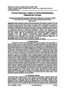
Figure 1. Chest X-ray of a 24-year-old woman reveals a cavitary lung lesion at the right upper lung (A). The chest CT scan demonstrates the thick walled cavity at the posterior segment of right upper lung (white arrowed) with a tree in bud pattern (black arrows) (B).
Figure 2. Chest X-ray of an 82-year-old man reveals homogenous consolidation at the right lower lung and the dense nodular lesions in both upper lungs (white arrowed) have not changed during 10 years (A). Chest CT scan demonstrates homogeneous consolidation with an air bronchogram (white arrow) and an old calcified pleural lesion (black arrow) (B).
segmental or lobar consolidation6, 7) and it occurs in any site of the lung6, 10). There have been many reports concerning the pathogenesis of postprimary TB in adults. Stead et al. proposed that postprimary TB was usually caused by reactivation of dormant 11) M. tuberculosis rather than by new exogenous reinfection . Yet
the recent reports that have employed mycobacterial genotyping techniques have suggested that exogenous reinfection was also a significant cause of postprimary TB in adults, and especially in 12-15) . But it is still an area with a high incidence of tuberculosis unknown if tuberculosis pneumonia in adults is caused by primary infection, endogenous reactivation or exogenous
232 The Korean Journal of Internal Medicine: Vol. 21, No. 4, December, 2006
Table 1. The final diagnostic methods for active pulmonary tuberculosis
Diagnostic methods
Group I Group II n=40 (%) n=16 (%)
Positive culture for M. tuberculosis (n= 31)
22(55%)
9(56.2%)
Typical histology (n=7)
3(7.5%)
Positive AFB* smear (n=12)
9(22.5%) 3(18.8%)
Clinical and radiological diagnosis (n=6)
4(25%)
6(15%)
0
AFB*, Acidfast bacillus
reinfection. Comparing the characteristics of tuberculous pneumonia with that of typical postprimary tuberculosis might be useful for understanding the pathogenesis of tuberculous pneumonia. Despite conducting a search of the related articles, we could not find any literature that compared tuberculous pneumonia with cavitary TB. In this study we analyzed the clinical and radiological characteristics of tuberculous pneumonia and cavitary TB, and we tried to determine the factors associated with the two characteristic radiological patterns.
location of the main lesion on chest X-rays or chest CT scans was classified as upper lung lesion (including the apical segment of the lower lobe), middle (including the lingular lobe) or lower lung lesion. We also assessed the presence of the following findings on radiographs 1) bronchogenic spread, which was defined as air space consolidation, a cavity or a tree with a bud pattern that was seen on chest X-rays or chest CT scans in another lobe other than the lobe with the main tuberculous lesion, 2) a tree with a bud pattern on CT scan was defined as centro-lobular branching linear structures, 3) hilar or mediastinal lymphadenopathy was defined as a lymph node larger than 1cm on the short axis on chest CT scan, 4) pleural effusion or 5) a previous TB scar (dense calcified pulmonary 2, 16) nodules, calcified lymph nodes or pleural thickening . Statistical analysis For comparison between cavitary TB and tuberuculous pneumonia, the Chi-square test or Fisher's exact test was used for the categorical variables and Student-t test was used for the continuous variables. Statistical significance was defined as a p-value < 0.05.
RESULTS MATERIALS AND METHODS Study Population We first reviewed the electronic hospital records of the patients diagnosed with active TB at Chungbuk National University Hospital from March 2003 to February 2006. Through reviewing the medical charts and radiographs, we enrolled the patients who met all of following criteria: 1) they had active pulmonary TB, 2) they were older than 15 years, 3) there was no history of prior active TB and 4) they had cavitary pulmonary TB or tuberculous pneumonia. We divided them into two groups: the cavitary TB group (Group I) and the tuberculous pneumonia group (Group II). Cavitary TB was defined as the presence of a gas-filled space surrounded by a discrete cavity wall in the lung parenchyma on a chest X-rays or a chest computed tomography (CT) scan (Figure 1). Tuberculous pneumonia was defined as the presence of homogeneous parenchymal consolidation on chest X-ray that was interpreted as bacterial pneumonia by a chest radiology specialist, and if chest CT scan was performed, the findings were also homogeneous parenchymal consolidation (Figure 2). Analysis of the clinical and radiological findings The demographic data, underlying comorbidities and laboratory data were compared between the two groups. The
Diagnosis of active pulmonary tuberculosis Forty patients with cavitary TB and sixteen patients with tuberculous pneumonia were enrolled in this study. Among the 56 cases, the final diagnosis of active pulmonary tuberculosis was made by the following methods (Table 1). The diagnosis in 31 patients was confirmed by positive culture for M. tuberculosis in the specimens (sputum, bronchial washing or pleural fluid). Among the patients without positive culture for M. tuberculosis, seven cases were confirmed by the typical histology (endobronchial mucosal biopsy, percutaneous pleural biopsy or percutaneous transthoracic lung biopsy). Twelve cases among the patients without positive culture for M.tuberculosis or typical histology had positive AFB smears of the sputum or the bronchial washing specimens. The remaining six cases were diagnosed as active pulmonary TB by the clinical and radiological findings; all of them had cavitary lesion and they improved after receiving antituberculous medication. Clinical characteristics and laboratory findings Fever was more frequent in the patients with tuberculous pneumonia, while hemoptysis was the more frequent presentation for cavitary TB patients. The duration of symptoms before visiting the hospital was shorter and the diagnosis after admission was more delayed for the patients with tuberculous pneumonia compared with those patients with cavitary TB, but
Ki Man Lee, et al : Clinical Investigation of Cavitary Tuberculosis and Tuberculous Pneumonia 233
Table 2. Clinical characteristics of the two groups
Table 3. Laboratory findings of the two groups
Parameter (Mean±SE)
Group I n=40 (%)
Group II n=16 (%)
p-value
Age (years)
43.8±3.0
59.4±4.2
.006
Gender, male: female
31:9
8:8
.058
Underlying comorbidity
11 (28%)
6 (38%)
.527
Cancer
1 (3%)
3 (19%)
.06
Diabetics
7
3
1.00
Long- term steroid use
3
0
.55
10(25%)
4(25%)
1.00
3.7±0.5
5.2±0.7
.086
Fever
20 (50%)
13(81%)
.032
Hemoptysis
9 (22%)
0
.048
Duration of symptom (days)
51.9±8.0
18.4±2.7