Romanian Journal of Oral Rehabilitation Vol. 5, No. 3, July - September 2013
CLINICAL ANALYSIS OF A DIGITAL METHOD FOR RADIOGRAPHIC ROOT CANAL LENGTH DETERMINATION Narcis M. Marcov*, Elena-Cristina G. Marcov “Carol Davila" University of Medicine and Pharmacy - București, Romania, Faculty of Dentistry, Department of Restorative Dentistry *Corresponding author:
Narcis Marcov, DMD, PhD, Lecturer e-mail:
[email protected], tel. +40.722.519.453
ABSTRACT Aim of the study The objective of this analysis was to investigate a digital method for radiographic working length determination. Material and methods 300 scanned periapical radiographs for 240 clinical cases with undergoing endodontic treatment had the working length digitally measured using a distance measurement program. A mathematical algorithm of calculus based on proportionality ratio between clinical and radiological dimensions of dental crown and root was used for each tooth. The data (before and after treatment) obtained for each analysed case from 10 observers were used to evaluate the accuracy of the method for each group of teeth. Results The mean determinations for initial and postoperative working length were in the range of the golden standard provided by the statistical data. The accuracy of the method varied for the pre and post treatment measurements in a direct correlation with the root particularities (degree of curvature, apex spatial orientation). Conclusions The results of this study suggest that the digital measurement tool combined with an approp riate algorithm of calculus can be used for clinical working length determination regardless the morphological endodontic particularities. Key words: digital radiography, working length, endodontics
on conventional periapical radiographs. This method can be changeling for excessively curved or with peculiar spatial orientation roots due to the subjectivity of the examiner [4-6]. Therefore, the aim of this study is to evaluate a digital method for radiographic working length determination in endodontic treated teeth with varied degrees of root curvature and without an initial marker.
INTRODUCTION Radiographic examination is an essential part of any endodontic treatment. It plays an important role in estimating and confirming the lengths of root canals before and during instrumentation. Cleaning, shaping and filling of the root canal system cannot be accomplished accurately unless the working length is correctly measured [1,2]. Working length is always measured in relation to the position of root apex because its precise location is a problem which causes countless endodontic failures [3]. There are several methods for determining the working length, but traditionally, the root length is determined by direct measurement
MATERIAL AND METHODS 300 scanned radiographs for 240 clinical cases with undergoing endodontic treatment were used to make digital measurements of the working length on the computers monitor with a program for distance measurement.
125
Romanian Journal of Oral Rehabilitation Vol. 5, No. 3, July - September 2013 The radiographs were taken in the Radiology Department of Universitary Hospital ”Prof.Dr. Dan Theodorescu” using a periapical semistandardized technique. Radiographs (with optimal degree of radiographic coverage and processing level) for anterior and posterior, upper and lower teeth, from adult patients and both genders were accepted. The digital measurement tool was provided by Xray Vision 3.7 (Apteryx Inc., Akron, OH, USA, 2010). The initial radiographs were not digitally enhanced and they were standardized scanned on a flatbed scanner. A mathematical algorithm of calculus based on proportionality ratio between clinical and radiological dimensions of dental crown and root was used for each tooth. The data regarding the pair of determinations (before and after treatment) obtained for each analyzed case from 10 observers (with different radiologic and clinical background) were used to evaluate the diagnostic accuracy of the method for each group of teeth. The quality of the odontometric determinations for each case was evaluated by a double initial (I) and final (II) analysis.
The ratio between DC.k and DRxk generated a correction factor that was used to determine the real length of the root canal (RL.k) by multiplication with the initial radiographically digitally measured length (RxL.k= WL). For each case the mathematical algorithm of calculus was : DC RL = ------------------- x RxL DRx The initial determination (ID) of RxL was measured considering two referral points: - cervical/ coronal landmarkthe radiographic separation line between the coronal and root endodontic territory (selected due to a variability of coronal integrity amongst the treated teeth) or the coronal point of access to the endodontic system; - apical landmark– the point detected as the radiographic projection of the physiologic apex (fig. 1). The digitally obtained working length Rx.L. was used during the endodontic treatment and after its completion the determinations accuracy was radiographic evaluated in the final analysis.
I. INITIAL ANALYSIS Individual comparative etalonation was made on each initial image for the graphic measurement tool by establishing some general landmarks: → the coronal part of the investigated tooth was clinically measured with a caliper and the obtained value was marked as DC (k) , were D = dimension, C = clinical, k = case number ; → the coronal part of the investigated tooth was radiographic measured with the digital program and the obtained value was marked as DRx (k), were D = dimension, Rx = radiographic, k = case number.
Figure 1. Initial determination
II. FINAL ANALYSIS New digital determinations of the working
126
Romanian Journal of Oral Rehabilitation Vol. 5, No. 3, July - September 2013 length were made on the posttreatment radiographic images with the already described individual comparative etalonation method and the final values obtained (FD) were compared with the initial determinations (ID) (fig.2).
All the determination were made for initial and final analysis, in a random selection in order to avoid observers subjectivity. The digital determinations were made automatically using the measurement program for points and segments and the clinical determinations for the coronal parts were made with a calliper with a precision of 0,1 mm. RESULTS The data obtained for initial and final determinations (for each case) were compared and after that, the information were statistically processed using an algebraic ratio to give a mean value (with a standard deviation of 0,5 mm) for each dental group anterior and posterior, upper and lower. Initial and final working length determinations (mm) are presented in table I and II.
Figure 2. Final determination
The observers used the zoom function and the isodensity determination program for identifying the apical landmark.
Table 1. Median Lower WL Mandible Central I Lateral I Canin 1PM 2 PM 1Molar M/D 12,6± 0,5 12,4 16,1 14,1 14,3 D14,2 ± 0,5 ID ±0,5 ± 0,5 ± 0,5 ± 0,5 M13,4 ± 0,5 12,5± 0,5 12,3 16 14 14,2 14 ± 0,5 FD ±0,5 ±0,5 ± 0,5 ± 0,5 13,3 ± 0,5 ΔD 0.992 0.991 0.993 0.992 0.993 0.985- 0.992
2MolarM/D 14 ± 0,5 13,3 ± 0,5 13.9 ± 0,5 13,2 ± 0,5 0.984- 0.992
Table 2. Median Upper WL
Maxilla Central I Lateral I Canin 13,5±0,5 12,6±0,5 17±0,5 ID FD
13,4±0,5
12,4±0,5
ΔD
0.996
0.984
1PM 13,7 ±0,5 16,9±0,5 13,5 ±0,5 0,994 0,985
2PM 1Molar 13,8 P 14,5±0,5 ±0,5 V11,3/11± 0,5 13,7 P14,3±0,5 ±0,5 V11,2/10,8±0,5 0,992 0,981-0,990
2 Molar P14±0,5 V10,7±0,5 P13,8 ±0,5 V 10,5±0,5 0,981-0,985
ΔD=FD /ID. The degree of acceptance as a equivalent determination was at a maxim variation of 0.01 which corresponds to a precision of determination of 10µ.
The meanings of the abbreviations are as follows: ID - initial determination; FD - final determination (mm); ΔD - variation of determination 127
Romanian Journal of Oral Rehabilitation Vol. 5, No. 3, July - September 2013 The variation of determination ΔD varied for all dental groups in a range of 0,981 to 0,996 (for maxilla 0,981-0,996 and for mandible 0,985-0,993). Initial working length determinations (ID) are slightly oversized in comparison with the posttreatment measurements (FD) but all results agree with the statistic data regarding the root length. Some specific observations can be made regarding the dental groups digitally measured. For the incisors group the digitally determined working length had the highest
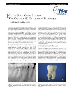
accuracy (up to 0.996) for the central upper (fig. 3) followed by the central lower and lateral lower due to their particular endodontic morphology with straight roots and well defined apex. The exception was the lateral maxilar incisors group with the lowest degree of accuracy ΔD = 0,984 because of the distooral root curvature in the apical third and the variety of apex location. In this case the digital odontometric method used a dot by dot measurement technique, the segmental method having no applicability (fig. 4).
Figure 3. ID and FD (11)
Figure 4. ID and FD (22)
The upper molars presented the widest endodontic variety and so, the digital measurements presents a wide range ΔD = 0,984-0,992 for the lower molars and ΔD = 0,981- 0,990 for the upper molars (fig. 5, 6). For the lower molars the segmental
measurement tool was used on the straight part of the root and the dot by dot addition measurement tool was used for the curved part until the detected radiographic apex was reached.
Figure 5. 16. Initial determination 128
Figure 6. 16. Final determination
Romanian Journal of Oral Rehabilitation Vol. 5, No. 3, July - September 2013 The different determinations can be explained by the misinterpretation of the mesial canals position and length due to the eccentrical projection. At the upper molars group the determinations were rather consistent for the oral and distobucal roots, major radiometric differences occurred for the mesiobucal root with extreme curvature and a high degree of overlapping with the oral root. At the opposite side, the curved canals final determinations may be shorten with up to 1mm comparative to initial determinations as the canal is straightened out by instrumentation.
2.
3.
4.
CONCLUSIONS 1. Radiographic digital techniques have a superior precision in identifying the radiologic apex in comparison with the classic film based visual method when they are used in combination with the selective image amplification and the
5.
histogram based on optical isodensity. Digital odontometry obtained with distances measuring programs has values comparable with the statistic determined root lengths and it eliminates the observers subjectivity based on different radiologic and clinical background. The radiographic digital working length determination has a quick learning curve and can be used in most cases regardless morphologic complexity and spatial particularities of the endodontic system. Digital odontometric technique has some limitations regarding the ortoradiality (especially for multiple root canals), but an individual digital approach for each root can easily overcome this problem. This digital method provides better results for narrow roots with a limited radiographic aspect on classic radiographs after an initial digital enhancement in order to get the correct working length.
REFERENCES 1 Marcov N., Popa B., Baştan E.C. Valoarea sistemelor digitale directe în determinările odontometrice. Revista Naţională de Stomatologie, 2002 vol V, nr. 3, pg.6-10. 2 Marcov N., Baştan E.C., Vârlan C.M., Vârlan V. Aprecierea odontometrică paraclinică a structurilor dentare cu morfologie radiculară particulară prin metode radiologice digitale directe. Revista Naţională de Stomatologie. 2002, vol V, nr.4, pg. 3-7. 3 Nisha Garg, Amit Garg. Textbook of Endodontics, 2010, Jaypee Brothers Medical Publishers 4 Orosco FA, Bernardineli N, Garcia RB, Bramante CM, Duarte MA, Moraes IG. In vivo accuracy of conventional and digital radiographic methods in confirming root canal working length determination by Root ZX. J Appl Oral Sci. 2012 Sep-Oct;20(5):522-5. 5 Alothmani, O.S., Friedlander, L.T., Monteith, B.D., Chandler, N.P Influence of clinical experience on the radiographic determination of endodontic working length. International Endodontic Journal 2013:46 (3), pp. 211-216 6 Woolhiser GA, Brand JW, Hoen MM, Geist JR, Pikula AA, Pink FE. Accuracy of film-based, digital, and enhanced digital images for endodontic length determination. Oral Surg Oral Med Oral Pathol Oral Radiol Endod. 2005 Apr;99(4):499-504.
129