Chinese-German J Clin Oncol
December 2014, Vol. 13, No. 12, P609–P613
DOI 10.1007/s10330-314-0014-8
Chylous ascites arises after chemotherapy of gastric signet ring cell carcinoma: A case report and review Kai Qin, Yi Cheng, Na Han, Jin Feng, Shiying Yu ( ) Department of Oncology, Tongji Hospital, Tongji Medical College, Huazhong University of Science and Technology, Wuhan 430030, China Received: 5 October 2014 / Revised: 28 October 2014 / Accepted: 20 November 2014 © Huazhong University of Science and Technology 2014 Abstract Chylous ascites, a rare clinical condition resulting from the disruption of the abdominal lymphatic system, usually diagnosed by paracentesis when the patients suffer ascites as primary symptom. The conditions, in which chylous ascites arise after chemotherapy of solid tumor, are rarely reported. In this paper we present a quite rare case of chylous ascites arising after chemotherapy of gastric signet ring cell carcinoma. Key words chylous ascites; gastric signet ring cell carcinoma; chemotherapy
Chylous ascites is a rare clinical condition resulting from the disruption of the abdominal lymphatic system caused by trauma or lymphatic obstruction. The diagnostic criteria of chylous ascites includes: the ascitic fluid is milky; the triglyceride level is greater than 200 mg/dL; aseptic; the positive results of chyle test and Sudan III test [1]. Multiple causes are responsible for chylous ascites, such as abdominal malignancy, liver cirrhosis, inflammation, congenital malformation, trauma, and miscellaneous disorders. Malignant diseases, as the most frequent cause in adults, are responsible for 17% of cases with atraumatic chylous ascites, in which lymphoma accounted for at least one third [2]. The chylous ascites is rarely caused by gastric malignancies, in which it is diagnosed by paracentesis when the patients suffer ascites as primary symptom [2]. Other conditions, such as chylous ascites arise after chemotherapy, are rarely reported. In this paper we present a quite rare case of chylous ascites arising after chemotherapy of gastric signet ring cell carcinoma. The clinical manifestations, treatment, prognosis of chylous ascites due to gastric malignancies were summarized based on literature search.
Case report A 31-year-old female patient was admitted to Gynecologic Oncology Ward in Tongji Hospital (Wuhan, China) with abdominal distention, right ovarian lesions and massive ascites revealed by ultrasound in January, 2014. She Correspondence to: Shiying Yu. Email:
[email protected]
was operated with an exploratory laparotomy, right oophorectomy and gastric omental biopsy. Intraoperative results: massive yellowish ascites with an enlarged right ovary (about 6 cm × 6 cm × 5 cm); multiple firm nodules in Dow cavity; great omentum wrapping around the transverse colon and gastric fundus; many palpable nodules on gastric body and transverse colon; multiple significantly enlarged retroperitoneal lymph nodes; normal appearance of uterus and left accessory. Histological results: gastric signet ring cell carcinoma; metastatic adenocarcinoma of the right ovary (possible gastrointestine-induced). Immunohistochemistrical results: CK7, Villin, CDX-2, TP, EMA, CK8/18 and CK19 were positive; CK20 and VEGF were scattered positive; Her-2, PR and ER were negative. Postoperative gastroscopy showed the distortion of gastric fundus, body and angle with bulky, stiff mucosal folds. The biopsy confirmed the adenocarcinoma of gastric body. Postoperative abdominal computed tomography (CT) scan revealed the thickened walls of stomach, rectum and urinary bladder; thickened great omentum; multiple enlarged retroperitoneal lymph nodes; ascites. Therefore the patient was diagnosed with gastric signet ring cell carcinoma, stage IV, with ascitic, peritoneal, great omental and pelvic metastasis. She received 2 cycles of chemotherapy with DCF regimen. The diuretic therapy and supportive care were given, too. The abdominal distention and pain were relieved after the treatments. The patient had a good quality of life. She was admitted to hospital again in March, 2014, with aggravated abdominal distension for one week. No complains of fever, nausea, vomiting, abdominal pain and
610
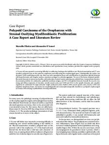
weight loss. The physical examination showed stable vital signs. The systemic examination was unremarkable. The abdomen was soft and distention. No rebound tenderness or tenderness was found, but shifting dullness was positive. Bowel sounds were audible. Blood analytical results: total leukocyte count, 9.43 × 10⁹ /μL; neutrophils, 72.4%; lymphocytes, 17.5%; hemoglobin, 136 g/L; platelet, 271 × 109 /L; sodium, 135.7 mmol/L; potassium, 4.53 mmol/L; urea, 5.81 mmol/L; creatinine, 53 μmol/L; glucose, 5.9 mmol/L; aspartate aminotransferase (AST), 15 U/L; aminotransferase (ALT), 19 U/L; total protein, 66.6 g/L; albumin, 38.6 g/L. Urinary analytical results: urinary protein (±). Abdominal computed tomography (CT) scan confirmed partial remission of enlarged retroperitoneal lymph nodes and thickened stomach wall, but massive ascites. Serum tumor markers results: carcino-embryonic antigen, 7.15 ng/mL; carbohydrate antigen 19-9, 2799.02 U/mL; carbohydrate antigen 72-4, 43.31 U/mL. Diagnostic abdominal paracentesis revealed 1200 mL of milky fluid in about 2 months after surgery (Fig. 1). Other ascitic characteristics: adenosine deaminase (ADA), 40 U/ L; triglycerides, 795 mg/dL; cholesterol, 104 mg/dL, carcinoembryonic antigen, > 1500 ng/mL; positive results of chyle test and Sudan III test. Cytology was positive for adenocarcinoma and the gram stain revealed no microorganisms. Therefore the ascites analysis suggested chylous ascites. In the first three days, the amount of drainage was about 1500–1800 mL/d after abdominocentesis, it had not decreased significantly until she received nutritional supportive therapy with a low-fat diet and medium-chain triglyceride supplementation, diuretics and octreotide treatment. As the disease evaluation was partial remission, she continued the chemotherapy with DCF regimen, with cisplatin intraperitoneal injection. The ascites reduced after 1 cycle and no reoccurrence had been observed. The chemotherapy continued.
http://zdlczl.chmed.net
Fig. 1 Milky fluid via abdominal paracentesis
Discussion Chylous ascites is a rare clinical condition. The incidence ranges from 1 in 20 000 to 1 in 187 000 [2]. One research from the Peking Union Medical College Hospital reported the incidence of chylous ascites was 6/100 000 in patients admitted to hospitals [3]. The ascites is the accumulation of a milky peritoneal fluid rich in triglycerides, due to the trauma or obstruction of thoracic or intestinal lymphatic vessels. The diagnostic criteria of chylous ascites includes: the presence of a milky and creamy ascitic fluid with a triglyceride content greater than 200 mg/dL, plus the positive results of chyle test and Sudan III test [3]. Multiple causes are responsible for chylous ascites, such as abdominal malignancy, liver cirrhosis, inflammation, congenital malformation, trauma, and miscellaneous disorders. Malignant diseases, as the most frequent cause
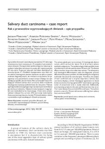
Fig. 2 Flow diagram of study selection process. P: Pubmed database; W: Wanfang database
in adults, are responsible for 17% of cases with atraumatic chylous ascites, in which lymphoma accounted for at least one third [2]. In this paper we present a quite rare case of chylous ascites arising after chemotherapy of gastric signet ring cell carcinoma, because of narrowing retroperitoneal lymph nodes. Two electronic databases (PubMed and Wanfang databases) were searched for the period from January 1950 to February 2014 (Fig. 2). The following MeSH terms were used: chyloascites, chylous ascites, chyloperitoneum and gastric tumor, gastric malignancy, gastric neoplasm, and gastric cancer. The references of the retrieved articles
Chinese-German J Clin Oncol, December 2014, Vol. 13, No. 12 Table 1
Case reports of chylous ascites in patients with gastric malignancies
Case No. Age (years) / sex 1
[4]
611
41/F
2 [5]
47/M
3 [6]
87/M
4 [7]
25/M
5 [8]
72/M
6 [9]
31/F
7 [10] 8 [11]
69/M 28/F
Our case
31/F
Diagnosis / stage
Clinical symptoms and signs
Gastric cancer / stage IV (bilateral ovary)
Abdominal tenderness, rebound tenderness, and guarding; afebrile; a palpable left supraclavicular node; CT scan showed bilateral ovarian tumors with minimal intraperitoneal fluid collection; laparoscope confirmed diffuse lymphatic ectasia and milky fluid discharge; there was no evidence of peritoneal metastasis Gastric signet ring cell carcinoma Abdominal distention, loss of weight, postprandial fullness, fatigue and anorexia; the abdominal ultrasound confirmed ascites and mild hepatomegaly; thrombocytopenia and disturbance of blood coagulation; paracentesis showed milky fluid rich in triglycerides (2225 mg/dL); CT revealed enlarged para-tracheal and mediastinal lymph nodes; gastroscopy showed infiltrative ulcerated gastric lesions with signs of bleeding Gastric diffuse large B cell lymphoma, stage I Loss of appetite, abdominal distension, hypoproteinemia, CT revealed ascites, ascitic biochemical analysis detected chylous ascites, endoscopy revealed nodular swelling of mucosa, swelling folds, and widespread erosions in the stomach Gastric Burkitt’s lymphoma Acute renal failure as a result of uric acid nephropathy and ascites (probably chylous) Gastric mucinous carcinoma, stage IV (ascites) Fatigue, anorexia, abdominal distension, weight loss; CT revealed ascites and liver cirrhosis; a milky ascitic fluid; the chyle test and Sudan III test were positive Gastric mucosa associated B cell lymphoma Abdominal distension, melena, shortness of breath; subxiphoid mass, pleural effusion and ascites; positive result of fecal occult blood test; gastroscopy showed gastric hyperplasia with ulcer Stomach carcinoma, stage IV Chylous ascites and chylothorax Gastric signet ring cell adenocarcinoma with Pleuritic chest pain; swollen right leg, left arm, and left endobronchial, mammary, ovarian, pleural, breast; previously subclavian vein thrombosis; bilateral pericardial, peritoneal, and osteal metastases pleural fluid; abdomen computed tomography revealed abundant ascites and right ovarian enlargement Gastric signet ring cell carcinoma, stage IV, Abdominal distention, a right ovarian tumor and abundant with ascitic, peritoneal, great omental and ascites; operated with an exploratory laparotomy, right pelvic metastasis oophorectomy and gastric omental biopsy; postoperative pathology and gastroscopy proved the diagnosis. She was admitted to hospital again because of aggravated abdominal distension. Diagnostic abdominal paracentesis revealed chylous ascites (to be continued)
were cross searched. The search was limited to studies in print publication with abstract available in English or Chinese. The clinical manifestations, treatment, prognosis of chylous ascites due to gastric malignancies were summarized (Table 1). Eight cases, plus our case, in which the patients suffering gastric malignancies were diagnosed with chylous ascites were found (Table 1). We summarized 9 cases of gastric malignancies with chylous ascites, 6 cases of gastric cancer, and 3 cases of lymphoma, in which there was five men, and four women. The mean age of onset was 47 years old. The pathological type of gastric cancer was adenocarcinoma and
signet ring cell carcinoma. Abdominal distention was the most common symptom, with 1 case of acute abdominal disease. Ascites was observed at the onset of the diseases, and its chylous character was confirmed by abdominal paracentesis. Daniel et al reported “bloating” as the main symptoms (81%) of atraumatic chylous ascites, and peritonitis was relatively infrequent (11%) [2]. Other chief complaints included acute renal failure, subxiphoid mass, and ovarian mass, all of which were related to the characteristics of gastric Krukenberg tumor and lymphoma. The cases 1–7 concluded the chylous ascites were usually diagnosed as the primary symptom by paracentesis.
612
http://zdlczl.chmed.net
Table 1 (Continued) Case reports of chylous ascites in patients with gastric malignancies Case No.
Cytology
Treatment of chylous ascites
[4]
Unknown
The TAH-BSO, a fat-free diet, additional chemotherapy
2 [5]
Unknown
3 [6] 4 [7]
Unknown Unknown
5 [8]
Positive
6 [9] 7 [10] 8 [11] Our case
Unknown Unknown Positive Positive
The patient did not have good response to total parenteral nutrition (TPN), somatostatin therapy and (MCT)-based diet transfusion support Chemotherapy with R-CHOP regiment Regression of stomach tumor after chemotherapy and the chylous effusion was observed Liver protective therapy, diuresis, anti-infection, supportive therapy and palliative operation Chemotherapy Unknown Unknown Nutritional supportive therapy with a low-fat diet, diuretics, octreotide and palliative chemotherapy
1
Prognosis No reoccurrence of ascites, additional chemotherapy continued The patient died on day 20 of hospitalization, because of DIC and respiratory failure Disease complete remission Tumor lysis syndrome; die of gastrointestinal bleeding Death on postoperative day 10 Unknown Unknown Unknown No reoccurrence of ascites, palliative chemotherapy continued
Note: CT: computed tomography; TAH-BSO: total abdominal hysterectomy, bilateral salpingooophorectomy; MCT: medium chain triglyceride; DIC: disseminated intravascular coagulation; R-CHOP: rituximab plus cyclophosphamide, doxorubicin, vincristine, and prednisone
However, our case showed yellowish, clear ascites at the beginning proved by the surgery. The diagnosis of gastric signet ring cell carcinoma was confirmed and the palliative chemotherapy was given. After two months, partial remission of enlarged retroperitoneal lymph nodes (Fig. 3) and thinning stomach wall were confirmed after 2 cycles of chemotherapy, while chylous ascites appeared. The causes of chylous ascites were very confused, it was important to rule out lymphatic injuries caused by surgery. Chylous ascites can occur early (around 1 week) after abdominal surgery because of disruption of the lymphatic vessels, or late (several weeks to months) because of adhesions or extrinsic compression of lymphatic vessels [12]. The history of bloating in our patient was about 1 week, characterized by acute onset in about 2 months after surgery, abdominal distension had no obvious relief after abdominal cavity drainage. Postoperative chyle leakage caused by invisible damage did not accord with the clinical symptoms, also can be ruled out. Because of the article described that: abdominal aorta, renal vein level is lymphatic relatively bulky and concentrated area, the damage in this area may be associated with lymphatic leakage formation [3]. Coincidentally, our patient had narrowed renal vein level retroperitoneal lymph nodes after chemotherapy. It may caused by chyle leakage as a result of narrowing retroperitoneal lymph nodes. To speak frankly, it would be complete lymphography to know the cause and location of chyle leakage. The studies of chylous ascites treatment are limited, addressing the best treatment regimens are hardly developed. One referred measure is a low-fat, low-sodium, high-protein diet with medium-chain triglyceride supplementation to reduce the production and flow of chyle.
Fig. 3 Abdominal CT (the white arrow) showed significantly narrowing retroperitoneal lymph nodes (a); but massive ascites appeared (Contrast with CT before chemotherapy; b)
Somatostatin therapy also remains an indefinite or second-line method in the treatment of chylous ascites. The possible mechanisms was not only decrease the intestinal absorption of fats, lower triglyceride concentration in the thoracic duct and attenuate lymph flow in the major lymphatic channels; but also decreases gastric, pancreatic
Chinese-German J Clin Oncol, December 2014, Vol. 13, No. 12
and intestinal secretions, inhibits motor activity of the intestine, slows the process of intestinal absorption and decreases splanchnic blood flow, which may further contribute to decreased lymph production. It has also been speculated that somatostatin improves chylous ascites by inhibition of lymph fluid excretion through specific receptors found in the normal lymphatic vessels of intestinal wall [13]. Diuretics and paracentesis are indicated to relieve symptoms and improve renal function. For the obstruction or rupture of lymphatic vessels, surgical procedure should be considered earlier, like in Case 1. Meanwhile, many authors believe that reasonable anti-cancer treatment can improve outcomes of malignant chylous ascites. The prognosis of chylous ascites mostly depends on the underlying pathological conditions causing lymphatic leakage. Press et al reported that 1 year mortality rate of chylous ascites was 71%, which caused by malignant tumor of up to 90% [1]. We summarized 9 cases: 2 cases of gastric adenocarcinoma were controlled well after treatment; 2 cases died of severe complications within few months; 1 case of lymphoma was cured after chemotherapy; 1 case of lymphoma might succumb to tumor lysis syndrome; 3 cases were unknown. Conclusion Chylous ascites, a rare presentation of ascites, may be diagnosed with ascites as the primary symptom by the method of abdominal paracentesis. However, in our paper we presented a quite rare case of chylous ascites associated with gastric signet ring cell carcinoma after chemotherapy. The reason might be chyle leakage as a result of narrowing retroperitoneal lymph nodes. The optimal management of chylous ascites was unknown, however, a low-fat, low-sodium, high-protein diet with mediumchain triglyceride supplementation, somatostatin, diuretics, paracentesis, and reasonable anti-cancer treatment were preferred to improve its prognosis. Conflicts of interest The authors indicated no potential conflicts of interest.
613
References 1. Press OW, Press NO, Kaufman SD. Evaluation and management of chylous ascites. Ann Intern Med, 1982, 96: 358–364. 2. Steinemann DC, Dindo D, Clavien PA, et al. Atraumatic chylous ascites: systematic review on symptoms and causes. J Am Coll Surg, 2011, 212: 899–905. 3. Wang XR. Review of domestic literature from the 247 patients with chylous ascites. J Clin Gastroenterol (Chinese), 2009, 21: 41–43. 4. Kang CM, Kim S, Kim BW, et al. Acute chylous peritonitis mimicking ovarian torsion in a patient with advanced gastric carcinoma. J Korean Med Sci, 2007, 22 Suppl: S164–S166. 5. De Mello RA, Gregório T, Cardoso T. Chylous ascites due to signet ring cell gastric adenocarcinoma. Niger J Clin Pract, 2012, 15: 487–490. 6. Yamamoto Y, Kumei S, Ogura T, et al. A case of primary gastric malignant lymphoma with chylous ascites. Nihon Shokakibyo Gakkai Zasshi (Japanese), 2013, 110: 1790–1796. 7. Kabat-Koperska J, Kutrzeba J, Chosia M. Acute kidney failure and ascites in Burkitt’s lymphoma of the stomach. Pol Arch Med Wewn (Polish), 2001, 105: 67–70. 8. Ma YY, Li HY, Li ZY, et al. A case of gastric mucinous adenocarcinoma with chylous ascites: a case report. Beijing Med J (Chinese), 2000, 22: 156. 9. Kang H, Huang YJ. A case of gastric malignant lymphoma with chylous ascites. Chin J Moder Med (Chinese), 2000, 10: 3. 10. Segal R, Waron M, Reif R, et al. Chylous ascites and chylothorax as presenting manifestations of stomach carcinoma. Isr J Med Sci, 1986, 22: 897–899. 11. Kayacan O, Karnak D, Ayşe Can B, et al. Gastric signet-ring cell adenocarcinoma presenting with left arm deep-vein thrombosis and bilateral chylothorax. Clin Appl Thromb Hemost, 2008, 14: 476–480. 12. Cárdenas A, Chopra S. Chylous ascites. Am J Gastroenterol, 2002, 97: 1896–1900. 13. Huang Q, Jiang ZW, Jiang J, et al. Chylous ascites: treated with total parenteral nutrition and somatostatin. World J Gastroenterol, 2004, 10: 2588–2591.
DOI 10.1007/s10330-314-0014-8 Cite this article as: Qin K, Cheng Y, Han N, et al. Chylous ascites arises after chemotherapy of gastric signet ring cell carcinoma: A case report and review. Chinese-German J Clin Oncol, 2014, 13: 609–613.