1262
ORIGINAL ARTICLE
Weakness Is the Primary Contributor to Finger Impairment in Chronic Stroke Derek G. Kamper, PhD, Heidi C. Fischer, MS, OTR/L, Erik G. Cruz, William Z. Rymer, MD, PhD ABSTRACT. Kamper DG, Fischer HC, Cruz EG, Rymer WZ. Weakness is the primary contributor to finger impairment in chronic stroke. Arch Phys Med Rehabil 2006;87:1262-9. Objective: To assess the relative contributions of several neurologic and biomechanic impairment mechanisms to overall finger and hand impairment in chronic hemiparetic stroke survivors. Design: Repeated-measures design. Setting: Clinical research laboratory. Participants: Thirty stroke survivors with chronic hemiparesis. Fifteen subjects had severe hand motor impairment and 15 had moderate impairment, as measured with the ChedokeMcMaster Stroke Assessment. Interventions: Not applicable. Main Outcome Measures: The biomechanic factors stiffness and resting flexion torque, together with the neurologic factors spasticity, strength, and coactivation, were quantified by using a custom hand manipulator, a dynamometer, and electromyographic recordings. Both passive and active rotations of the metacarpophalangeal joints of the fingers were examined. Results: Although subjects in the severely impaired group exhibited statistically greater passive stiffness and resting flexion torque than their moderately impaired counterparts (P⬍.05), the overall effect of these biomechanic changes appeared small in relation to the deficits attributable to neurologic changes such as spasticity and, especially, weakness. In fact, weakness in grip strength and isometric extension accounted for the greatest portion of the variance between the 2 groups (2⫽.40 and 2⫽.23, respectively). Conclusions: Thus, deficits in hand motor control after stroke seem to derive mainly from weakness, which may be attributable to the loss of descending corticospinal pathway activation of motoneurons. Key Words: Hand; Human; Muscle spasticity; Muscle weakness; Rehabilitation; Stroke. © 2006 by the American Congress of Rehabilitation Medicine and the American Academy of Physical Medicine and Rehabilitation
From the Sensory Motor Performance Program, Rehabilitation Institute of Chicago, Chicago, IL (Kamper, Fischer, Cruz, Rymer); Department of Biomedical Engineering, Illinois Institute of Technology, Chicago, IL (Kamper); Jesse Brown VA Medical Center Lakeside CBOC, Chicago, IL (Rymer); and Department of Physical Medicine and Rehabilitation, Northwestern University Feinberg School of Medicine, Chicago, IL (Rymer). Supported by the Department for Veterans Affairs (merit review grant no. 537D46088). No commercial party having a direct financial interest in the results of the research supporting this article has or will confer a benefit upon the author(s) or upon any organization with which the author(s) is/are associated. Reprint requests to Derek Kamper, PhD, Sensory Motor Performance Program, Rehabilitation Institute of Chicago, Ste 1406, 345 E Superior St, Chicago, IL 606011, e-mail:
[email protected]. 0003-9993/06/8709-10667$32.00/0 doi:10.1016/j.apmr.2006.05.013
Arch Phys Med Rehabil Vol 87, September 2006
HRONIC HEMIPARESIS IS A common outcome of stroke. Roughly one third of all people who experience a stroke will C have some residual impairment of the upper extremity. Hand 1-3
function is routinely compromised, with impairment of finger extension being the most common contributor.4 Questions remain, however, regarding the origins of these deficits. The stroke produces an initial flaccid paresis, which is gradually replaced by hypertonicity in the muscles flexing the fingers5-7 and a stereotypically flexed resting hand posture. Certain muscle groups are more likely than others to achieve some degree of recovery, such that force production and movement in the flexion direction, for example, is relatively better than that in extension or abduction and adduction.8 These secondary changes to the initial stroke presentation have been attributed to a number of mechanisms. Although some researchers have emphasized biomechanic alterations, such as contracture,9 muscle atrophy,10 and increased muscle stiffness,11,12 other researchers have focused on neurologic changes such as spasticity,5,13,14 excessive coactivation of flexors and extensors,15,16 and reduced reciprocal inhibition.17,18 The relative contributions of the various mechanisms to the overall hand impairment have not been fully evaluated. This knowledge is important because treatment of 1 mechanism may be very different, or even in conflict with, treatment for another mechanism. As an example, questions remain as to whether perceived changes in joint stiffness after stroke have a primarily mechanical9,12 or neurologic7,19 origin. Proposed treatment of the former, with bracing or musculotendon lengthening,20 may not be beneficial if the source is neurologic. Alternatively, treatment of heightened dynamic joint stiffness with drugs reducing muscle excitation, such as botulinum toxin21 or baclofen,22 may not be efficacious if mechanical tissue changes are primarily responsible. Thus, in our present study, we sought to determine the extent to which each potential mechanism contributes to the overall deficits in motor control. Reflex properties and voluntary muscle activation of both hands were evaluated in 30 stroke survivors with chronic hemiplegia. Based on clinical evaluations, subjects were evenly divided between those with severe and moderate impairment. We hypothesized that the group with severe impairment would show more evidence of neurologically based mechanisms (spasticity, coactivation of flexors and extensors, weakness) but not mechanically based changes (contracture, passive stiffness) than the group with moderate impairment. METHODS Participants Thirty subjects with chronic hemiparesis subsequent to stroke participated in the study. Subjects were recruited through fliers and from a registry of voluntary participants, both approved by the institutional review board of Northwestern University. All subjects had to have a clinical history of a single stroke occurring at least 9 months before testing (range, 10mo–21y). Subjects were selected on the basis of severity of hand motor impairment, as assessed by a research occupational therapist
1263
WEAKNESS AND FINGER IMPAIRMENT IN STROKE, Kamper Table 1: Subject Characteristics Group
Age (y)
Time Since CVA (y)
FMA*
Right-/Left-Hand Impairment
Impairment of Dominant/Nondominant Arm
Severely impaired Moderately impaired
57⫾12 59⫾11
6.8⫾5.4 5.9⫾4.2
29⫾12 45⫾10
6/9 9/6
5/10 9/6
NOTE. Values are mean ⫾ standard deviation (SD). Abbreviations: CVA, cerebrovascular accident; FMA, Fugl-Meyer Assessment. *FMA score represents the total score for the upper-extremity section (/66).
using the stage of hand section of the Chedoke-McMaster Stroke Assessment (CMSA).23 Further inclusion criteria included passive supination of the forearm into a neutral posture and the presence of motor activation in the finger flexors. All subjects gave informed consent according to the Declaration of Helsinki. Fifteen of the subjects were classified as having severe hand impairment (CMSA stage of hand section, stage 2 or 3), whereas the other 15 subjects were classified as having moderate hand impairment (stage 4 or 5). The CMSA consists of a 7-stage rating scale corresponding to 7 stages of motor recovery, which follow a stereotypical course according to Brunnstrom’s theory.24 Thus, the severely impaired group showed some flexion activity (active or reflexive) but limited finger extension. The moderately impaired group exhibited significant control of mass finger extension and flexion but had difficulty with controlled movement of individual digits or joints. Anatomic magnetic resonance images revealed that hemispheric lesions for both subject groups were widely distributed, with all subjects having a mixture of cortical and subcortical infarcts. The side of the body with predominant impairment varied across subjects (table 1). The upper-extremity score for the Fugl-Meyer Assessment (FMA)25 was also obtained for each subject (see table 1 for a summary of group characteristics). There were no significant differences in either age or time since the stroke between the groups (P⬎.3). Protocol The less-affected (unimpaired) hand served as the control for the more-affected (impaired) hand for each subject. Although deficits in the unimpaired hand have been reported,26,27 we believe that the ability to exactly match age, hand dimensions, lifestyle, and numerous other factors permits a better comparison than attainable with data from neurologically intact, agematched subjects for the outcome variables of interest in this study. Thus, subjects participated in 2 separate testing sessions, one for each hand. Each subject completed the same protocol, with the trials run in the same order. Grip strength was first recorded using a dynamometer.a Assistance was provided if needed to position the digits on the device. Maximum grip forces were recorded sequentially for both hands. A rotary actuator was used for the rest of the trials.5,6,28 The wrist was placed in a cast such that both wrist and forearm were maintained in neutral positions of flexion and extension and pronation and supination, respectively. The palm was positioned perpendicular to a tabletop, aligning the metacarpophalangeal (MCP) joints of all 4 fingers with the shaft of a servomotor. The fingers were coupled to the shaft through a custom vacuum pillow, as shown elsewhere.5,28 Thus, rotation of the motor shaft produced equivalent rotation of the MCP joints of all 4 fingers simultaneously and vice versa. Range of MCP joint rotation was chosen to cover the full comfortable range for each subject, as permitted by the experimental device. Fixed extension and flexion limits were used throughout each
experimental session, with the same limits applied to both the impaired hand and unimpaired hand for a given subject. Across all subjects, the range of MCP joint rotation was 55.1°⫾10.1°. MCP torque,b angle,c and angular velocityd were measured throughout the trials. Muscle activity was recorded by using active surface electromyographic electrodes.e Differential electrodes were placed over the flexor digitorum superficialis (FDS) to sample an extrinsic flexor muscle, extensor digitorum communis (EDC) to sample an extrinsic extensor muscle, and the first dorsal interosseous (FDI) to sample an intrinsic muscle. The experimental trials consisted of a series of either voluntary contractions or imposed MCP rotations, intended to examine spasticity, muscle coactivation, strength, and passive stiffness. Spasticity, a velocity-dependent stretch reflex,29 was assessed by using the servomotor to impose constant-velocity rotation of the MCP joints from the flexion limit to the extension limit. The fingers were held at the extension limit for 2 seconds before being rotated back to the flexion limit. Subjects remained declaratively relaxed throughout the trial. Two speeds were used: a speed slow enough not to produce a stretch reflex (10°/s) and 1 fast enough to trigger a stretch reflex (300°/s).5,28 Three trials were run at each speed. For 6 additional trials, 3 at 300°/s and 3 at 10°/s, a short cutaneous stimulus was applied to the dorsal surface of the hand immediately before the initiation of stretch through surface electrodes.f The magnitude of the pulse was adjusted to a level just below pain threshold. A train of 10 pulses evenly spaced 20-ms apartg began 300ms before the start of the stretch, such that the stimulation period ended 100ms before the stretch. This sequence of trials was repeated with stimulation applied to the palmar surface of the hand. Maximum voluntary isometric MCP extension and flexion torques were then recorded. Each subject was asked to push the proximal phalanges against the vacuum pillow entrapping the fingers whereas the motor maintained a neutral (0° of finger extension) MCP posture. Three trials were performed for both extension and flexion. All trials were followed by 1-minute rest periods. Because of limitations with the servomotor and torque cell, only isometric extension could be maximally performed with the unimpaired hand. Lastly, small perturbations were applied to the MCP joints to determine the passive characteristics of the fingers. Subjects were asked to relax while the motor imposed a pseudorandom binary sequence (PRBS) of ⫾2° steps over a 10second period.30-32 The PRBS perturbations were applied at a series of operating points, spaced 10° apart, spanning the available passive MCP range of motion. The same operating points were used for both hands in a given subject. Analysis Grip strength was characterized by the maximum force achieved. The force values obtained were averaged across the 2 testing sessions for each hand. The grip strength for the impaired hand was then normalized by the grip strength for the unimpaired hand. Arch Phys Med Rehabil Vol 87, September 2006
1264
WEAKNESS AND FINGER IMPAIRMENT IN STROKE, Kamper
Torque, velocity, and angle data for the other trials were low-pass filtered at 150Hz with a fourth-order Butterworth filter, before sampling at 500Hz through an analog-to-digital board.h The data were subsequently digitally low-pass filtered at 10Hz by using a 30-order finite impulse response filter. Raw electromyographic data were band-pass filtered between 10 to 225Hz before sampling. The signals were then rectified and low-pass filtered at 10Hz to generate envelopes for the electromyographic activity. Envelope amplitudes were normalized by the maximum value recorded during the experimental session. Flexor spasticity was quantified by using torque measures rather than electromyographic activity because imposed rotation of the MCP joints may trigger a reflex in a number of muscles. The peak torque, referenced from the initial resting torque, was calculated for the period from the start of rotation to the end of the hold phase. The mean peak torque for the slower (10°/s) trials was subtracted from the mean peak torque for the faster (300°/s) trials to obtain the velocity-dependent torque. Because passive tissue characteristics may increase the resistance to stretch for increasing stretch velocities, we attempted to estimate the magnitude of this effect by measuring the velocitydependent torque for the unimpaired hand. This torque was subtracted from the velocity-dependent torque of the impaired hand to yield the reflex torque. The peak reflex torque was used as the measure of spasticity. The delay between the start of stretch and the onset of any muscle response was quantified for each fast trial. Baseline levels of electromyographic activity were recorded for the period prior to initiation of the stretch, and the standard deviation (SD) of this activity was determined. The time between stretch initiation and the point in which muscle activity first exceeded the baseline value by 3 SDs was designated as the reflex delay for a given muscle. The mean reflex delay was obtained by averaging these values for each subject. Isometric strengths in flexion and in extension were estimated by measuring the torque generated while the MCP joints were held in the neutral position. The maximum torque value for each trial was obtained by averaging across a 100-ms window centered at the point of maximum torque in the specified (flexion or extension) direction. Muscle coactivation was also assessed during the isometric flexion and extension trials. For each isometric extension trial, the maximum extensor electromyographic value was found by averaging across a 100-ms window centered about the point of maximum EDC activity. FDS and FDI activity were then averaged across the same window as the EDC electromyographic activity. Electromyographic activity rather than torque was used to determine the point of maximal activity to account for trials in which subjects were unable to generate an extension torque. The maximum muscle activity for each flexion trial was obtained by averaging all 3 electromyographic signals across a 100-ms window centered at the point of maximum flexion torque. Coactivation analyses were then performed on the ratio of muscle activity during intended flexion or extension trials. Passive stiffness was computed from the trials of PRBS perturbations. Each trial record was segmented into a series of steps from flexion to extension based on the angular velocity signal. Least-squares estimation was used to fit a parametric model to the recorded system response. One of 2 models was used, depending on the detection of an electromyographic reflex response. A reflex was determined to have occurred if the level of the FDS or FDI electromyographic activity exceeded baseline activity by more than 3 SDs after the initiation of extension. Before the reflex, or if no reflex was detected, a passive second-order inertia (I)⫺damping (B)⫺stiffness (K) Arch Phys Med Rehabil Vol 87, September 2006
model was assumed.30 The parameters were estimated through multiple linear regression using the measured angle, angular velocity, and torque signals and the computed angular acceleration:
⫽ I¨ ⫹ B˙ ⫹ K( ⫺ 0) ⫹ s
(1)
where is the measured torque; s is the static offset torque; I is the inertia coefficient; B is the damping coefficient; K is the stiffness coefficient; is the MCP angle; ˙ is the MCP angular velocity; and ¨ is the MCP angular acceleration. The full parametric model, which includes terms representing the reflex contribution to the recorded torque, was fit to the portion of the step after reflex initiation using nonlinear leastsquares estimation:
⫽ I¨ ⫹ B˙ ⫹ K( ⫺ 0) ⫹ Br˙ ⫹ Kre⫺
冉 冊 * 共 ⫺ 兲 ⫹ , t ⱖ t t⬘ ⫺ ␣ 2
0
s
reflex
(2)
where Br is the reflex damping coefficient, Kr is the reflex stiffness coefficient, ␣ is the 30ms, is the 60ms, treflex is the time to reflex initiations, and t= is the t⫺treflex. The gaussian reflex stiffness term, along with the values of ␣ and , were chosen to mimic the twitch response for a mixed muscle.33 The optimized values for I, B, K, Br, and Kr were averaged across each perturbation within a trial. The variance accounted for in the torque signal by the model was computed for each trial. Statistics To test the impact of the different mechanisms on functional impairment, a doubly multivariate analysis of variance (ANOVA) was performed by using SPSS software.i The following dependent variables were used in the analysis: the normalized grip strength, the peak isometric extension (ISO_EXT), the peak spastic reflex torque (SPAS), the flexor:extensor activity ratio during voluntary extension, the extensor:flexor activity ratio during voluntary flexion, dynamic stiffness (K), and static stiffness (OFF_TRQ). The independent variable was subject impairment group. Where the multivariate analysis of variance results proved significant according to the Wilks value, subsequent univariate ANOVA testing of each dependent variable was performed. The 2 values were computed to estimate the variance explained by each dependent variable. The impact of cutaneous stimulation on the magnitude of the spastic reflex response was assessed independently. Reflex torques obtained from trials with palmar and dorsal stimulation and without stimulation were compared through a repeated-measures ANOVA. Finally, to test for an association with global arm impairment, the outcome measures listed above were included in a multiple linear regression versus the upper-extremity FMA score. Model variables were included in a stepwise fashion, with an entry criterion of P less than .05 and a deletion criterion of P greater than .10. RESULTS We found that there was an orderly relation between overall impairment severity, evaluated with the CMSA, and several quantitative measures of impairment sources, as revealed by the multivariate analysis (Wilks ⬍.001). Subsequent univariate testing of the dependent variables showed most of the
WEAKNESS AND FINGER IMPAIRMENT IN STROKE, Kamper
1265
Table 2: Univariate Results Group Means Dependent Variable
GRIP (% unimp) ISO_ EXT (% unimp) SPAS (Nm) K (Nm/°) CO_FLX OFF_TRQ (Nm) CO_EXT
Severe
Moderate
P
2
22⫾12 45⫾16 ⬍.001 .399 10⫾16 33⫾26 .007 .232 0.88⫾0.61 0.36⫾0.37 .008 .226 0.011⫾0.016 ⫺0.003⫾0.013 .016 .191 1.06⫾0.78 0.53⫾0.46 .032 .153 0.28⫾0.25 0.12⫾0.17 .053 .128 0.83⫾0.35 0.65⫾0.61 .325 .035
NOTE. Values are mean ⫾ SD. Abbreviations: CO_EXT, extensor:flexor activity ratio during voluntary flexion; CO_FLX, flexor:extensor activity ratio during voluntary extension; GRIP, grip strength; ISO_EXT, peak isometric extension; K, dynamic stiffness; OFF_TRQ, static stiffness; SPAS, peak spastic reflex torque; % unimp, percentage of value for unimpaired hand.
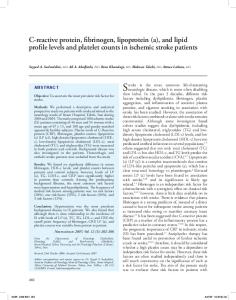
potential impairment mechanisms tested were significantly different for the 2 subject groups (table 2). For example, the peak spastic reflex torque was more than twice as great for the severely impaired subjects as for the moderately impaired subjects (fig 1). The threshold delay until the initiation of reflex electromyographic activity was shorter for the severely impaired group, although the difference was marginally insignificant (61⫾23ms vs 79⫾27ms, P⫽.06). Preconditioning of the stretch reflex with cutaneous stimulation to either the palmar or dorsal surface of the hand had no effect on the magnitude of the spastic stretch reflex (P⫽.53). Abnormal muscle activation patterns were also seen during voluntary isometric extension of the MCP joints. During voluntary extension, the more severely impaired subjects exhibited a much greater ratio of normalized flexor to extensor activity for the impaired hand (P⫽.032) but not for the unimpaired hand (.31⫾.17 vs .33⫾.29, respectively) (fig 2). The higher ratio resulted primarily from smaller extensor activation rather than greater flexor activation. There was no significant difference in extensor coactivation during voluntary flexion between the 2 groups (P⫽.32).
Fig 1. The difference in the reflex torque response to stretch between the 2 hands for the severely impaired and moderately impaired subjects. Error bars represent 1 SD. The response was larger for the severely impaired subjects (P