British Journal of Rheumatology 1998;37:1054–1059
CHLAMYDIA TRACHOMATIS ANTIBODY DETECTION AND DIAGNOSIS OF REACTIVE ARTHRITIS S. BAS and T. L. VISCHER Division of Rheumatology, Department of Internal Medicine, University Hospital, Geneva, Switzerland
SUMMARY Objective. To investigate whether determining the presence of serum or synovial fluid (SF ) IgG and IgA of anti-Chlamydia antibodies with two recent commercially available enzyme-linked immunosorbent assays (ELISA) using synthetic peptides or recombinant antigen could be helpful to detect possible Chlamydia trachomatis (CT )-involved disease in rheumatological patients without evidence of urogenital CT infection. Methods. The prevalence of such antibodies was determined in samples from patients with well-defined disease, i.e. CT sexually acquired arthritis and from patients with other inflammatory arthropathies unrelated to CT. Results. When considering IgG and/or IgA anti-MOMP or anti-LPS antibodies, a sensitivity of 100% was obtained for serum and SF samples, but with a low specificity. A sensitivity and a specificity equal or close to 80% were observed for the SF IgG anti-MOMP antibodies. Conclusion. Clinically, the most appropriate determination was the SF IgG anti-MOMP antibodies. This commercially available ELISA test could be useful for the diagnosis of probable CT reactive arthritis. K : Sexually acquired reactive arthritis, Chlamydia trachomatis, Antibodies, Enzyme-linked immunoassay, Synovial fluid.
R arthritis is characterized by asymmetrical mono/oligoarthritis, following a urogenital or intestinal infection. Chlamydia trachomatis (CT ) is often responsible for the urogenital infection, even in the absence of direct clinical symptoms [1–3]. The diagnosis of C. trachomatis sexually acquired reactive arthritis (CT-SARA), defined as arthritis with evidence of urogenital CT infection, has recently been improved by the development of different commercially available kits used to detect CT nucleic acids in urogenital smears and urine from symptomatic or silently infected patients. However, these tests are not useful if arthritis develops after the initial phase of infection, when the bacteria are not detectable any more. In such cases, serological methods in combination with anamnesis, clinical picture and course of disease could provide a strong indication for probable CT-SARA. To this end, we have developed and evaluated an enzyme-linked immunosorbent assay ( ELISA) using Chlamydia elementary bodies as antigens [4]. The main problems were the low sensitivity of this ELISA and the fact that it could also detect antibodies against Chlamydia pneumoniae or Chlamydia psittaci. As anti-C. pneumoniae antibodies are highly prevalent in the adult population [5, 6 ], the relatively low specificity might be attributed to the presence of these antibodies. Since then, two apparently sensitive commercially available ELISA tests, using synthetic peptides or recombinant antigen, have been developed. One uses synthetic peptides derived from species-specific epitopes in the variable domain IV of the major outer membrane protein (MOMP) of CT and not homolo-
gous to C. pneumoniae MOMP (Labsystems Research Laboratory, Helsinki, Finland). The other is based on an exclusively Chlamydia-specific recombinant fragment of the total lipopolysaccharide (LPS) (3-deoxy-manno-2-octulopyranosonic acid or 3-Kdo) (Medac GmbH, Hamburg, Germany). This antigen, genus specific, allows the detection of antibodies against C. trachomatis, C. pneumoniae and C. psittaci. We therefore used these commercially available ELISA tests for two main purposes. First, to differentiate the CT responses (determined with the MOMP antigen) from the responses against any Chlamydia species (determined with the LPS antigen). Secondly, to estimate whether these tests could allow the identification of some probable CT-SARA cases among patients defined as SARA (without evidence of urogenital CT infection) or undifferentiated mono/oligoarthritis. Their performances were evaluated in terms of sensitivity and specificity by comparing the serum and synovial fluid (SF ) responses of samples from patients with well-defined disease, i.e. CT-SARA, with samples from patients with other inflammatory arthropathies unrelated to CT. Since it is well known that antibodies to CT are present in a large number of cases with no clinical or microbiological evidence of chlamydial infection [7, 8], we also examined serum samples from healthy blood donors. PATIENTS AND METHODS Patients Serum and SF samples came from our collection and have been kept for various times at −70°C (after centrifugation for 10 min at 1600 g for SF ). The diagnosis was made at the time of sample collection. All the patients were questioned about possible genitourinary infection (urethritis, dysuria, discharge).
Submitted 23 January 1998; revised version accepted 28 May 1998. Correspondence to: S. Bas, Research Laboratory, Division of Rheumatology, University Hospital, 1211 Geneva 14, Switzerland.
© 1998 British Society for Rheumatology 1054
BAS AND VISCHER: CHLAMYDIA TRACHOMATIS ANTIBODIES IN ARTHRITIS
Serum and SF samples came from individuals distributed in the following groups. 1. CT-SARA (19 sera and 13 SF ): asymmetrical mono/oligoarthritis with urethritis and evidence of urogenital CT infection [three had a positive urethral/endocervical Chlamydia antigen detection by direct immunofluorescence, 14 had a positive urethral/endocervical Chlamydia culture, two had a positive urethral/endocervical CT DNA amplification with the Amplicor test of Roche Diagnostic Systems Inc. (Branchburg, NJ, USA)]; HLA-B27: nine positive and six negative patients, four not determined. 2. SARA (17 sera and nine SF ): asymmetrical mono/ oligoarthritis with urethritis, but without proof or evidence for CT infection (nine had a negative urethral/endocervical Chlamydia detection, in eight patients no search was made for various reasons); HLA-B27: three positive and four negative patients, 10 not determined. 3. Undifferentiated rheumatoid factor-negative mono/ oligoarthritis (58 sera and 30 SF ): 1–4 joints, asymmetrical, mainly lower extremities; absence of evidence for another defined rheumatic disease; HLA-B27: four positive and 22 negative patients, 32 not determined. 4. Other inflammatory arthropathies (19 sera and 10 SF ): rheumatoid factor-positive rheumatoid arthritis (RA) (n = 4), gout (n = 6), septic arthritis (n = 5), Lyme arthritis (diagnosis confirmed by Western blot) (n = 3), Salmonella enteritidis reactive arthritis (n = 1). The urethral/endocervical Chlamydia detection was not performed for these patients and was presumed to be negative. 5. Healthy blood donors (31 sera).
1055
Median age (yr), range and percentage of female individuals are given in Tables I and II. Methods The ELISA kits detecting the IgG and IgA anti-CT MOMP antibodies were purchased from Labsystems Research Laboratory (Helsinki, Finland). The ELISA kits detecting the IgG, IgM and IgA anti-Chlamydia LPS antibodies were purchased from Medac GmbH (Hamburg, Germany). The assays and calculations were performed according to the manufacturer’s instructions. Briefly, for the anti-MOMP assays, the optical density (OD) values were measured at a wavelength of 450 nm and the cut-offs calculated from the OD values obtained for positive controls. For the antiLPS assays, the OD were measured at 405 nm (reference wavelength 490 nm) and the cut-offs calculated from the OD values obtained for negative controls. We considered as >cut-off, values interpreted as positive according to the Medac instructions, i.e. � cutoff + 10%. These kits therefore give qualitative rather than quantitative results. Calculations Sensitivity, specificity, positive predictive value, negative predictive value, false-positive and falsenegative rates were calculated as described previously [4]. Statistics The percentages of positive samples obtained for groups 1 (CT-SARA) and 4 (inflammatory arthropathies unrelated to CT ) were compared using Fisher’s exact test.
TABLE I Percentage of positive sera for anti-Chlamydia antibodies Anti-MOMP Group
Age* (yr)
Anti-LPS
IgG
IgA
IgG
1. Chlamydia trachomatis sexually acquired reactive arthritis (n = 19)
26 (32%) 18–55
79**
53
89**
2. Uroarthritis (n = 17)
40 (29%) 22–62
53
59
3. Idiopathic mono/ oligoarthritis (n = 58)
43 (43%) 16–77
29
4. Inflammatory arthropathies (n = 19)
39 (32%) 17–46
5. Healthy blood donors (n = 31)
28 (29%) 20–39
IgM IgA
IgG and/or IgA anti-MOMP (no IgG and/or IgA anti-LPS )
IgG and/or IgA anti-LPS (no IgG and/or IgA anti-MOMP)
IgM anti-LPS and IgG and/or IgA anti-MOMP
5
68
5
21
5
76
12
82
0
18
0
50
71
12
48
5
22
9
42
37
58
5
42
5
26
0
42
32
52
3
23
13
23
0
*Median (percentage of female patients) range. **P < 0.05; Fisher’s exact test between groups 1 and 4.
20 40
0 0
RESULTS Distribution of serum and SF anti-Chlamydia antibodies in the four diagnostic groups of patients and in healthy blood donors The number of patients with IgG anti-MOMP antibodies was the highest for the CT-SARA group (77–79% for both serum and SF ), but 56% of the uroarthritis patients were also found to be positive for SF IgG anti-MOMP antibodies, whereas only 20% of the control group of patients (with inflammatory arthropathies unrelated to CT ) were found to be positive for SF IgG ( Tables I and II ). The number of patients with serum IgA anti-MOMP antibodies was the highest for the uroarthritis group (56–59%), but 53–54% of the CT-SARA patients were also found to be positive ( Tables I and II ). When the obtained OD were reported for each group of individuals, high values (>1.7 for IgG and >0.8 for IgA) were observed in all groups of individuals tested, except in the group of patients with inflammatory arthropathies unrelated to CT ( Tables I and II, Figures 1–4). Sensitivity, specificity, positive predictive value, negative predictive value, false-positive and falsenegative rates obtained for the different antiChlamydia antibody assays and their combinations The sensitivity was better for anti-LPS than for antiMOMP antibodies; the serum IgG anti-LPS value was the best. The specificity was always better for antiMOMP than for anti-LPS antibodies, and was generally better in SF than in serum; the best result (90%) was obtained for the SF IgA anti-MOMP antibodies ( Table III ).
50 30 *Median (percentage of female patients) range. **P < 0.02; Fisher’s exact test between groups 1 and 4.
50 20 Serum SF 40 (50%) 17–45 4. Inflammatory arthropathies (n = 10)
30 10
10 0
60 50
0 0
7 3 27 30 63 33 17 7 Serum SF 44 (30%) 17–77 3. Idiopathic mono/oligoarthritis (n = 30)
33 17
7 7
43 30
3 10
0 0 33 22 0 0 78 56 44 56 Serum SF 40 (33%) 22–62 2. Uroarthritis (n = 9)
56 44
22 0
89 56
23 23 8 15 77 62 0 0 85 85** 54 46 77 77** Serum SF 26 (31%) 18–55 1. Chlamydia trachomatis sexually acquired reactive arthritis arthritis (n = 13)
IgG IgG Group
Age* (yr)
IgA
IgM
IgA
IgG and/or IgA anti-LPS (no IgG and/or IgA anti-MOMP) IgG and/or IgA anti-MOMP (no IgG and/or IgA anti-LPS ) Anti-LPS Anti-MOMP
TABLE II Percentage of positive serum and synovial fluid samples for anti-Chlamydia antibodies
0 0
BRITISH JOURNAL OF RHEUMATOLOGY VOL. 37 NO. 10
IgM anti-LPS and IgG and/or IgA anti-MOMP
1056
Percentage of individuals with antibodies recognizing only one of the two tested antigens A small percentage (between 0 and 15%) of patients and healthy blood donors have IgG and/or IgA antiMOMP antibodies, but no IgG and/or IgA anti-LPS antibodies ( Tables I and II ). The proportion of individuals with IgG and/or IgA anti-LPS antibodies, but no IgG and/or IgA antiMOMP antibodies, varied between 18 and 40%, depending on the group and the type of sample (serum or SF ) considered ( Tables I and II ). DISCUSSION A laboratory marker of disease can be a useful aid when it is commonly found in a particular disease (sensitivity) and seldom in other diseases (specificity). With regard to CT-SARA, the specificity of any test will always be lowered by the fact that CT infection is common in the adult population. In this study, we found that 42% of our healthy blood donors have serum IgG anti-MOMP antibodies. Moreover, depending on the antigen used in the test (CT specific or not), the specificity will also be lowered by crossreacting antibodies. Thus, when LPS (a genus-specific epitope) is used as antigen, both specific and crossreactive antibodies (anti-C. psittaci and, particularly,
BAS AND VISCHER: CHLAMYDIA TRACHOMATIS ANTIBODIES IN ARTHRITIS
1057
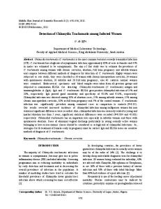
F. 1.—Serum IgG anti-MOMP antibodies.
F. 2.—Serum IgA anti-MOMP antibodies.
F. 3.—Synovial fluid IgG anti-MOMP antibodies.
anti-C. pneumoniae) can be detected. Consequently, the sensitivity is higher for anti-LPS than for antiMOMP antibodies, but the specificity is lower. Nevertheless, the SF IgG anti-MOMP antibody determination gave a sensitivity and a specificity equal or close to 80% and could be clinically useful. The ELISA kits used in this study allow determination of the patients with only anti-CT antibodies from the anti-MOMP antibody results. The patients with
only anti-C. pneumoniae antibodies are assumed to be those with anti-LPS but no anti-MOMP antibodies; those with both types of antibodies cannot be determined. However, it seems that anti-LPS antibodies are not detected in some individuals with anti-MOMP antibodies. This observation can be explained if both types of antibodies are not produced with the same delay after infection or if the expression of LPS is preferentially downregulated.
1058
BRITISH JOURNAL OF RHEUMATOLOGY VOL. 37 NO. 10
F. 4.—Synovial fluid IgA anti-MOMP antibodies. TABLE III Sensitivity, specificity, positive predictive value, negative predictive value, false-positive and false-negative values obtained for the different anti-Chlamydia antibody assays and their combinations Sensitivity (%)
Specificity (%)
Predictive value (+ ) (%)
Predictive value (− ) (%)
False-positive rate (%)
False-negative rate (%)
Serum antibodies* IgG anti-MOMP IgA anti-MOMP IgG anti-LPS IgA anti-LPS IgG and/or IgA anti-MOMP or anti-LPS
79 53 89 68 100
58 63 42 58 21
65 59 61 62 56
73 57 80 65 100
42 37 58 42 79
21 47 11 32 0
Synovial fluid antibodies† IgG anti-MOMP IgA anti-MOMP IgG anti-LPS IgA anti-LPS IgG and/or IgA anti-MOMP or anti-LPS
77 46 85 62 100
80 90 70 50 40
83 86 79 62 68
73 56 78 50 100
20 10 30 50 60
23 54 15 38 0
Isotype(s) considered
*Positive patients were 19 patients with Chlamydia trachomatis sexually acquired reactive arthritis and negative patients were four patients with rheumatoid arthritis, six patients with gout, five patients with septic arthritis, three patients with Lyme arthritis and one patient with Salmonella enteritidis reactive arthritis. †Positive patients were 13 patients with Chlamydia trachomatis sexually acquired reactive arthritis and negative patients were two patients with rheumatoid arthritis, three patients with gout, one patient with septic arthritis, three patients with Lyme arthritis and one patient with Salmonella enteritidis reactive arthritis.
As we previously observed [4], a substantial proportion of CT-SARA patients do not have detectable anti-CT antibodies. Indeed, if only anti-MOMP antibodies are reflecting CT infection, 21% of CT-SARA and 18% of CT-urethritis (data not shown) do not have IgG or IgA anti-MOMP antibodies. Similarly, carriage of C. pneumoniae without a demonstrable serological response has also been reported [9]. The significance of these observations is not known. Among rheumatological patients with possible CT-involved disease, a substantial number of uroarthritis patients had serum and SF IgG and/or IgA anti-MOMP antibodies, therefore confirming a probable CT infection. For the group of patients with idiopathic mono/oligoarthritis, only the percentage of positive samples for SF IgA anti-MOMP antibodies appeared to be slightly higher than the one found for the group of patients with inflammatory arthropathies unrelated to CT. However, when the results were expressed in OD values, a semi-quantitative approach, a number of samples had absorbance higher than those
of control patients. Nevertheless, a higher prevalence could be assumed in this group of patients since latent CT genitourinary tract infection was reported in patients with undiagnosed oligoarthritis [1–3] and CT DNA was found in 11 out of 27 SF samples from patients with seronegative mono/oligoarthritis [10]. This low number of positive samples can be related to the fact that 21% of well-characterized CT-SARA patients have no serum IgG or IgA anti-MOMP antibodies. This could be explained by in vitro studies suggesting that, once chlamydial infection becomes chronic, there is a state of persistent infection in which shedding of infectious elementary bodies is curtailed and expression of MOMP and LPS is downregulated [11–14]. CT may be hidden in synovial tissue or elsewhere without an obvious serological response. In conclusion, the use of synthetic peptide or recombinant antigen improves the sensitivity compared to a test using whole elementary bodies as antigen [4]. However, the anti-LPS assays, which are not species specific, appear less useful than the anti-MOMP assays.
BAS AND VISCHER: CHLAMYDIA TRACHOMATIS ANTIBODIES IN ARTHRITIS
These ELISA tests allow a non-invasive screening procedure which provides important information about previous contact with Chlamydia and could be reliable diagnostic tools in probable CT-SARA. Positive results are only suggestive, not definitely diagnostic, and should always be interpreted in relation to history, clinical picture and the course of disease. The determination of SF IgG anti-MOMP antibody could be particularly useful since a sensitivity and a specificity of roughly 80% were observed. The specificity reached 90% with the determination of SF IgA anti-MOMP antibody. Nevertheless, some CT-SARA patients had no detectable antibodies and this deficit could be related to an impaired clearance of CT. Our results also show that a high percentage of uroarthritis patients had anti-MOMP antibodies, indicating a probable CT-SARA. For idiopathic mono/oligoarthritis patients, the percentages of positive serum samples were comparable to those obtained for control patients, but a higher absorbance was observed for a number of samples and for these cases a CT-SARA could probably be considered too. Nevertheless, a major disadvantage of these tests is that they are only qualitative or, at best, semi-quantitative. A The technical assistance of Ursula Spenato is grateful acknowledged. This study was supported by the Fonds National Suisse de la Recherche Scientifique (grant 32-47299.96).
4.
5.
6. 7. 8.
9.
10.
11.
12.
R 1. Weyand CM, Goronzy JJ. Clinically silent infections in patients with oligoarthritis: results of a prospective study. Ann Rheum Dis 1992;51:253–8. 2. Kvien TK, Glenna˚s A, Melby K et al. Reactive arthritis: incidence, triggering agents and clinical presentation. J Rheumatol 1994;21:115–22. 3. Wollenhaupt HJ, Kolbus F, Weissbrodt H et al. Manifestations of Chlamydia induced arthritis in patients
13.
14.
1059
with silent versus symptomatic urogenital chlamydial infection. Clin Exp Rheumatol 1995;13:453–8. Bas S, Cunningham T, Kvien TK, Glenna˚s A, Melby K, Vischer TL. The value of isotype determination of serum antibodies against Chlamydia for the diagnosis of Chlamydia reactive arthritis. Br J Rheumatol 1996; 35:542–7. Karvonen M, Tuomilehto J, Pitka¨niemi J, Naukkarinen A, Saikku P. Chlamydia pneumoniae IgG antibody prevalence in south-western and eastern Finland in 1982 and 1987. Int J Epidemiol 1994;23:176–84. Kuo CC, Jackson LA, Campbell LA, Grayston JT. Chlamydia pneumoniae ( TWAR). Clin Microbiol Rev 1995;8:451–61. Schachter J. Chlamydial infections. N Engl J Med 1978;298:428–35. Meijer CJ, Calame JJ, de Windt EJ et al. Prevalence of Chlamydia trachomatis infection in a population of asymptomatic women in a screening program for cervical cancer. Eur J Clin Microbiol Infect Dis 1989;8:127–30. Hammerschlag MR, Chirgwin K, Roblin PM et al. Persistent infection with Chlamydia pneumoniae following acute respiratory illness. Clin Infect Dis 1992;14: 178–82. Bas S, Griffais R, Kvien TK, Glenna˚s A, Melby K, Vischer TL. Amplification of plasmid and chromosome Chlamydia DNA in synovial fluid of patients with reactive arthritis and undifferentiated seronegative oligoarthropathies. Arthritis Rheum 1995;38:1005–13. Beatty WL, Byrne GI, Morrison RP. Morphologic and antigenic characterization of interferon c-mediated persistent Chlamydia trachomatis infection in vitro. Proc Natl Acad Sci USA 1993;90:3998–4002. Beatty WL, Morrison RP, Byrne GI. Persistent chlamydiae: from cell culture to a paradigm for Chlamydia pathogenesis. Microbiol Rev 1994;58:686–99. Beatty WL, Morrison RP, Byrne GI. Immunoelectronmicroscopic quantitation of differential levels of chlamydial proteins in cell culture model of persistent Chlamydia trachomatis infection. Infect Immun 1994; 62:4059–62. Beatty WL, Morrison RP, Byrne GI. Reactivation of persistent Chlamydia trachomatis infection in cell culture. Infect Immun 1995;63:199–205.