Journal of Medical Microbiology (2004), 53, 999–1005
DOI 10.1099/jmm.0.45702-0
Characterizing uncommon Burkholderia cepacia complex isolates from an outbreak in a haemodialysis unit Andrea V. Souza,1 Cla´udia R. Moreira,1 Jacyr Pasternak,2,3 Maria de Lurdes Hirata,2 Denise Alves Saltini,2 Viviane Cristina Caetano,2 Suely Ciosak,2 Fa´tima M. Azevedo,1 Patricia Severino,1 Peter Vandamme4 and Vanda D. Magalha˜es1 Correspondence
1
Centro de Pesquisa Experimental, Instituto de Ensino e Pesquisa Albert Einstein - Av. Albert Einstein 627, 05651-901, Sa˜o Paulo, SP, Brazil
2
Infection Control Service, Hospital da Beneficeˆncia Portuguesa de Sa˜o Paulo, Sa˜o Paulo, Brazil
3
Infection Control Service, Hospital Israelita Albert Einstein, Sa˜o Paulo, Brazil
4
Laboratorium voor Microbiologie, Faculteit Wetenschappen, Universiteit Gent, Gent, Belgium
Vanda D. Magalha˜es
[email protected]
Received 15 April 2004 Accepted 2 July 2004
An outbreak of bacteraemia in a haemodialysis unit where 65 episodes of infection involved 35 outpatients is reported. Burkholderia cepacia complex was the agent most frequently recovered from blood. Thirty-three environmental and clinical isolates of B. cepacia complex were characterized by whole-cell protein electrophoresis and recA-RFLP profile. Fourteen isolates were genomovar I and 16 isolates were not classifiable by their recA-RFLP pattern. Ribotyping, random amplification of polymorphic DNA (RAPD) and integron profile were used to explore the clonality of the isolates, and revealed multiple strain genotypes. Four ribotypes and RAPD types and three integron patterns were identified. The water supply was identified as the source of the outbreak, and inappropriate cleaning and a leak in the reverse osmosis tubing connection were the probable causes of contamination. B. cepacia complex was still recovered from blood of patients even after apparently adequate measures were taken and water quality standards were met, suggesting that higher standards for water quality should be adopted in haemodialysis units. The genomovars recovered here were distinct from those commonly reported for cystic fibrosis isolates.
INTRODUCTION Patients receiving long-term haemodialysis are at increased risk of bloodstream infections, usually due to repeated vascular access. Advances in aseptic techniques have reduced the risk of infection in these patients, but outbreaks continue to occur, accounting for 12–38 % of mortality in patients with chronic renal disease (Roth & Jarvis, 2000; Vanherweghem et al., 1991). Inadequate catheter care, contamination of water supply, defects in membrane integrity and reprocessed dialysers have all been implicated in outbreaks in haemodialysis units. Haemodialysis fluids and equipment support the growth of hydrophilic bacteria and contamination of them is directly related to the quality of the water supply. Although the water supply for haemodialysis is not required to be sterile, the number of bacteria present must fall within specific stan-
dards to reduce the risk of bloodstream infection (Roth & Jarvis, 2000). Therefore, monitoring of water quality in a haemodialysis setting is mandatory. Members of the Burkholderia cepacia complex are taxonomically complex, but only B. cepacia complex from the respiratory tract of cystic fibrosis patients has been extensively studied (Coenye et al., 2001; Vermis et al., 2002). Species in this group cannot be reliably identified by phenotypic characteristics and have been classified into at least nine genomic species or genomovars (Vandamme et al., 2002). Here we describe extensive molecular characterization of uncommon B. cepacia isolates recovered from blood and environmental samples in a haemodialysis setting. The data unequivocally implicated the water supply as a source of infection.
METHODS Patients and haemodialysis setting. The haemodialysis unit at the
Abbreviation: RAPD, random amplification of polymorphic DNA.
Hospital da Beneficeˆncia Portuguesa in Sa˜o Paulo, Brazil, provides long-
Downloaded from www.microbiologyresearch.org by IP: 37.44.207.85 On: Fri, 20 Jan 2017 07:56:50
45702 & 2004 SGM Printed in Great Britain
999
A. V. Souza and others
term haemodialysis for approximately 79 patients/month using 10 stations. Vital signs and body temperature are always monitored during dialysis procedure. The dialysis machines (Baxter) are disinfected daily, and dialyser coils (cellulose acetate hollow-fibre) are reprocessed mechanically and reused, individually, up to 12 times as established by the regulating agencies that care for public health in Brazil. Chlorinetreated municipal water from reservoir 1 passes through sand filters, activated charcoal filters, deionizers and reverse osmosis machines. Treated water is maintained in reservoir 2 from where it is distributed to dialysis machines. Water quality is assessed monthly by collecting 100 ml samples from different points of both reservoirs: after each filter passage, after reverse osmosis, before dialysis machines and dialysates. Faecal coliforms, heterotrophic bacteria and the amount of endotoxins are monitored. Epidemiological investigation was carried out as a cohort study of chronic renal failure patients being dialysed three times a week. A case was defined as any long term haemodialysis patient with an episode of bloodstream infection documented with a positive blood culture.
using primers directed to the conservative regions flanking those elements (39CS and 59CS) as described by Le´vesque & Roy (1993). Three millilitres of overnight culture were washed and resuspended in 1 ml distilled water from which 5 ìl was immediately used in PCR. Fifty microlitres of the reaction mixture contained 200 ìM dNTP, 50 pmol each primer, 13 PCR buffer with additional MgCl2 to the final concentration of 3 mM, 2.5 U Taq DNA polymerase. Reaction mixture constituents were supplied by Amersham Biosciences. Gene Amp 9600 PCR System thermal cycler (Perkin Elmer Applied Biosystems) was programmed, after a modification of the protocol proposed by Le´vesque & Roy (1993), for 10 min at 95 8C, followed by 12 cycles of 1 min at 94 8C, 1 min at 55 8C and 5 min at 72 8C, 12 cycles of 1 min at 94 8C, 1 min at 55 8C and 6 min at 72 8C and 12 cycles of 1 min at 94 8C, 1 min at 55 8C and 7 min at 72 8C. After electrophoresis at 5 V/cm for 2 h on 1.2 % agarose gels, PCR products were stained with ethidium bromide and photographed under UV transillumination.
RESULTS
Bacterial isolation, culture and identification. Isolates were recov-
ered from blood of patients undergoing haemodialysis treatment who presented with symptoms of bacteraemia (chills and fever). Ten millilitres of blood from the venous return, collected after wiping the venous port with povidine/alcohol, were inoculated into an enriched medium (Bactec NR 660). Water culture was done by standard methods using a filtration technique (Millipore) and spread on nutrient agar (3 % beef extract, 5 % peptone) for quantitative culture at room temperature for 7 days. Phenotypic identification and antibiotic susceptibility tests were carried out in the Microscan system (Baxter). Identification of B. cepacia complex bacteria was confirmed by wholecell protein electrophoresis (Pot et al., 1994) and recA restriction fragment length polymorphism (RFLP) analysis (Mahenthiralingam et al., 2000; Vandamme et al., 2002). Genomic DNA extraction. Genomic DNA was extracted using a
modified method described by Mekalanos (1983). Briefly, an overnight culture was washed with 50 mM Tris/HCl (pH 8.0), 50 mM EDTA, and resuspended in 500 ìl of the same buffer. The cell suspension was treated with 20 ìl lysozyme (10 mg ml 1 ; Sigma) for 30 min at 37 8C, followed by incubation with 25 ìl proteinase K (20 mg ml 1 ; Invitrogen) and 20 ìl SDS (10 %) for another 30 min at 55 8C. DNA was purified by phenol/chloroform extraction, precipitated with 3 M NaCl (0.1 volume) and absolute ethanol (2.5 volume) and was resuspended in 100 ìl deionized water. Ribotyping. Ribotyping was carried out as previously described by
Gruner et al. (1993), using the endonuclease PvuII for genomic DNA digestion. Restriction fragments were separated by 0.8 % agarose gel electrophoresis in 0.53 TBE buffer (89 mM Tris, 89 mM boric acid, 2 mM EDTA, pH 8.3) for 18 h at 1 V/cm. Southern blots were performed as described by Sambrook & Russell (2001) and the plasmid pKK3535 (Brosius et al., 1981), carrying the Escherichia coli rrn operon, was used as a probe. The plasmid was linearized with BglII and labelled by the digoxigenin system (DIG DNA labelling and detection kit; Roche Molecular Biochemicals). DNA fingerprinting patterns were visually analysed and each ribotype pattern was designated R followed by a number. RAPD. Fifty nanograms of total B. cepacia DNA was used in a 50 ìl PCR
with primer 272 (59-AGCGGGCCAA) as previously described by Mahenthiralingam et al. (1996). Amplification products were photographed under UV transillumination after electrophoresis on agarose gel (2 % in 13 TAE, 40 mM Tris-acetate, 1 mM EDTA buffer) and staining with ethidium bromide.
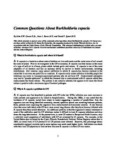
Outbreak investigation Seventy-nine chronic renal patients visit the haemodialysis unit of Hospital Beneficeˆncia Portuguesa three times per week. Between May 2001 and December 2002, there were 65 bacteraemia infection episodes involving 35 outpatients. The species isolated from blood during this period are listed in Table 1. B. cepacia complex was the most recurrent microorganism isolated and two of the involved patients also had B. cepacia complex isolated from dialysate and from arterial wound (Table 2). A retrospective review of patients undergoing dialysis in the unit showed that only three episodes of bacteraemia had occurred during the preceding year (Fig. 1).
Table 1. Bacterial species isolated from bloodstream of haemodialysis patients Bacterial species
No. isolates
Achromobacter xylosoxidans Acinetobacter lwoffii Agrobacterium radiobacter B. cepacia Chryseobacterium meningosepticum Chryseobacterium indologenes Enterobacter cloacae Enterobacter intermedius Moraxella sp. Pseudomonas aeruginosa Ralstonia paucula Ralstonia pickettii Staphylococcus aureus Staphylococcus auricularis Staphylococcus epidermidis Stenotrophomonas maltophilia Streptococcus milleri Vibrio sp.
1 2 1 28 1 1 2 1 2 3 3 3 1 2 2 10 1 1
Integron detection. The presence of integrons was investigated by
1000
Downloaded from www.microbiologyresearch.org by IP: 37.44.207.85 On: Fri, 20 Jan 2017 07:56:50
Journal of Medical Microbiology 53
Uncommon B. cepacia complex in haemodialysis
Table 2. Description of B. cepacia complex isolates analysed ND,
Not determined.
Isolates IEP174 IEP175 IEP176 IEP177 IEP178 IEP179 IEP180 IEP181 IEP182 IEP183 IEP184 IEP185 IEP186 IEP187 IEP188 IEP189 IEP190 IEP191 IEP192 IEP193 IEP194 IEP195 IEP196 IEP197 IEP198 IEP199 IEP200 IEP201 IEP202 IEP203 IEP204 IEP205 IEP206 IEP207
Patients 1 2 3 4 5 6 Reservoir Reservoir Reservoir Reservoir Reservoir 6 7 4 8 9 7 10 11 12 13 Reservoir Reservoir Reservoir 14 15 Reservoir Reservoir Reservoir Reservoir Reservoir Reservoir Reservoir Reservoir
Material
Date of collection
Ribotyping
RAPD
Integron
recA-RFLP
Blood Blood Blood Blood Blood Blood Water Water Water Water Water Dialysate Blood Blood Blood Blood Blood Blood Blood Blood Arterial wound Water Water Water Blood Blood Water Water Water Water Water Water Water Water
18/07/2001 18/07/2001 18/07/2001 18/07/2001 28/07/2001 04/08/2001 15/08/2001 20/08/2001 20/08/2001 22/08/2001 19/08/2001 21/08/2001 10/09/2001 11/09/2001 17/09/2001 17/09/2001 17/09/2001 25/09/2001 25/09/2001 25/09/2001 20/11/2001 05/12/2001 05/12/2001 05/12/2001 26/03/2002 14/05/2002 14/05/2002 14/05/2002 14/05/2002 14/05/2002 14/05/2002 14/05/2002 14/05/2002 14/05/2002
R1 R1 R1 R1 R1 R2 R2 R2 R2 R2 R2 R2 R1 R1 R1 R1 R1 R1 R1 R1 R2 R2 R2 R2 R4 R3 R3 R3 R1 R2 R2 R2 R2 R2
A A A A A B B B B B B B A A A1 A A1 A A A B B B B D C C C A B B B B B
I1 I1 I1 I1 I1 I2 I2 I2 I2 I2 I2 I2 I1 I1 I1 I1 I1 I1 I1 I1 I2 I2 I2 I2 I4 I3 I3 I3 I1 Ø Ø Ø Ø Ø
AT AT AT AT AT K K K K K K K AT AT AT AT AT AT AT
12
No. isolates
10 8 6 4 2
May
20 00 Jul 2 000 Sep 2000 Nov 2000 Jan 2 001 Mar 2001 May 20 01 Jul 2 001 Sep 2001 Nov 2001 Jan 2 002 Mar 2002 May 20 02 Jul 2 002 Sep 2002 Nov 2002 Jan 2 003 Mar 2 003
0
Fig. 1. Epidemiological curve of bacteremia outbreak in a haemodialysis unit. B. cepacia complex isolates are represented by bars in black. All the other genera are represented by white bars. http://jmm.sgmjournals.org
ND
K K ND ND
Se27 AT AT AT AT K K K K K
The haemodialysis procedure, including equipment disinfection, and the whole water system were reviewed. No backflow was detected in the machines and nurses were following good practices for dialysis set-up. Water quality monitoring exceeded limits of the American Association of Medical Instrumentation (AAMI) standards for heterotrophic bacteria on four occasions. In July 2001, one point of reservoir 2 yielded 700 c.f.u. ml 1 and a sample point before dialysis machine 1 produced 330 c.f.u. ml 1 . In August 2001, reservoir 2 yielded 720 c.f.u. ml 1 and in February 2002, a sample before another dialysis machine showed 1650 c.f.u. ml 1 . All other water samples showed less than 200 c.f.u. ml 1 (upper limit by AAMI) during the outbreak. Faecal coliforms were not detected. B. cepacia was isolated from the water sample in July 2001. In August 2001, reservoir 2 and all pipes and tubing were
Downloaded from www.microbiologyresearch.org by IP: 37.44.207.85 On: Fri, 20 Jan 2017 07:56:50
1001
A. V. Souza and others
replaced. Nevertheless, B. cepacia continued to be isolated from blood and from water on six further occasions, although water quality monitoring showed less than 200 c.f.u. ml 1 of heterotrophic bacteria (Table 2). The probable source of contamination was finally identified in December 2002 when a loose connection in the reverse osmosis tubing was detected, leading to a leak that could allow the entrance of bacteria into the water system. Repair of this particular part of the system stopped the outbreak.
1
2
3
4
5
6
7
8
9
10
11
Identification of B. cepacia complex isolates B. cepacia complex formed a homogeneous group. All B. cepacia isolates gave the same antibiogram pattern, being multi-resistant but fully susceptible to sulfa/trimethoprin. Thirty-three B. cepacia isolates, from 17 clinical and 16 environmental samples, were available for further characterization. A B. cepacia complex isolate (IEP198) recovered from the bloodstream of a patient admitted to another ward during the outbreak period was included as a control in the study. Isolates were confirmed as B. cepacia complex by whole-cell protein electrophoresis (data not shown) and genomovar status was identified by HaeIII-recA RFLP analysis (Mahenthiralingam et al., 2000). A recA amplicon of the expected size was generated for each isolate and two distinct RFLP patterns were obtained (Table 2). Fourteen isolates, three from patients and 11 from water, were identified as type K which corresponds to genomovar I (Vermis et al., 2002) and 16 isolates were designated AT (unknown genomovar), 13 from patients and three from water. The control strain (IEP 198) was designated Se27, a unique profile not seen before. Molecular typing of B. cepacia complex isolates by ribotyping and RAPD The B. cepacia complex isolates were characterized by ribotyping and RAPD. Three ribotypes were identified from epidemiologically linked patients and from water. Thirteen (76 %) isolates from blood were ribotype 1 and this strain was also found once in water. Ribotype 2, found in blood, dialysate and arterial wound, was the most frequent genotype in water samples from reservoirs (13/16, 81 %). Ribotype 3 was recovered from the bloodstream of a patient and, on the same day, was also found in water (Table 2 and Fig. 2). RAPD fingerprinting resulted in five to 10 bands over a size range of 0.3–5 kb. Each assay was repeated twice to assure its reproducibility. Although some minor variations were noted between experiments, by computing only the main amplified bands, four different patterns were consistently obtained (Table 2 and Fig. 3). All isolates, apart from two, identified as R1 presented the A pattern for RAPD. IEP188 and IEP190 showed a one band difference when compared to strains classified as A, which suggests a close relationship between them, and they were classified as pattern A1. All R2 isolates 1002
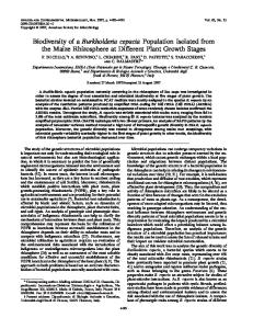
Fig. 2. DNA fingerprints of B. cepacia determined by ribotyping. Lanes: 1, IEP201; 2, IEP199; 3, IEP198; 5, IEP194; 6, IEP185; 7, IEP180; 8, IEP202; 9, IEP187; 10, IEP174; 4 and 11, º HindIII molecular size standards. Lanes 8, 9 and 10, ribotype 1; lanes 5, 6 and 7, ribotype 2; lanes 1 and 2, ribotype 3; lane 3, control strain, ribotype 4.
were identified as pattern B and all R3 showed RAPD pattern C. The control strain IEP198 yielded unique patterns both for RAPD (D) and ribotyping (R4). Integron profiles of B. cepacia complex isolates Among the epidemiologically linked isolates, three different profiles of integrons were obtained. Three bands at positions 0.8, 0.85 and 1.4 kb were detected in R1 isolates (data not shown). Eleven R2 isolates from clinical and water samples showed a single band of 0.8 kb (I2). Five B. cepacia complex from water, all isolated on the same day and ribotyped as R2, had no detectable integron structure (Table 1). Isolates identified as R3 contained a 0.7 kb integron. The control strain was distinct.
DISCUSSION The B. cepacia complex has been recognized as an emerging opportunistic pathogen, usually related to respiratory infection of cystic fibrosis patients (Crowley et al., 2002). However, characterization of strains to genomovar level of B. cepacia complex causing infections in renal settings has not been extensive. Bacteraemia caused by B. cepacia in patients undergoing peritoneal dialysis has been described, but subtyping was not performed to confirm strain relationships (Berkelman et al., 1982; Flaherty et al., 1993). An outbreak among chronic renal patients in Thailand was traced to diluted chlorhexidine-cetrimide solution contaminated with the same strain as was isolated from the bloodstream (Kaitwatcharachai et al., 2000). Contaminated reverse osmosis membranes were also responsible for a polyclonal B. cepacia complex bacteraemia in chronic renal patients in
Downloaded from www.microbiologyresearch.org by IP: 37.44.207.85 On: Fri, 20 Jan 2017 07:56:50
Journal of Medical Microbiology 53
Uncommon B. cepacia complex in haemodialysis 1
2
3
4
5
6
7
8
9 10 11 12
13 14 15 16 17 18 19 20 21 22
Fig. 3. DNA fingerprints of B. cepacia determined by RAPD assay. Lanes: 1, IEP185; 2, IEP186; 3, IEP187; 4, IEP188; 5, IEP189; 6, IEP190; 7, IEP191; 8, IEP192; 9, IEP193; 10, IEP194; 11, IEP195; 13, IEP196; 14, IEP197; 15, IEP198; 16, IEP199; 17, IEP200; 18, IEP201; 19, IEP202; 20, IEP203; 12 and 21, DNA size standard (º EcoRI/HindIII + pBR322 HaeIII). Lanes 2, 3, 5, 7, 8, 9, and 19, pattern A; lanes 4 and 6, pattern A1; lanes 1, 10, 11, 13, 14, and 20, pattern B; lane 15, control strain, pattern D; lanes 16, 17 and 18, pattern C; lane 22, negative amplification control.
another outbreak in Brazil (Magalha˜es et al., 2003). In the latter study, Burkholderia cenocepacia (formerly known as genomovar III) and Burkholderia vietnamiensis were identified by recA-RFLP. The present report describes the investigation of bloodstream infection of patients undergoing long-term dialysis. B. cepacia complex was the most frequently isolated microorganism and recA-RFLP pointed to a polyclonal nature of the outbreak, which was confirmed by other molecular typing data. The results are consistent with bacterial diversity found in environmental samples (Salles et al., 2002). Data on the distribution of genomovars of B. cepacia complex have been mostly restricted to isolates from cystic fibrosis patients (Crowley et al., 2002; Mahenthiralingam et al., 2002), where genomovar III is consistently the most common, followed by genomovars II (37.8 %) and I (26 %) (Vermis et al., 2002). The identification of genomovar I isolates in this study represents, to our knowledge, the first time it has been confirmed in a haemodialysis setting. Another unassigned recA-RFLP profile (AT) was identified in the majority of isolates and further taxonomic analysis, including DNA–DNA hybridization, is required. Our data suggest that types of B. cepacia complex involved in bacteraemia are different from those isolated from the respiratory tract of cystic fibrosis patients. Nevertheless, widespread genomovar III and genomovar V (B. vietnamiensis) has been previously identified in another haemodialysis setting (Magalha˜es et al., 2003). Ribotyping and RAPD have been successfully applied to molecular epidemiology studies of B. cepacia (Bingen et al., 1993; Brisse et al., 2000; Okazaki et al., 1999). RAPD correctly grouped epidemiologically related B. cepacia strains from a large collection according to previous data obtained by PFGE and ribotyping (Mahenthiralingam et al., 1996; Bingen et al., 1993; Brisse et al., 2000). Investigating a nosocomial outbreak, Okazaki et al. (1999) concluded that RAPD offered an http://jmm.sgmjournals.org
alternative discriminatory method to PFGE, providing results within a shorter time and with less complexity than the latter. B. cepacia isolates that showed identical ribotypes in this study were considered a single clone. RAPD patterns agreed with ribotyping results, except for two isolates IEP188 and IEP190, which showed a single band difference in the RAPD profile. In the light of ribotyping and integron data, we considered both isolates as part of the R1 clone. A previous work suggested that ribotyping could provide reliable identification of B. cepacia complex genomovars (Brisse et al., 2000). Here, all R2 strains were identified as genomovar I. R1 and R3 strains showed novel recA-RFLP patterns and further study may reveal heterogeneity among them, as observed for other B. cepacia isolates of novel RFLP type (unpublished observations). Integrons are plastic genetic elements that acquire and lose gene cassettes related mostly to antibiotic resistance. Horizontal transfer of integrons is mediated by plasmids and transposons (Stokes & Hall, 1989). Different bacterial genera can exhibit the same integron content and, conversely, a genetically unique strain can show a different integron profile (Severino & Magalha˜es, 2002). Recently, they have been used as strain markers for investigation of outbreaks (Severino & Magalha˜es, 2003; Martin et al., 2001). The rationale behind the use of this new molecular epidemiological tool is that isolates of a clonal origin, in a limited period of time, would not have enough time to modify their integron content. The integron profile of the B. cepacia isolates showed good correlation with the other typing methods and only isolates recovered from a reservoir on 14 May 2002, which were classified as R2, did not show inserted cassettes in their integron structure. They could represent part of a B. cepacia R2 population that lost the resistance cassettes in the absence of selective pressure. The epidemiological investigation and typing data linked the bacteraemia episodes to water contamination. The probable
Downloaded from www.microbiologyresearch.org by IP: 37.44.207.85 On: Fri, 20 Jan 2017 07:56:50
1003
A. V. Souza and others
source of contamination was related to inadequate cleaning procedures that left leaking connections of the reverse osmosis tubing. Environmental biofilm-forming bacteria or micro-organisms present in cleaning solutions could have entered the water system through this opening. Tubing connections are known to be critical segments of the system and biofilm formation is recognized as a risk for haemodialysis patients (Man et al., 1998; Dasgupta, 2002). The counts of bacteria in water are influenced by the microbiological methods used. In this study the measurement of water contamination followed optimized AAMI recommendations for bacterial counts. During the outbreak period, in four instances more than 200 c.f.u. ml 1 were detected, but only once was B. cepacia identified. Unfortunately, this isolate was not available for molecular typing. Paradoxically, other B. cepacia isolated from the reservoir occurred in water samples that met the AAMI standards. In all instances when B. cepacia isolates recovered from water were available for typing, they were linked to bloodstream infection episodes. In another outbreak of bacteraemia of a polyclonal nature most water samples were also within the limits of AAMI guidelines (Magalha˜es et al., 2003). The microbiological standard for dialysate and water purity in haemodialysis has been a matter of discussion (Lonnemann, 2000; Tielemans et al., 2001). While AAMI recommends an upper limit of 200 c.f.u. ml 1 in haemodialysis water, the Deutsche Arbeitgemeinschaft fu¨r Klinische Nephrologie (DafKN) from the German Renal Society recommends a maximum of 100 c.f.u. ml 1 (Lonnemann, 2000). In the light of the outbreak described here and considering the increased risk of chronic renal patients, we suggest that higher water quality standards should be adopted in haemodialysis units, as proposed by DafKN.
REFERENCES Berkelman, R. L., Godley, J., Weber, J. A., Anderson, R. L., Lerner, A. M., Petersen, N. J. & Allen, J. R. (1982). Pseudomonas cepacia peritonitis
associated with contamination with automatic peritoneal dialysis machines. Ann Intern Med 96, 456–458. Bingen, E. H., Weber, M., Derelle, J., Brahimi, N., Lambert-Zechovsky, N. Y., Vidailhet, M., Navarro, J. & Elion, J. (1993). Arbitrarily primed
polymerase chain reaction as a rapid method to differentiate crossed from independent Pseudomonas cepacia infections in cystic fibrosis patients. J Clin Microbiol 31, 2589–2593. Brisse, S., Verduin, C. M., Milatovic, D. & 7 other authors (2000).
Distinguishing species of the Burkholderia cepacia complex and Burkholderia gladioli by automated ribotyping. J Clin Microbiol 38, 1876–1884. Brosius, J., Ullrich, A., Raker, M. A., Gray, A., Dull, T. J., Gutell, R. R. & Noller, H. F. (1981). Construction and fine mapping of recombinant
plasmids containing the rrnB ribosomal RNA operon of E. coli. Plasmid 6, 112–118.
epidemiology of cystic fibrosis-linked Burkholderia cepacia complex isolates from three national referral centres in Ireland. J Appl Microbiol 92, 992–1004. Dasgupta, M. K. (2002). Biofilms and infection in dialysis patients.
Semin Dial 15, 338–346. Flaherty, J. P., Garcia-Houchins, S., Chudy, R. & Arnow, P. M. (1993).
An outbreak of Gram-negative bacteremia traced to contaminated Orings in reprocessed dialyzers. Ann Intern Med 119, 1072–1078. Gruner, E., Kropec, A., Huebner, J., Altwegg, M. & Daschner, F. (1993).
Ribotyping of Pseudomonas aeruginosa strains isolated from surgical intensive care patients. J Infect Dis 167, 1216–1220. Kaitwatcharachai, C., Silpapojakul, K., Jitsurong, S. & Kalnauwakul, S. (2000). An outbreak of Burkholderia cepacia bacteremia in hemodialysis
patients: an epidemiologic and molecular study. Am J Kidney Dis 36, 199–204. Le´vesque, C. & Roy, P. H. (1993). PCR analysis of integrons. In Diagnostic Molecular Microbiology: Principles and Applications, pp. 590–594. Edited by D. H. Persing, T. H. Smith, F. C. Tenover & T. J. White. Washington, DC: American Society for Microbiology. Lonnemann, G. (2000). The quality of dialysate: an integrated
approach. Kidney Int Suppl 76, S112–S119. Magalha˜es, M., Doherty, C., Govan, J. R. W. & Vandamme, P. (2003).
Polyclonal outbreak of Burkholderia cepacia complex bacteraemia in haemodialysis patients. J Hosp Infect 54, 120–123. Mahenthiralingam, E., Campbell, M. E., Foster, J., Lam, J. S. & Speert, D. P. (1996). Random amplified polymorphic DNA typing of Pseudo-
monas aeruginosa isolates recovered from patients with cystic fibrosis. J Clin Microbiol 34, 1129–1135. Mahenthiralingam, E., Bischof, J., Byrne, S. K., Radomski, C., Davies, J. E., Av-Gay, Y. & Vandamme, P. (2000). DNA-based diagnostic
approaches for identification of Burkholderia cepacia complex, Burkholderia vietnamiensis, Burkholderia multivorans, Burkholderia stabilis, Burkholderia cepacia genomovars I and III. J Clin Microbiol 38, 3165–3173. Mahenthiralingam, E., Baldwin, A. & Vandamme, P. (2002). Burkhol-
deria cepacia complex infection in patients with cystic fibrosis. J Med Microbiol 51, 533–538. Man, N. K., Degremont, A., Darbord, J. C., Collet, M. & Vaillant, P. (1998). Evidence of bacterial biofilm in tubing from hydraulic pathway
of hemodialysis system. Artif Organs 22, 596–600. Martin, M. C., Gonza´lez-Hevia, M. A., Alvarez-Riesgo, J. A. & Mendoza, M. C. (2001). Salmonella serotype Virchow causing salmonellosis in a
Spanish region. Characterization and survey of clones by DNA fingerprinting, phage typing and antimicrobial resistance. Eur J Epidemiol 17, 31–40. Mekalanos, J. J. (1983). Duplication and amplification of toxin genes in
Vibrio cholerae. Cell 35, 253–263. Okazaki, M., Watanabe, T., Morita, K. & 10 other authors (1999).
Molecular epidemiological investigation using a randomly amplified polymorphic DNA assay of Burkholderia cepacia isolates from nosocomial outbreaks. J Clin Microbiol 37, 3809–3814. Pot, B., Vandamme, P. & Kersters, K. (1994). Analysis of electro-
phoretic whole-organism protein fingerprints. In Modern Microbial Methods. Chemical Methods in Prokaryotic Systematics, pp. 493–521. Edited by M. Goodfellow & A. G. O’Donnell. Chichester, UK: Wiley. Roth, V. R. & Jarvis, W. R. (2000). Outbreaks of infection and/or
Coenye, T., Vandamme, P., Govan, J. R. W. & LiPuma, J. J. (2001).
pyrogenic reactions in dialysis patients. Semin Dial 13, 92–96.
Taxonomy and identification of the Burkholderia cepacia complex. J Clin Microbiol 39, 3427–3436.
Salles, J. F., De Souza, F. A. & van Elsas, J. D. (2002). Molecular method
Crowley, D., Daly, M., Lucey, B. & 7 other authors (2002). Molecular
1004
to assess the diversity of Burkholderia species in environmental samples. Appl Environ Microbiol 68, 1595–1603.
Downloaded from www.microbiologyresearch.org by IP: 37.44.207.85 On: Fri, 20 Jan 2017 07:56:50
Journal of Medical Microbiology 53
Uncommon B. cepacia complex in haemodialysis
Sambrook, J. & Russell, D. W. (2001). Molecular Cloning: a Laboratory
Manual. 3rd edn. Cold Spring Harbor, NY: Cold Spring Harbor Laboratory. Severino, P. & Magalha˜es, V. D. (2002). The role of integrons in the
dissemination of antibiotic resistance among clinical isolates of Pseudomonas aeruginosa from an intensive care unit in Brazil. Res Microbiol 153, 221–226. Severino, P. & Magalha˜es, V. D. (2003). Integrons as tools for
Vandamme, P., Henry, D., Coenye, T., Nzula, S., Vancanneyt, M., LiPuma, J. J., Speert, D. P., Govan, J. R. W. & Mahenthiralingam, E. (2002). Burkholderia anthina sp. nov. and Burkholderia pyrrocinia, two
additional Burkholderia cepacia complex bacteria, may confound results of new molecular diagnostic tools. FEMS Immunol Med Microbiol 33, 143–149.
epidemiological studies. Clin Microbiol Infect 10, 156–162.
Vanherweghem, J. L., Tielemans, C., Goldman, M. & Boelaert, J. (1991). Infections in chronic hemodialysis patients. Semin Dial 4,
Stokes, H. W. & Hall, R. M. (1989). A novel family of potentially mobile
240–244.
DNA elements encoding a site-specific gene-integration function: integrons. Mol Microbiol 3, 1669–1683. Tielemans, C., Hoenich, N. A., Levin, N. W., Lonnemann, G., Favero, M. S. & Schiffl, H. (2001). Are standards for dialysate purity in hemodialysis
insufficiently strict? Semin Dial 14, 328–336.
http://jmm.sgmjournals.org
Vermis, K., Coenye, T., Mahenthiralingam, E., Nelis, H. J. & Vandamme, P. (2002). Evaluation of species-specific recA-based PCR tests for
genomovar level identification within the Burkholderia cepacia complex. J Med Microbiol 51, 937–940.
Downloaded from www.microbiologyresearch.org by IP: 37.44.207.85 On: Fri, 20 Jan 2017 07:56:50
1005