Kidney International, Vol. 58, Suppl. 77 (2000), pp. S-59–S-66
CELL CYCLE AND APOPTOSIS
Cell cycle regulation in diabetic nephropathy GUNTER WOLF Department of Medicine, Division of Nephrology and Osteology, University of Hamburg, Hamburg, Germany
Cell cycle regulation in diabetic nephropathy. Renal hypertrophy is one of the earliest abnormalities of diabetic nephropathy. Although selected cell populations, such as tubulointerstitial fibroblasts, may undergo sustained proliferation in the diabetic environment, most renal cells such as mesangial cells are arrested in the G1-phase of the cell cycle after actively leaving G0phase and some self-limited early proliferation. High glucose, transforming growth factor-beta (TGF-), angiotensin II, and probably other factors induce inhibitors of cyclin-dependent kinases (CDK) including p21Cip1 and p27Kip1. These CDK-inhibitors bind to and inactivate G1-phase cyclin/CDK complexes. The consequence is a lack in kinase activity, underphosphorylation of the retinoblastoma gene protein, and a failure to initiate the G1-S-phase transit. The half-life of CDK-inhibitors may also be increased by serine phosphorylation mediated through activated MAP kinases. Treatment of diabetic rats with angiotensin-converting enzyme inhibitors attenuates glomerular hypertrophy and abolishes the glomerular expression of the CDK-inhibitors p16INK4 and p27Kip1, thus indicating that the cell cycle arrest can be therapeutically influenced. Cell cycle proteins may also be involved in these molecular events, leading to a limited degree of tubular apoptosis, which is a feature of diabetic nephropathy. Although not definitively proven, accumulating evidence suggests that early hypertrophy of renal cells may act as pacemaker for subsequent irreversible structural changes, such as glomerulosclerosis and tubulointerstitial fibrosis. Therefore, a better understanding of altered processes of cell cycle regulation is necessary to develop novel therapeutic strategies to prevent diabetic nephropathy. The recent observation that glomerular hypertrophy and proteinuria do not develop in diabetic p21Cip1 knockout mice indicates that this approach is feasible.
Diabetic nephropathy encompasses a complex of structural changes including renal hypertrophy, thickening of basement membranes, and progressive glomerular and tubulointerstitial accumulation of extracellular matrix components [1–3]. At least in the early phases of diabetic nephropathy, glomerular and tubulointerstitial infiltration of immunocompetent cells such as macrophages/ monocytes occurs [4]. Furthermore, local proliferation of both macrophages and myofibroblasts likely contributes to the tubulointerstitial infiltrate observed in diaKey words: diabetic nephropathy, hypertrophy, cell cycle regulation, p27kip1, fibrosis, progression of renal disease.
2000 by the International Society of Nephrology
betic nephropathy [4–6]. Glomerular and tubular hypertrophy is one of the earliest structural alterations of diabetic nephropathy [1, 7, 8]. Ultrasound investigations in humans document increased overall renal size at the time of diagnosis of type 1 diabetes [9]. Although one cannot be certain about the previous duration of the diabetic milieu in an individual patient before diagnosis, increased renal size is nevertheless one of the earliest organ alterations observed during the course of diabetes. Furthermore, a correlation has been found between sonographically determined kidney size and renal function over 8 years of follow-up such that large kidneys in patients with type 1 diabetes and normal serum creatinine was a morphologic marker for subsequent deterioration of renal function [9]. Active growth of nephrons is largely responsible for increased renal size, but hemodynamic mechanisms, such as hyperfiltration and hyperperfusion that leads to kidney hyperemia, as well as osmotic changes also contribute to enlargement of the kidneys [1, 10–14] A controversial discussion continues over the years about whether changes in glomerular hemodynamics or adaptive growth changes of the kidney are more important for the progression of diabetic nephropathy [14–16]. This disputed point, however, seems to have been resolved because hemodynamic changes and renal growth are intimately connected and may represent two faces of the same coin. Hemodynamic changes, such as mechanical stretch or alterations in laminar flow, induce synthesis of specific growth factors, activate distinct signal transduction pathways, and stimulate growth and extracellular matrix production in glomerular cells [17–19]. Many of these changes are amplified in hyperglycemic situations [1, 19]. Proteinuria, which is increased in glomerular hyperdynamic states, influences activation, growth, and matrix synthesis of tubular cells fundamentally [20, 21]. Conversely, induction of renal growth, mediated by the diabetic environment, may also eventually alter glomerular hemodynamics, for example by an increase in capillary filtration surface [1]. Finally, all vasoactive hormones (e.g., angiotensin II [ANG II] or endothelin) exert profound growth stimulatory actions on many renal cells, particularly in the presence of high glucose [22–25].
S-59
S-60
Wolf: Cell cycle and diabetes
Expansion of the glomerular mesangium occurs within a few years of the onset of insulin-dependent diabetes mellitus and correlates closely with the deterioration in glomerular function [1, 7, 8]. Pathologic alterations of the tubulointerstitium such as tubulointerstitial fibrosis and tubular atrophy are also closely linked to the deterioration of renal function in patients with diabetes of both types 1 and 2 [2, 3]. Hypertrophy of the proximal tubule mainly accounts for the increase in kidney size in diabetes because glomeruli alone account for less than 10% of the kidney’s volume [1, 3]. Although it can be debated whether initial adaptive renal growth, as observed in the diabetic state, is causally linked to the irreversible later changes such as glomerulosclerosis and tubulointerstitial fibrosis, evidence is accumulating that suggests that this is indeed the case [1, 9]. Given that the growth response of every cell is dictated by the mechanisms of cell cycle regulation, an understanding of these molecular processes is essential for the development of novel therapeutic strategies to prevent the development of diabetic nephropathy. RENAL GROWTH IN DIABETES The particular growth response of a distinct renal cell (proliferation vs. hypertrophy) during the diabetic state depends on its intrinsic genetic program, which is specific for the cell type, and the presence of growth factors in the local environment [1, 10, 11]. Although all cells have the same cell cycle machinery, a distinct growth factor may cause different growth responses in the kidney depending on the specific cell type. For example, transforming growth factor- (TGF-), a key factor in the development of diabetic nephropathy, induces hypertrophy of mesangial, tubular cells, but also stimulates proliferation of tubulointerstitial fibroblasts [12, 26–29]. In addition, ANG II exerts proliferative effects on some renal cells (mesangium cells, fibroblasts, distal tubular cells) but also mediates hypertrophy of proximal tubules [22, 25, 30–32]. These discriminatory effects are probably caused by variations in growth factor receptor expression and coupling of these receptors to certain signal transduction pathways [1]. Some of the multiple hormones, cytokines, and growth factors [33–39], which have been implicated in renal growth associated with diabetic nephropathy, are shown in Table 1. Continuous production and release of many of these growth factors during the diabetic state cause not only renal growth as an integral early part of diabetic nephropathy but may be also ultimately explain why patients with diabetes have a higher incidence of renal cancer [40]. One of the more recently characterized factors is leptin, traditionally not considered to be a growth factor [36, 37]. Leptin is produced by adipocytes and reduces food intake through interacting with specific hypothala-
Table 1. Growth factors and peptides implicated in renal growth during diabetes mellitus Factor
Reference
Transforming growth factor- Platelet-derived growth factor- Insulin-like growth factor-I Insulin Hepatocyte growth factor Tumor necrosis factor-␣ Fibroblast growth factor Vascular endothelial growth factor Leptin Angiotensin II Endothelin 1
12, 72 10 34 10, 11 35 11 11 38 36 19, 23 39
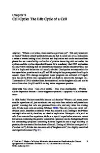
mic receptors [37]. However, we discovered that leptin stimulates proliferation and synthesis of TGF- in glomerular endothelial cells in vitro and in vivo [36]. Furthermore, leptin infusion in normal rats leads ultimately to proteinuria and segmental glomerular sclerosis [36]. Preliminary evidence also suggests that leptin increases TGF- receptor type 2 expression on mesangial cells indicating a paracrine cross-talk between glomerular endothelial and mesangial cells (abstract; Han et al, J Am Soc Nephrol 10:681A, 1999). Inasmuch as hyperleptinemia is part of the metabolic syndrome in type 2 diabetes [36, 37], increased leptin concentrations could indeed modulate growth of glomerular cells in diabetic nephropathy. In addition to increased protein synthesis, a cell cycle– independent decrease in proteinase activity leading to an attenuated turnover of structural and extracellular matrix proteins likely contributes to diabetic hypertrophy of the kidney [41–45]. Inhibition of such proteases may be caused by multiple factors including high glucose, advanced glycated end products (AGEs), ANG II, and TGF- [41, 44–46]. CELL CYCLE REGULATION The growth of each cell is determined by different phases called the cell cycle (Fig. 1). The period between two mitoses, which defines the somatic cell cycle, is called the interphase. Renal cells, under normal conditions, have a very low turnover rate of less than 1% [1, 47, 48]. These dormant cells can be considered to rest in a G0phase and have left the active processes of the cell cycle (Fig. 1). After stimulation with a growth factor or cytokine, dormant cells actively enter the G1-phase in which cells increase their size and stimulate protein and mRNA synthesis to prepare for DNA replication. Immediate early genes, encoding various transcription factors, are also activated in the early G1-phase [48]. If conditions to pass the restriction point are satisfied, cells enter the S-phase after a short lag. During S-phase, the double helix unfolds, DNA strands are separated, stabilized, and DNA is replicated by polymerases. At the end of the S-
Wolf: Cell cycle and diabetes
Fig. 1. The different phases of the cell cycle. Quiescent cells are withdrawn form the cell cycle in G0-phase. After stimulation by growth factors, cells reenter the cycle at G1 and prepare for DNA synthesis. After passing the restriction point in late G1, cells are committed to enter the S-phase where DNA replication occurs. in G2, cells further prepare for mitosis (M-phase) where cells divide. Cell cycle progression is principally controlled by the kinase activity of cyclin/CDK-complexes.
phase, the total content of DNA doubles to the fully replicated value of 4n. Cells prepare for mitosis in the G2-phase then enter mitosis (M-phase), which itself is composed of several distinct steps. After cell division, cells may start another cell cycle with reentering the G1phase or may, alternatively, withdraw from the active cell cycle into the G0-phase (Fig. 1). Progression through the cell cycle and transition between different phases is controlled by a series of protein kinases [48–54]. These active protein kinases are holoenzymes composed of two subunits: cyclins and their partner cyclin-dependent kinases (CDK). Some cyclin/CDK complexes act only in specific cell phases whereas others are more widely distributed [52]. Regulation of these cyclin/CDK complexes in the kidney has been reviewed [48, 51, 53, 54]. In brief, cyclins have short half-lives (less than 60 min) and their protein levels fluctuate throughout the cell cycle with increasing synthesis and subsequent degradation by the ubiquitin-proteasome complex [51, 53]. Transcription and synthesis of various cyclins may be negatively or positively controlled through specific growth factors. In contrast, CDKs are constitutively expressed, and their activity depends on binding to cyclins and phosphorylation. Cyclins accumulate during specific phases of the cell cycles and bind, after reaching a critical concentration, to their putative CDK partners. Subsequent to complex phosphorylation and dephosphorylation steps whose upstream effectors remain in-
S-61
completely characterized, the cyclin/CDKs heterodimers are activated and they exert kinase activity [48, 52]. For example, a critical substrate for cyclin D/CDK4,6 complexes that control G1-S-phase transit, is the protein product of the retinoblastoma gene (pRB). On phosphorylation by active cyclin D/CDK4,6 kinases, hyperphosphorylated pRB releases the transcription factor E2F that binds to the promoter regions of multiple target genes which are essential for further cell cycle progression [48]. Kinase activity of cyclin/CDK complexes is also negatively regulated by small proteins called CDK-inhibitors (CKI). Two families of CKIs can be grouped according to structural homology [48, 51]. Although common sense may suggest that an overall increase in CKIs directly inactivates cyclin/CDK complexes by simple binding to those complexes and thus interfering with their kinase activity, the situation is likely much more complex. For example, the CKI p21Cip1 can inhibit cyclin/CDK complexes, but it may alternatively function as assembly factor cyclin D/CDK4 heterodimers [53]. CKIs can be also redistributed between different cyclin/CDK complexes [48]. A decrease in synthesis of a specific cyclin may lead to release of CKIs, which then could bind and inhibit other cyclin/CDKs complexes [51]. Finally, recent evidence suggests that peptide fragments of CKIs, released after ubiquitin-mediated proteolysis, could actually act as downstream activators of cyclin/CDK complexes facilitating cell cycle progression [48]. Cell cycle events in diabetic nephropathy As depicted in Fig. 2 for mesangial cells, high glucose in vitro as well as the diabetic milieu in vivo result in a biphasic growth response [12, 27]. Initially, the degree of proliferation is limited, followed by cell cycle arrest and hypertrophy [12, 27]. Mesangial cells exposed to high glucose actively enter the cell cycle as demonstrated by expression of immediate early genes such as c-fos, c-jun, and Egr-1 [55–57]. After one or two complete rounds of cell-cycle progression with completion of mitosis, cells are arrested in the G1-phase and undergo hypertrophy [12]. This change in phenotype is mediated by TGF- because a neutralizing anti-TGF- antibody prevented the late inhibitory effects of high glucose on hypertrophy of mesangial cells [12]. Cell cycle regulation of mesangial cells in diabetes To gain a better insight into the molecular mechanisms surrounding this high glucose–mediated G1-phase arrest, we and others investigated the regulation of CKIs. Incubation of mouse mesangial cells, in the absence of other factors for 48–96 h, in culture medium with high glucose stimulated p27Kip1 protein expression but did not influence mRNA abundance [58]. These effects were independent of the osmolarity of the medium. High glucose–
S-62
Wolf: Cell cycle and diabetes
Fig. 2. Development of mesangial cell growth in diabetic nephropathy. Experiments in diabetic animals as well as cell culture studies of mesangial cells exposed to ambient high glucose indicate a biphasic growth response. There is an early, self-limited degree of proliferation indicating reentry and progression through the cell cycle. This proliferation is followed by sustained hypertrophy of mesangial cells being associated with an increase in synthesis and deposition of extracellular matrix proteins. Eventually, end-stage diabetic nephropathy with glomerular sclerosis, tubular atrophy, tubulointerstitial fibrosis, and irreversible loss of renal function will evolve.
stimulated expression of p27Kip1 involved the activation of PKC and partly depended on induction of TGF- [58]. p27Kip1 protein, induced by high glucose, mainly bound to CDK2 but not to CDK4 [58]. p27Kip1 antisense, but not missense, oligonucleotides inhibited high glucose–stimulated total protein synthesis and converted the hypertrophy into a proliferative phenotype suggesting G1-phase exit [58] We extended these studies to diabetic db/db mice, a model of type 2 diabetes [59]. Glomerular p27Kip1 protein, but not mRNA expression, was strongly enhanced in diabetic db/db mice compared with nondiabetic db/⫹ littermates [59]. Immunohistochemical studies revealed that this stimulated expression was due to an increase in mesangial and endothelial staining for p27Kip1 [59]. Primary cultures of mesangial cells from db/⫹ and db/db revealed a low p27Kip1 expression when cultured in normal glucose–containing medium [59]. However, increasing the glucose concentration of the medium induced p27Kip1 expression in both cell lines and arrested the cells in the G1-phase. Glomerular p27Kip1 protein also increased in the murine streptozotocin type 1 diabetes [59]. These data clearly indicate that the stimulated mesangial expression of p27Kip1 in db/db mice compared with normoglycemic littermates is caused by high glucose and not by additionally genetic effects [59]. Moreover, ANG II also induces p27Kip1 expression in renal cells [60, 61]. This mechanism may contribute to p27Kip1 expression in diabetic nephropathy in vivo because the intrarenal renin-angiotensin system is activated during diabetes [19, 23]. We have obtained preliminary evidence that mitogen-activated protein (MAP)-kinases directly phosphorylates p27Kip1 and increases its stability (abstract; Wolf et al, J Am Soc Nephrol 10:465 A, 1999). Because MAP kinases are activated in cultured mesangial cells exposed to high glucose and in glomeruli from diabetic rats [62–64], it is possible that phosphorylation of p27Kip1 partly contributes to the induced expression observed in the diabetic environment. To further investi-
gate a functional role of p27Kip1 in high glucose–mediated mesangial hypertrophy, we compared the growth behavior of mesangial cells cultured from p27Kip1⫹/⫹ and ⫺/⫺ mice. In contrast to p27Kip1 wild-type cells, high glucose did not increase hypertrophy in p27Kip1 ⫺/⫺ mesangial cells. Reconstitution of p27Kip1 expression in ⫺/⫺ cells using an inducible expression system led to G1-phase arrest. However, mesangial hypertrophy was only observed when p27Kip1 was induced in high glucose, suggesting that p27Kip1-mediated G1-phase arrest is a necessary prerequisite, but this alone was not sufficient for the development of hypertrophy (abstract; Wolf et al, J Am Soc Nephrol 10:692A, 1999). In accordance with this observation, Kuna and coworkers studied the expression of p21Cip1 in experimental diabetic nephropathy [65]. They found a progressive increase in mesangial cells that stained positive for p21Cip1 at days 3 and 9 after induction of diabetes with streptozotocin compared with normoglycemic control mice [65]. This increase in p21Cip1 was associated with glomerular hypertrophy. Furthermore, cellular hypertrophy, induced in cultured mesangial cells exposed to high ambient glucose concentration, was also associated with an increase in p21Cip1 protein expression, whereas the levels of p57Kip2, another member of the CIP/KIP family of CKIs, did not change [65]. The most direct evidence for p21Cip1 as a pivotal regulator of glomerular hypertrophy comes from studies of Shankland’s group who induced experimental type 1 diabetes in p21Cip1 knockout mice [66]. The glomerular tuft area increased significantly in diabetic p21Cip1 ⫹/⫹ mice at day 60 compared with controls indicating glomerular hypertrophy [66]. Despite increased glomerular TGF- mRNA expression, diabetic p21Cip1 ⫺/⫺ mice did not develop glomerular hypertrophy [66]. Moreover, diabetic p21Cip1 ⫹/⫹ mice, but not p21Cip1 ⫺/⫺ mice, become proteinuric compared with nondiabetic controls [66]. Interestingly, tubular but not glomerular proliferation was increased in diabetic p21Cip1
Wolf: Cell cycle and diabetes
⫺/⫺ mice [66]. These data provide overwhelming evidence that p21Cip1 is necessary for the development of diabetic hypertrophy and that a loss of this CKI prevents the functional consequences of glomerular hypertrophy such as proteinuria [66]. It is well known that angiotensin-converting enzyme (ACE) inhibitor treatment prevents glomerular hypertrophy in diabetes mellitus [15, 67, 68]. We recently tested the effects of enalapril on glomerular expression of CKIs in BBdp rats, an autoimmune model of type 1 diabetes [69]. Glomerular expression of p16INK4, p21Cip1, and p27Kip1 were all stimulated in BBdp rats compared with findings in nondiabetic BBdr animals [69]. Enalapril treatment for 3 weeks reduced the glomerular expression of p16INK4 and p27Kip1 but not of p21Cip1 [69]. The ACE inhibitor treatment also prevented renal hypertrophy, but had, in the dosage used, no effect on systolic blood pressure or glucose concentration [69]. These data demonstrate that ACE inhibitor treatment attenuates glomerular hypertrophy in diabetes by interfering with the expression of selected CKI [69]. Although it remains unclear whether these effects are due to ANG II itself or are caused by normalization of glomerular hemodynamics, these findings provide strong evidence that modulation of renal cell cycle events is feasible in diabetes mellitus. Cell cycle regulation of tubulointerstitial cells in diabetes The role of tubular hypertrophy in the progression of diabetic nephropathy has been indicated [2, 3, 70]. Ziyadeh and coworkers were the first to describe that high glucose induces hypertrophy of cultured proximal tubular cells, a response mediated by autocrine TGF- [71, 72]. In the early 1990s, Fine and colleagues provided evidence that TGF- transforms a mitogenic stimulus into hypertrophy [73]. At that time, TGF- was not even known under its current name [73]. Franch and associates investigated more recently the cell cycle mechanism underlying this TGF--mediated hypertrophy in tubular cells and found that these cells actively enter the G1-phase from G0, but do not progress further [74]. The molecular mechanisms underlying this G1-phase arrest is an inhibition of cyclin E/CDK2 kinase activity without influencing cyclin D/CDK4 complexes [74, 75]. Because overall synthesis of cyclin E or CDK 2 does not change after TGF- treatment, it is likely that induction of CKI such as p57Kip2 occurs and subsequent binding to the heterodimers is responsible for the attenuated cyclin E/CDK2 kinase activity [75]. This inhibition of cyclin E/CDK2 kinase maintains pRB in its underphosphorylated form preventing cell cycle progression [74, 75]. Similar mechanisms with an activated cyclin D, but with inhibition of cyclin E kinase complexes, have been described in tubular cells in the streptozotocin model at day 10 [52]. How-
S-63
ever, it has been alternatively described in nonrenal systems that TGF- decreases expression of CDK4 and also induces the CDK4-specific inhibitor p15INK4b [76, 77]. These changes disrupt binding of CKIs such as p27Kip1 to cyclin D/CDK4 complexes leading to a redistribution and binding to cyclin E/CDK2 with kinase inhibition of these complexes [76, 77]. High glucose and/or TGF- cause sustained proliferation of some renal cell types such as tubulointerstitial fibroblasts [26, 78, 79]. Moreover, other cells such as glomerular endothelial cells extensively progress through the cell cycle despite autocrine synthesis and secretion of TGF- [36]. Indeed, an increase in glomerular capillaries was found diabetic rats [80]. Why TGF- fails to induce G1-phase arrest in these cells remains currently unclear, but this may be due to changes in TGF- receptor surface expression, signal transduction pathways, or specific changes in cell cycle regulation in these cells. Thus, although most renal cells undergo hypertrophy in diabetes mellitus, limited cell populations within the kidney may proliferate [1, 78, 79]. Apoptosis as a cell cycle–dependent mechanism Although apoptosis of glomerular cells seems not to be a relevant feature of diabetic nephropathy [65, 66], apoptosiss of tubular cells increases [81]. This increase in apoptosis is associated with a decrease in bcl-2 expression and an increase in bax gene expression, which predisposes to cell death by binding to and inhibiting bcl-2 [81]. An increase in apoptosis, forced onto tubular cells by the diabetic environment, may partly explain tubular atrophy, a feature of advanced diabetic nephropathy [2, 3, 82]. Recent evidence suggest that cell cycle regulatory proteins are involved in apoptotic mechanisms [48, 82– 85]. Studies have shown that cells typically exit the cell cycle in late G1-phase during apoptosis. It appears that CKIs may be important in preventing apoptosis because apoptosis is markedly increased in nephritic p27Kip1 ⫺/⫺ mice compared with control p27Kip1⫹/⫹ animals [83]. Moreover, apoptosis induced by growth factor deprivation or cycloheximide is markedly increased in p27Kip1 ⫺/⫺ cells and is prevented by reconstituting p27Kip1 levels [84]. Carboxyl-termini of p21Cip1 and p27Kip1 are truncated by caspases in nonrenal cells [85]. Thus, cell cycle regulatory proteins may also be directly involved in apoptosis. A unifying cell cycle regulatory model for mesangial and tubular cell hypertrophy in diabetic nephropathy is proposed in Fig. 3. Cells subjected to the diabetic environment actively enter the G1-phase and may complete one or two mitoses. These cells then become growth arrested in late G1-phase and undergo hypertrophy. The induction of CKIs such as p21Cip1 and p27Kip1 is pivotal for this arrest. High glucose and other factors such as ANG II and AGEs induce TGF-. TGF- in turn stimulates the expression of p21Cip1 and p27Kip1. Other CKIs
S-64
Wolf: Cell cycle and diabetes
Fig. 3. Central role of CKI inhibitors p21Cip1 and p27Kip1 in high glucose-induced G1-phase arrest. High glucose as well as other factors (ANG II, AGEs, mechanical stretch) all induce TGF-. TGF- in turn stimulates the expression of the CDK-inhibitors p21Cip1 and p27Kip1. The high glucose-mediated induction of p27Kip1 is to some extent independent of TGF-. High glucose additionally activates MAP kinases which phosphorylate and may stabilize p27Kip1. Both CDK-inhibitors, likely in concert, mediate G1-phase arrest by binding to and inhibiting G1-phase cyclin/CDK complexes. Moreover, it has been shown that TGF- leads to a downregulation of cyclin D as well as induction of p16INK4 with a potential liberation of p27Kip1 that could now bind to cyclin E and further reinforce the G1-phase arrest (→ ⫽ stimulation, - - -䉴 ⫽ inhibition).
(p16INK4, p57Kip2) may play additional roles [69, 75]. The high glucose-mediated induction of p27Kip1 is to some extent independent of TGF-. High glucose activates MAP kinases, which, in turn, phosphorylate and may further stabilize p27Kip1. Both CDK-inhibitors, likely in concert, mediate G1-phase arrest by binding to and inhibiting G1-phase cyclin/CDK complexes. Moreover, it has been shown that TGF- leads to a down-regulation of cyclin D as well as induction of p16INK4 with a potential liberation of p27Kip1 that could now bind to cyclin E and further reinforce the G1-phase arrest. CONCLUSION Renal growth is one of the earliest features of diabetic nephropathy [1]. Although some distinct cell populations such as tubulointerstitial fibroblast exhibit proliferation, most renal cells including mesangial and tubular cells are growth arrested in the G1-phase of the cell cycle, after an initial limited degree of proliferation, and undergo cellular hypertrophy. Accumulating data over the last few years have provided convincing evidence that this hypertrophy requires the induction of specific cell cycle proteins such as CKIs. The CKIs p21Cip1 and p27Kip1 are induced by high glucose (and possibly other factors of the diabetic milieu), and bind to cyclin E/CDK2,4 complexes and inhibit their kinase activity. The result is the underphosphorylation of pRB with retention of transcription factor E2F and a failure to exit G1-phase. Diabetic renal hypertrophy thus serves as a paradigm that hypertrophy is principally an active process with complex cell cycle regulatory processes. Elliot P. Joslin (1869–1962), the Bostonian pioneer of diabetic treatment, lamented in 1950, after a half-century
of personal experience in treating patients with diabetes mellitus, the excessive incidence of renal disease in diabetes mellitus [86]. In the last sentence of the article summarizing his lifework, Joslin imagined: “that the next 50 years hold more of promise than the last” [86]. This hope has unfortunately not been fulfillled. Although the mortality of patients with type 1 diabetes declined over the past years, the spectrum of diabetes has been expanded by type 2 diabetes [87], a disease largely unknown to Joslin. The exponentially increasing number of patients entering chronic dialysis programs with renal failure resulting from type 2 diabetes in the face of dwindling economic resources and the exceptionally high mortality rates of these patients receiving dialysis necessitates a better insight into early pathophysiologic processes of diabetic nephropathy to prevent the progression of disease [87]. Hypertrophy of renal cells is an early abnormality of diabetic nephropathy and is caused by changes in cell cycle regulation forced onto renal cells through the diabetic milieu. Although not definitively proven, accumulating evidence suggests that this early hypertrophy may act as pacemaker for the subsequent irreversible structural changes such as glomerulosclerosis and tubulointerstitial fibrosis. It is imaginable that prevention of hypertrophy by modifying cell cycle events in diabetes mellitus may prevent progression of diabetic nephropathy. In this regard, “size does really matter” [88]. ACKNOWLEDGMENTS Original work from the author is supported by the Deutsche Forschungsgemeinschaft (Wo 460/2–4, and a Heisenberg scholarship). I thank my friend and long-time collaborator Fuad N. Ziyadeh, MD (University of Pennsylvania, Philadelphia, Pennsylvania), for a critical reading of this manuscript and helpful discussions.
Wolf: Cell cycle and diabetes Reprint requests to Gunter Wolf, M.D., Division of Nephrology and Osteology, Pavilion 61, Martinistrae 52, D-20246 Hamburg, Germany. E-mail:
[email protected]
REFERENCES 1. Wolf G, Ziyadeh FN: Molecular mechanisms of diabetic renal hypertrophy. Kidney Int 56:393–405, 1999 2. Ziyadeh FN, Goldfarb S: The renal tubulointerstitium in diabetes mellitus. Kidney Int 39:464–475, 1991 3. Gilbert RE, Cooper ME: The tubulointerstitium in progressive diabetic kidney disease: more than an aftermath of glomerular injury. Kidney Int 56:1627–1637, 1999 4. Yang NS, WU LL, Nikolic-Paterson DJ, Ng YY, Yang WC, Mu W, Gilbert RE, Cooper ME, Atkins RC, Lan HY: Local macrophage and myofibroblast proliferation in progressive renal injury in the rat remnant kidney. Nephrol Dial Transplant 13:1967– 1974, 1998 5. Wolf G, Thaiss F: Hyperglycemia-pathophysiological aspects at the cellular level. Nephrol Dial Transplant 10:1109–1112, 1995 6. Sharma K, Ziyadeh FN: Biochemical events and cytokine interactions linking glucose metabolism to the development of diabetic nephropathy. Semin Nephrol 17:80–92, 1997 7. MacLeod JM, White KE, Tate H, Bilous RW: Efficient morphometric analysis of glomerular mesangium in insulin-dependent diabetic patients with early nephropathy. Kidney Int 51:1624–1628, 1997 8. Osterby R: Glomerular structural changes in type I (insulin-dependent) diabetes mellitus: causes, consequences, and prevention. Diabetologia 35:803–812, 1992 9. Baumgartl HJ, Sigl G, Banholzer P, Halsbeck M, Standl E: On the prognosis of IDDM patients with larger kidneys. Nephrol Dial Transplant 13:630–634, 1998 10. Abboud HE: Growth factors and diabetic nephropathy: an overview. Kidney Int 52 (Suppl 60):S3–S6, 1997 11. Flyvbjerg A, Gronbaek H, Bak M, Nielsen B, Christiansen T, Hill C, Logan A, Orskov H: Diabetic kidney disease: the role of growth factors. Nephrol Dial Transplant 13:1104–1107, 1998 12. Wolf G, Sharma K, Chen Y, Ericksen M, Ziyadeh FN: High glucose-induced proliferation in mesangial cells is reversed by autocrine TGF-. Kidney Int 42:647–656, 1992 13. Wolf G: Molecular mechanisms of renal hypertrophy: role of p27Kip1. Kidney Int 56:1262–1265, 1999 14. Zatz R, Meyer TW, Rennke HG, Brenner BM: Predominance of hemodynamic rather than metabolic factors in the pathogenesis of diabetic glomerulopathy. Proc Natl Acad Sci USA 82:5963–5967, 1985 15. Zatz R, Dunn BR, Meyer TW, Anderson S, Rennke HG, Brenner BM: Prevention of diabetic glomerulopathy by pharmacological amelioration of glomerular capillary hypertension. J Clin Invest 77:1925–1930, 1986 16. Mogensen CE: Microalbuminuria, blood pressure and diabetic renal disease: origin and development of ideas. Diabetologia 42: 263–285, 1999 17. Ingram AJ, Ly H, ThAi K, Kang MJ, Scholey JW: Mesangial cell signaling cascades in response to mechanical strain and glucose. Kidney Int 56:1721–1728, 1999 18. Riser BL, Cortes P, Yee J, Shraba AK, Asano K, RodriguezBarbero A, Narins RG: Mechanical strain and high glucose induced alterations in mesangial cell collagen metabolism: role of TGF-. J Am Soc Nephrol 9:827–836, 1998 19. Wolf G, Ziyadeh FN: The role of angiotensin II in diabetic nephropathy: emphasis on nonhemodynamic mechanisms. Am J Kidney Dis 29:153–163, 1997 20. Remuzzi G, Ruggenenti P, Benigni A: Understanding the nature of renal disease progression. Kidney Int 51:1–15, 1997 21. Brunskill NJ: Molecular interactions between albumin and proximal tubular cells. Exp Nephrol 6:491–495, 1998 22. Wolf G: Vasoactive factors and tubulointerstitial injury. Kidney Blood Press Res 22:62–70, 1999 23. Kennefick TM, Anderson S: Role of angiotensin II in diabetic nephropathy. Semin Nephrol 17:441–447, 1997 24. Gomez-Garre D, Ruiz-Ortega M, Ortego M, Largo R, Lopez-
25.
26.
27. 28.
29. 30.
31. 32. 33. 34. 35. 36.
37. 38.
39. 40.
41. 42. 43. 44.
45.
S-65
Armada MJ, Plaza JJ, Gonzalez E, Egido J: Effects and interactions of endothelin-1 and angiotensin II on matrix protein expression and synthesis and mesangial cell growth. Hypertension 27:885– 892, 1996 Wolf G, Neilson EG, Goldfarb S, Ziyadeh FN: The influence of glucose concentration on angiotensin II-induced hypertrophy of proximal tubular cells in culture. Biochem Biophys Res Commun 176:902–909, 1991 Han DC, Isono M, Hoffman BB, Ziyadeh FN: High glucose stimulates proliferation and collagen type I synthesis in renal cortical fibroblasts: mediation by autocrine activation of TGF-. J Am Soc Nephrol 10:1891–1899, 1999 Young BA, Johnson RJ, Alpers CE, Eng E, Gordon K, Floege J, Couser WG: Cellular events in the evolution of experimental diabetic nephropathy. Kidney Int 47:935–944, 1995 Ziyadeh FN, Sharma K, Ericksen M, Wolf G: Stimulation of collagen gene expression and protein synthesis in murine mesangial cells by high glucose is mediated by autocrine activation of transforming growth factor-. J Clin Invest 93:536–542, 1994 Sharma K, Ziyadeh FN: Hyperglycemia and diabetic kidney disease: the case for transforming growth factor- as a key mediator. Diabetes 44:1139–1146, 1995 Wolf G, Ziyadeh FN, Helmchen U, Zahner G, Schroeder R, Stahl RAK: ANG II is a mitogen for a murine cell line isolated from medullary thick ascending limb of Henle’s loop. Am J Physiol 268:F940–F947, 1995 Wolf G, Haberstroh U, Neilson EG: Angiotensin II stimulates the proliferation and biosynthesis of type I collagen in cultured murine mesangial cells. Am J Pathol 140:95–107, 1992 Wolf G: Molecular mechanisms of angiotensin II in the kidney: emerging role in the progression of renal disease beyond haemodynamics. Nephrol Dial Transplant 13:1131–1142, 1998 Nakamura T, Fukui M, Ebihara I, Osada S, Nagaoka I, Tomino Y, Koide H: mRNA expression of growth factors in glomeruli from diabetic rats. Diabetes 42:450–456, 1993 Flybjerg A: Role of growth hormone, insulin-like growth factors (IGFs) and IGF-binding proteins in the renal complications of diabetes. Kidney Int 52 (Suppl 60):S12–S19, 1997 Liu Y, Tolbert WM, Sun AM, Dworkin LD: in vivo and in vitro evidence for increased expression of HGF receptor in kidney of diabetic rats. Am J Physiol 271:F1202–F1210, 1996 Wolf G, Hamann A, Han DC, Helmchen U, Thaiss F, Ziyadeh FN, Stahl RAK: Leptin stimulates proliferation and TGF- expression in renal glomerular endothelial cells: potential role in glomerulosclerosis. Kidney Int 56:860–872, 1999 Ballermann BJ: A role for leptin in glomerulosclerosis? Kidney Int 56:1154–1155, 1999 Cooper ME, Vranes D, Youssef S, Stacker SA, Cox AJ, Rizkalla B, Casley DJ, Bach LA, Kelly DJ, Gilbert RE: Increased renal expression of vascular endothelial growth factor (VEGF) and its receptor VEGFR-2 in experimental diabetes. Diabetes 48:2229–2239, 1999 Benigni A, Colosio W, Brena C, Bruzzi I, Bertani T, Remuzzi G: Unselective inhibition of endothelin receptors reduces renal dysfunction in experimental diabetes. Diabetes 47:450–456, 1998 Lindblad P, Chow WH, Chan J, Bergstro¨m A, Wolk A, Gridley G, McLaughlin JK, Nyre´n O, Adami HO: the role of diabetes mellitus in the aetiology of renal cell cancer. Diabetologia 42:107– 112, 1999 Song RH, Singh AK, Leehey DJ: Decreased glomerular proteinase activity in the streptozotocin diabetic rat. Am J Nephrol 19:441– 446, 1999 Teschner M, Schaefer RM, Svarnas A, Heidland U, Heidland A: Decreased proteinase activity in isolated glomeruli of streptozotocin diabetic rats. Am J Nephrol 9:464–469, 1989 Olbricht CJ, Geissinger B: Renal hypertrophy in streptozotocin diabetic rats: role of proteolytic lysosomal enzymes. Kidney Int 11:966–972, 1992 Scherberich JE, Wolf G, Albers C, Nowack A, Stuckhardt C, Schoeppe W: Glomerular and tubular membrane antigens reflecting cellular adaptation in human renal failure. Kidney Int 36 (Suppl 27):S38–S51, 1989 Thaiss F, Wolf G, Assad A, Zahner G, Stahl RAK: Angiotensin-
S-66
46.
47. 48. 49. 50. 51. 52. 53. 54. 55.
56. 57. 58.
59. 60. 61.
62. 63.
64.
65.
66.
67.
Wolf: Cell cycle and diabetes
ase: A gene expression and enzyme activity in isolated glomeruli of diabetic rats. Diabetologia 39:275–280, 1996 Ling H, Vavakas S, Schaefer L, Schnittler HJ, Schaefer RM, Heidland A: Angiotensin II-induced cellular hypertrophy: potential role of impaired proteolytic activity in cultured LLC-PK1 cells. Nephrol Dial Transplant 10:1305–1312, 1995 Wolf G: Cellular mechanisms of tubule hypertrophy and hyperplasia. Miner Electrolyte Metab 21:303–316, 1995 Shankland SJ, Wolf G: Cell cycle regulatory proteins in renal disease. Am J Physiol 278:F515–F529, 2000 Terada Y, Inoshita S, Nakashima O, Kuwahara M, Sasaki S, Marumo F: Cyclins and the cyclin-kinase system—their potential roles in nephrology. Nephrol Dial Transplant 13:1913–1916, 1998 Preisig PA, Franch HA: Renal epithelial cell hyperplasia and hypertrophy. Semin Nephrol 15:327–340, 1995 Shankland SJ: Cell-cycle control in renal disease. Kidney Int 52:294–308, 1997 Preisig P: A cell cycle-dependent mechanism of renal tubule epithelial cell hypertrophy. Kidney Int 56:1193–1198, 1999 Scho¨cklmann HO, Lang S, Sterzel RB: Regulation of mesangial cell proliferation. Kidney Int 56:1199–1207, 1999 Shankland SJ, Al’Douahji M: Cell cycle regulatory proteins in glomerular disease. Exp Nephrol 7:207–211, 1999 Wolf G, Heeger PS, Neilson EG: Proto-oncogenes as targets of hormone and growth-factor actions in the kidney, in Hormones, Autocoids, and the Kidney, edited by Goldfarb S, Ziyadeh FN. New York, Churchill Livingstone, 1991: 111–139 Shankland SJ, Scholey JW: Expression of growth-related protooncogenes during diabetic renal hypertrophy. Kidney Int 47:782– 788, 1995 Kreisberg JI, Radnik RA, Ayo SH, Garoni J, Saikumar P: High glucose elevates c-fos and c-jun transcripts and proteins in mesangial cell cultures. Kidney Int 46:105–112, 1994 Wolf G, Schroeder R, Ziyadeh FN, Thaiss F, Zahner G, Stahl RAK: High glucose stimulates expression of p27Kip1 in cultured mouse mesangial cells: relationship to hypertrophy. Am J Physiol 273:348–356, 1997 Wolf G, Schroeder R, Thaiss F, Ziyadeh FN, Helmchen U, Stahl RAK: Glomerular expression of p27Kip1 in diabetic db/db mouse: role of hyperglycemia. Kidney Int 53:869–879, 1998 Wolf G, Stahl RAK: Angiotensin II-stimulated hypertrophy of LLC-PK1 cells depends on the induction of the cyclin-dependent kinase inhibitor p27Kip1. Kidney Int 50:2112–2119, 1996 Hannken T, Schroeder R, Stahl RAK, Wolf G: Angiotensin II-mediated expression of p27Kip1 and induction of cellular hypertrophy in renal tubular cells depend on the generation of oxygen radicals. Kidney Int 54:1923–1933, 1998 Kang MJ, Wu X, Ly H, Thai K, Scholey JW: Effect of glucose on stress-activated protein kinase activity in mesangial cells and diabetic glomeruli. Kidney Int 55:2203–2214, 1999 Simm A, Mu¨nch G, Seif F, Schenk O, Heidland A, Richter H, Vamvakas S, Schinzel R: Advanced glycation endproducts stimulate the MAP-kinase pathway in tubulus cell line LLC-PK1. FEBS Lett 440:481–484, 1997 Haneda M, Araki SI, Togawa M, Sugimoto T, Isono M, Kikkawa R: Activation of mitogen-activated protein kinase cascade in diabetic glomeruli and mesangial cells cultured under high glucose conditions. Kidney Int 52 (Suppl 60):S66–S69, 1997 Kuan CJ, Al-Douahji M, Shankland SJ: The cyclin kinase inhibitor p21WAF1, Cip1 is increased in experimental diabetic nephropathy: potential role in glomerular hypertrophy. J Am Soc Nephrol 9:986– 993, 1998 Al-Douahji M, Brugarolas J, Brown PAJ, Stehman-Breen CO, Alpers CE, Shankland SJ: The cyclin kinase inhibitor p21WAF1/CIP1 is required for glomerular hypertrophy in experimental diabetic nephropathy. Kidney Int 56:1691–1699, 1999 Sassy-Prigent C, Heudes D, Jouquey S, Auberval D, Belair
68. 69. 70. 71.
72. 73.
74. 75.
76. 77. 78. 79. 80. 81.
82. 83. 84. 85.
86. 87. 88.
MF, Michel O, Hamon G, Bariety J, Bruneval P: Morphometric detection of incipient glomerular lesions in diabetic nephropathy in rats. Protective effects of ACE inhibition. Lab Invest 73:64–71, 1995 Anderson S: Effects of angiotensin-converting enzyme inhibitors in experimental diabetes. J Am Soc Nephrol 1:S51–S54, 1990 Wolf G, Wenzel U, Ziyadeh FN, Stahl RAK: ACE inhibitor treatment reduces glomerular p16INK4 and p27Kip1 expression in diabetic BBdp rats. Diabetologia 42:1425–1432, 1999 Wolf G, Neilson EG: Molecular mechanisms of tubulointerstitial hypertrophy and hyperplasia. Kidney Int 39:401–420, 1991 Ziyadeh FN, Snipes ER, Watanabe M, Alvarez R, Goldfarb S, Haverty TP: High glucose induces cell hypertrophy and stimulates collagen gene transcription in proximal tubule. Am J Physiol 259:F704–F714, 1990 Rocco MV, Chen Y, Goldfarb S, Ziyadeh FN: Elevated glucose stimulates TGF- gene expression and bioactivity in proximal tubule. Kidney Int 41:107–114, 1992 Fine LG, Holley RW, Nasri H, Badie-Dezfoooly B: BSC-1 growth inhibitor transforms a mitogenic stimulus into a hypertrophic stimulus for renal proximal tubular cells: relationship to Na⫹/H⫹ antiport activity. Proc Natl Acad Sci USA 82:6163–6166, 1985 Franch HA, Shay JW, Alpern RJ, Preisig PA: Involvement of pRB family in TGF-dependent epithelial cell hypertrophy. J Cell Biol 129:245–254, 1995 Liu B, Preisig PA: TGF--mediated hypertrophy in rat renal epithelial cells involves inhibiting pRB phosphorylation by preventing activation of cdk2/cyclin E kinase. Am J Physiol 277:F186–F194, 1999 Ewen ME, Sluss HK, Whitehouse LL, Livingston DM: TGF beta inhibition of cdk4 synthesis is linked to cell cycle arrest. Cell 74:1009–1020, 1993 Reynisdottir I, Polyak K, Iavarone A, Massague´ J: Kip/Cip and Ink4 Cdk inhibitors cooperate to induce cell cycle arrest in response to TGF-beta. Genes Dev 9:1831–1845, 1995 Rach R, Rytter No¨rgaard JO: Renal enlargement: comparative autoradiographic studies of 3H-thymidine uptake in diabetic and uninephrectomized rats. Diabetologia 25:280–287, 1983 Romen W, Takahashi A: Autoradiographic studies on the proliferation of glomerular and tubular cells of the rat kidney in early diabetes. Virchows Arch Cell Pathol 40:339–345, 1982 Nyengaard JR, Rasch R: The impact of experimental diabetes mellitus in rats on glomerular capillary number and sizes. Diabetologia 36:189–194, 1993 Ortiz A, Ziyadeh FN, Neilson EG: Expression of apoptosisregulatory genes in renal proximal tubular epithelial cells exposed to high ambient glucose and in diabetic kidneys. J Invest Med 45:50–56, 1997 Lorenzi M, Caaglicro E, Toledo S: Glucose toxicity for human endothelial cells in culture: delayed replication, disturbed cell cycle, and accelerated death. Diabetes 34:621–627, 1985 Ophascharoensuk V, Fero NL, Hughes J, Roberts JM, Shankland SJ: The cyclin-kinase inhibitor p27Kip1 safeguards against inflammatory injury. Nat Med 4:575–580, 1998 Hiromura K, Pippin JW, Fero ML, Roberts JM, Shankland SJ: Modulation of apoptosis by the cyclin-dependent kinase inhibitor p27Kip1. J Clin Invest 103:597–604, 1999 Levkau B, Koyama H, Raines EW, Clurman BE, Herren B, Orth K, Roberts JM, Ross R: Cleavage of p21 (Cip1/Waf1) and p27 (Kip1) mediates apoptosis in endothelial cells through activation of CDK2: role of a caspase cascade. Mol Cell 1:553–563, 1998 Joslin EP: A half-century experience in diabetes mellitus. BMJ May 13:1095–1098, 1950 Ritz E, Orth SR: Nephropathy in patients with type 2 diabetes mellitus. N Engl J Med 341:1127–1133, 1999 Al-Awqati Q, Preisig PA: Size does matter: will knockout of p21WAF1/CIP1 save the kidney by limiting compensatory renal growth? Proc Natl Acad Sci USA 96:10551–10553, 1999