JOURNAL OF VIROLOGY, Sept. 2006, p. 9159–9170 0022-538X/06/$08.00⫹0 doi:10.1128/JVI.00422-06 Copyright © 2006, American Society for Microbiology. All Rights Reserved.
Vol. 80, No. 18
CD80 and CD86 Control Antiviral CD8⫹ T-Cell Function and Immune Surveillance of Murine Gammaherpesvirus 68 Shinichiro Fuse,1 Joshua J. Obar,1 Sarah Bellfy,1 Erica K. Leung,2 Weijun Zhang,1 and Edward J. Usherwood1* Department of Microbiology and Immunology, Dartmouth Medical School, Lebanon, New Hampshire 03756,1 and Honours Biology & Pharmacology Program, McMaster University, Hamilton, Ontario, Canada L8S 4L82 Received 1 March 2006/Accepted 27 June 2006
The interactions between CD80 and CD86 on antigen-presenting cells and CD28 on T cells serve as an important costimulatory signal in the activation of T cells. Although the simplistic two-signal hypothesis has been challenged in recent years by the identification of different costimulators, this classical pathway has been shown to significantly impact antiviral humoral and cellular immune responses. How the CD80/CD86-CD28 pathway affects the control of chronic or latent infections has been less well characterized. In this study, we investigated its role in antiviral immune responses against murine gammaherpesvirus 68 (MHV-68) and immune surveillance using CD80/CD86ⴚ/ⴚ mice. In the absence of CD80/CD86, primary antiviral CD8ⴙ T-cell responses and the induction of neutralizing antibodies were severely impaired. During long-term immune surveillance, the virus-specific CD8ⴙ T cells were impaired in IFN-␥ production and secondary expansion and exhibited an altered phenotype. Surprisingly, a low level of viral reactivation in the lung was observed, and this effect was independent of CD28 and CTLA-4. Thus, CD80 and CD86, signaling through CD28 and possibly another unidentified receptor, are required for optimal immune surveillance and antiviral immune responses to murine gammaherpesvirus. well-characterized signals mediated by CD80/CD86, several studies have indicated that CD80/CD86 may transduce signals independent of CD28 and CTLA-4 (28, 57). The role of CD80/CD86-CD28 costimulation in the initiation of antiviral T-cell responses has been studied in several infection models. Dependency on this costimulatory signal varies according to the identity of the virus. Primary CD8⫹ T-cell responses in CD28⫺/⫺, CD80/CD86⫺/⫺, or CTLA-4-immunoglobulin (Ig)-treated mice are greatly attenuated after infection with influenza virus, herpes simplex virus type 1, or vesicular stomatitis virus (VSV) (4, 12, 29, 44), and responses to lymphocytic choriomeningitis virus (LCMV) are slightly reduced in CD28⫺/⫺ or CTLA-4 transgenic mice (2, 8, 42, 59). A decrease in the frequency of virus-specific memory CD8⫹ T cells was also observed in the LCMV and influenza virus models. Furthermore, the signal is critical for the production of neutralizing antibodies (Abs), mainly due to its requirement for the generation of germinal centers and thus class switch recombination (5, 29, 44, 59). Despite the well-characterized role of the CD80/CD86CD28 axis in acute infections, its role in chronic viral infections is poorly studied. This question is particularly interesting because in human Epstein-Barr virus and cytomegalovirus infections, virus-specific CD8⫹ T cells show dynamic regulation of CD28 expression during differentiation (25), indirectly suggesting the role of this interaction in shaping the T-cell response against these viruses. In support of the idea that signaling through CD28 is important in chronic infections, infection of CD28⫺/⫺ mice with high-titer chronic LCMV (clone 13) resulted in an impaired memory CD8⫹ T-cell frequency and increased viral titer (8). Furthermore, Kemball et al. recently reported that CD80/CD86 blockade along with blocking CD40L-CD40 interactions during priming lead to a signifi-
Costimulation through the CD80/CD86-CD28 pathway is a key event in the induction of both humoral and cellular immune responses. CD80 and CD86 are upregulated on mature antigen-presenting cells (APCs) and bind to CD28 on the T cell, transducing a crucial second signal for T-cell activation along with the T-cell receptor (7). CD28 signaling, thought to be mediated through the phosphatidylinositol 3-kinase–protein kinase B (Akt) pathway and growth factor-receptor-bound protein 2 (Grb2), results in increased interleukin-2 (IL-2) production, upregulation of CD25 (the IL-2 receptor ␣ chain), entry of the T cell into the cell cycle, and enhanced T-cell survival through the upregulation of the antiapoptotic molecule Bcl-XL (1, 49). Engagement of CD28 mediates recruitment of lipid rafts to the immunological synapse, which lowers the activation threshold of the T cell and amplifies the magnitude of the overall T-cell response (1). Once T cells are activated, they upregulate expression of cytotoxic T-lymphocyte antigen 4 (CTLA-4), another receptor for CD80/CD86. CTLA-4 negatively regulates T-cell responses by the following mechanisms: sequestering CD80 and CD86 away from CD28 by its high affinity to these molecules; recruiting phosphatases such as Src homology region 2 domain-containing phosphatase 1 (SHP-1) and -2, thereby, dephosphorylating downstream signals of the TCR; and transducing a signal into the APCs, resulting in the induction of indoleamine 2,3-dioxygenase and the catabolism of tryptophan, creating an inhibitory environment for the T cell (13, 18, 19). In addition to these
* Corresponding author. Mailing address: Department of Microbiology and Immunology, 1 Medical Center Dr., Lebanon, NH 03756. Phone: (603) 650-7730. Fax: (603) 650-6223. E-mail: edward.j.usherwood @dartmouth.edu. 9159
9160
FUSE ET AL.
cantly reduced antiviral CD8⫹ T-cell response and increased viral burden in a murine polyomavirus model (23). Murine gammaherpesvirus 68 (MHV-68) initially replicates in the lung after intranasal infection and then establishes a latent infection. The latent infection is primarily in B lymphocytes (41) but also occurs in macrophages (53), dendritic cells (15), and lung epithelial cells (39). During the persistent phase of infection, there is believed to be only very-low-level expression of viral antigens, unlike other commonly studied chronic virus infection models such as persistent strains of LCMV (54). Therefore, this model is very relevant for understanding immune responses in human gammaherpesvirus infections such as EBV and human herpesvirus 8. In addition, data obtained with this model may be relevant for other human and animal persistent infections where the virus is present at only very low levels. Here we report that CD80 and CD86 control antiviral immune responses and immune surveillance of MHV-68 through CD28-dependent and -independent signals. We show that in the absence of either CD80/CD86 or CD28, the primary virusspecific CD8⫹ T-cell response and Ab response were significantly impaired. Furthermore the antiviral memory CD8⫹ T cells were impaired in their ability to produce gamma interferon (IFN-␥) and proliferate upon secondary antigenic challenge and were altered in phenotype, suggesting impaired differentiation. In addition, our data point to a CD28-independent CD80/CD86 signal that is required for effective immune surveillance against MHV-68 reactivation. These results describe a previously undefined role of CD80/CD86 in controlling antigammaherpesvirus CD8⫹ T-cell responses and in immune surveillance to the virus.
MATERIALS AND METHODS Mice, virus, and reagents. MHV-68 virus (clone G2.4) was originally obtained from A. A. Nash (University of Edinburgh, Edinburgh, United Kingdom). Virus was propagated and the titer was determined as previously described (40). A recombinant vaccinia virus expressing the open reading frame 61524–531 (ORF61524–531)/Kb epitope (rVV-ORF61) of MHV-68 was obtained from Peter Doherty (St. Jude Children’s Research Hospital, Memphis, TN) (36). A recombinant gammaherpesvirus 68 strain containing a frameshift mutation in ORF73 (FS73) and the revertant virus (FS73R) were provided by Stacey Efstathiou (University of Cambridge, Cambridge, United Kingdom) (17). For MHV-68 infections, mice were infected intranasally with 400 PFU under anesthesia with 2,2,2-tribromoethanol. For vaccinia virus challenge, mice were given 106 PFU of rVV-ORF61 intraperitionally. C57BL/6 mice were purchased from The National Cancer Institute (Bethesda, MD). CD80/CD86⫺/⫺ mice on the C57BL/6 background were obtained from Lloyd Kasper (Dartmouth Medical School, Hanover, NH), and CD28⫺/⫺, CD80⫺/⫺, and CD86⫺/⫺ mice were obtained from The Jackson Laboratory (Bar Harbor, ME) and bred in the Dartmouth-Hitchcock Medical Center mouse facility. The Animal Care and Use Program of Dartmouth College approved all animal experiments. Anti-CTLA-4 monoclonal antibody (MAb), clone UC10-4F10-11, was purified from culture supernatants of hybridoma obtained from the American Type Culture Collection (ATCC no. HB304). The T-Gel purification kit (Pierce Biotechnology, Rockford, IL) was used for MAb purification. Two hundred micrograms of the MAb was injected intraperitoneally every 2 to 3 days. Tissue preparation. Single-cell suspensions of spleens and mediastinal lymph nodes (MLNs) were prepared by passing through cell strainers. Lungs were injected with 2 ml of minimal essential medium containing 417.5 g/ml Liberase CI and 200 g/ml DNase I (both obtained from Roche, Indianapolis, IN), minced with scissors, and then incubated for 30 min at 37°C and passed through cell strainers. Suspensions were resuspended in 80% isotonic Percoll and subsequently overlaid with 40% isotonic Percoll. Samples were then centrifuged at
J. VIROL. 400 ⫻ g for 25 min at 4°C, and the cells at the 80%/40% interface were collected, washed, and counted. MHC/peptide tetramer, antibody staining, and flow cytometric analysis. Major histocompatibility complex (MHC)/peptide tetramers for the ORF61524–531/Kb (TSINFVKI) and ORF6487–495/Db (AGPHNDMEI) epitopes conjugated to allophycocyanin were obtained from the NIH Tetramer Core Facility (Emory University, Atlanta, GA). Cells were stained for 1 h at room temperature in the dark as previously described (32). Cells were further stained with PerCP-conjugated anti-CD8␣ (clone 53-6.7) and antibodies against the following surface markers: fluorescein isothiocyanate (FITC)-conjugated anti-CD44 (IM7), FITCconjugated anti-CD62L (MEL-14), FITC-conjugated anti-CD27 (LG.7F9), FITC-conjugated anti-CD69 (H1.2F3), phycoerythrin (PE)-conjugated antiCD127 (A7R34), PE-conjugated anti-CD122 (SH4), and PE-conjugated antiCD25 (PC61). Staining and analysis were done as previously described (32). Intracellular cytokine staining. B cells were removed from splenocytes by panning for 1 h on plates coated overnight with 100 g/ml goat anti-mouse IgG/IgM (Jackson ImmunoResearch Laboratories, West Grove, PA). The Bcell-depleted splenocytes were incubated with 1 g/ml of the appropriate peptide plus 10 U/ml IL-2 and 10 g/ml brefeldin A in complete medium at 37°C for 5 h. Cells were stained with anti-CD8 antibody and anti-CD44 antibody and then fixed and rendered permeable before staining with allophycocyanin-conjugated anti-IFN-␥ (XMG1.2) and PE-conjugated anti-tumor necrosis factor alpha (TNF-␣; MP6-XT22), anti-IL-2 (JES6-5H4), or anti-granzyme B (clone 100) as described previously (46). Analysis was performed on a FACSCalibur flow cytometer using CellQuest software (BD Immunocytometry Systems, Mountain View, CA). BrdU staining. Proliferation of virus-specific CD8⫹ T cells in vivo was measured by bromodeoxyuridine (BrdU) staining. Latently infected mice with MHV-68 were given water containing 0.8 mg/ml BrdU (Fisher Scientific, Hampton, NH) for 5 days. Splenocytes and lung lymphocytes were stained with both the ORF61524–531/Kb or ORF6487–495/Db tetramer and anti-CD8␣ then stained with anti-BrdU Ab according to the BrdU flow kit protocol (BD Pharmingen). In vivo cytotoxicity assay. The in vivo cytotoxicity assay was performed as previously described (31). Briefly, naı¨ve C57BL/6 splenocytes were pulsed with the ORF61524–531 peptide or no peptide and were labeled with 0.5 M CFSE (Molecular Probes, Eugene, OR) or 10 M CellTracker orange (Molecular Probes), respectively. Cells were mixed at a 1:1 ratio, and 2 ⫻ 107 cells were injected intravenously. One day later, mice were sacrificed and collected spleen cell suspensions were incubated with 20 g/ml 7-amino actinomycin D (Sigma-Aldrich) for 15 min at room temperature in the dark to label dead cells. Cells were analyzed by flow cytometry, and specific lysis was calculated using the following formulas: ratio ⫽ (no. CFSE-labeled cells/no. CellTracker orange-labeled cells) and % of specific lysis ⫽ [1 ⫺ (ratio of naive/ratio of infected) ⫻ 100]. Virus neutralization assay. Serum was collected from mice latently infected with MHV-68. Six threefold dilutions of serum were made in duplicate, starting at a 1/50 dilution. The diluted serum samples were mixed with 40 to 50 PFU of MHV-68 and incubated on ice for 1 h. Four hundred microliters of the mixture was then added to NIH 3T3 monolayers, which were then incubated at 37°C for 1 h. Two milliliters of medium containing carboxymethyl cellulose was added, and the plates were cultured for 6 days at 37°C. Then monolayers were fixed with methanol for 15 min and stained with Giemsa stain for at least 4 h, and plaques were counted microscopically. The dilution (fold) of serum which resulted in a 50% reduction in the number of plaques compared to the no-serum control was calculated. CD4ⴙ IFN-␥ ELISPOT assay. The IFN-␥ enzyme-linked immunospot (ELISPOT) assays to quantify CD4⫹ T-cell responses were done as previously described (10). Briefly, CD4⫹ cells were purified from MHV-68-infected spleens using a CD4⫹ enrichment kit (Stemcell Technologies). Purity was approximately 80 to 90%. A total of 105 purified cells were added to the first well in triplicate and were diluted at threefold dilutions for four wells. APCs were prepared by infecting naı¨ve splenocytes with 1 PFU/cell at 37°C for 1 h and subsequently irradiating the cells. A total of 5 ⫻ 105 APCs were added in each well. Cells were incubated at 37°C for 36 to 48 h and were developed and analyzed as previously described (46). Data were expressed as the number of IFN-␥-producing CD4⫹ T cells, correcting for the exact purity of the sample. Standard plaque assay and in vitro amplification. Infectious virus titers in the lungs were determined by standard plaque assays as previously described (40). To allow amplification of viral titers, a 1/10 dilution of lung homogenates was added to NIH 3T3 cell monolayers in duplicate and the cells were cultured for 6 days without the addition of carboxymethyl cellulose. An additional 1 ml of medium was added at day 2. At day 6, cells and supernatants were collected,
VOL. 80, 2006
CD80/CD86 COSTIMULATION AND GAMMAHERPESVIRUS LATENCY
9161
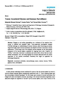
FIG. 1. Normal splenomegaly but enhanced recruitment of mononuclear cells in the lung during persistent MHV-68 infection. Number of total lymphocytes in the spleen (A), MLN (B), and lung (C and D) were counted at indicated time points. Each point represents data from an individual animal (A, C, and D). LNs were pooled from three to four mice, and the average cell number was calculated (B). Representative data from two to four experiments are shown. *, P ⬍ 0.05; NS, not significant.
freeze-thawed to release the virus replicating inside the cells, and spun down, and virus titers in the supernatants were measured using a standard plaque assay. Quantitative PCR for viral transcripts. Latent viral DNA was quantified by quantitative fluorescent (QF)-PCR for the ORF50 gene as previously described (47). Statistical analysis. P values were calculated using Student’s t test unless stated otherwise. P ⬍ 0.05 was considered significant.
RESULTS Impaired primary expansion of antiviral CD8ⴙ T-cell response in the absence of CD80/CD86. It has been reported in several viral infection models, such as LCMV, influenza virus, or VSV, that CD80/CD86-CD28 costimulation significantly impacts the magnitude and function of the antiviral CD8⫹ T-cell response (4, 8, 12, 29, 42, 44, 59). CD8⫹ T cells play a crucial role in the control of initial replication of MHV-68 and contribute to long-term immune surveillance (14, 24). Therefore, we performed detailed analysis of the anti-MHV-68 CD8⫹ T-cell response in CD80/CD86⫺/⫺ mice. First, to characterize the total cellular response, the numbers of total lymphocytes in the spleens, MLNs, and lungs were enumerated at different time points after infection with MHV68. One of the hallmarks of MHV-68 infection is the induction of splenomegaly around 2 to 3 weeks postinfection. As shown in Fig. 1A, normal splenomegaly was observed in CD80/ CD86⫺/⫺ mice, and the total number of splenocytes was not affected at any time point. Total lymphocyte numbers in the MLNs were comparable to those in B6 mice at day 10 but were decreased by ⬃2-fold at day 14 (Fig. 1B). The absence of
CD80/CD86 did not affect the number of lymphocytes recruited to the lungs at days 10 and 14 (Fig. 1C). However, the number of lung-infiltrating lymphocytes was increased during long-term infection in CD80/CD86⫺/⫺ mice (Fig. 1D). In order to assess the effect of CD80/CD86 deficiency on the magnitude of the antiviral CD8⫹ T-cell response, the kinetics of CD8⫹ T-cell responses specific for the two major epitopes in C57BL/6 mice, ORF61524–531 and ORF6487–495, were quantified using MHC/peptide tetramers (Fig. 2). At the peak of the CD8⫹ T-cell response, the numbers of virus-specific CD8⫹ T cells in the lung were significantly reduced compared to those in B6 controls (Fig. 2A and B). The ORF61524–531-specific response was significantly reduced at day 10 postinfection (Fig. 2A). At day 14, the responses against both epitopes were significantly lower: an ⬃2-fold reduction (Fig. 2A and B). In the spleen, a more severe reduction was observed (Fig. 2C and D). The numbers of ORF6487–495-specific CD8⫹ T cells were reduced ⬃3-fold at day 10 postinfection, although this reduction was not statistically significant. At day 14, the numbers of virus-specific CD8⫹ T cells were significantly reduced for both epitopes: ⬃4-fold for ORF61524–531 and ⬃3-fold for ORF6487–495 (Fig. 2C and D). The significant decrease in the number of virus-specific cells in both organs was due to lower frequencies, not fewer numbers of cells per organ (Fig. 1A and C) (data not shown). In the MLNs, the CD8⫹ T-cell response to both epitopes was reduced at day 14 (Fig. 2E), and this was due to a decrease in the total number of lymphocytes (Fig. 1B). Data from Fig. 1 and 2 reveal that CD80/CD86 costimulation sig-
9162
FUSE ET AL.
J. VIROL.
FIG. 2. Impaired primary CD8⫹ T-cell responses against MHV-68 in the absence of CD80/CD86. The number of CD8⫹ T cells specific for the ORF61524–531/Kb (A and C) and ORF6487–495/Db (B and D) epitopes in the lung (A and B) or the spleen (C and D) were measured at indicated time points by staining with the corresponding MHC/peptide tetramer, anti-CD8, and anti-CD44. Each point represents data from an individual mouse: open circles, B6 controls; filled circles, CD80/CD86⫺/⫺ mice. Horizontal bars indicate mean values. Representative data from three independent experiments are shown. *, P ⬍ 0.05. (E) ORF61524–531- and ORF6487–495-specific CD8⫹ T-cell responses in the MLN at days 10 and 14 postinfection. LNs were pooled from three to four mice, and the average numbers of cells per LN were calculated. Representative data from three experiments are shown.
nificantly influences the magnitude of the primary CD8⫹ T-cell response against MHV-68 infection. In the spleens, virus-specific CD8⫹ T cells during long-term infection, days 42 and 97 postinfection, were comparable to those of B6 controls (Fig. 2C and D). However, there was a significantly increased number of ORF61524-531-specific CD8⫹ T cells in the lungs of CD80/CD86⫺/⫺ mice at day 68 postinfection (Fig. 2A). This was due to an increased number of lung-infiltrating lymphocytes present in CD80/CD86⫺/⫺ mice (Fig. 1D). Altered phenotype and function of MHV-68-specific CD8ⴙ T cells in CD80/CD86ⴚ/ⴚ mice. The numbers of virus-specific CD8⫹ T cells were maintained during persistent infection despite the reduced numbers at the peak of the response (Fig. 2A to E). We speculated that the absence of CD80/CD86 costimulation during priming would result in a functionally defective memory CD8⫹ T-cell population, despite the similarities in frequencies and numbers. Because CD8⫹ T-cell function frequently correlates with its phenotype (25, 54), we first analyzed tetramer-positive CD8⫹ T cells for the activation/memory markers CD44, CD62L, CD127 (IL-7R␣), CD122 (IL-15R), CD27, CD25, and CD69. CD27 expression by ORF61524–531-
specific CD8⫹ T cells in the spleen, measured by cells positive as well as the mean fluorescent intensity (MFI), was significantly reduced in CD28⫺/⫺ and CD80/CD86⫺/⫺ mice (see Fig. 3A). This was not observed in the lungs because the majority of virus-specific memory CD8⫹ T cells in the lungs were low for CD27 expression (data not shown), which is in agreement with the phenotype of memory CD8⫹ T cells in peripheral organs (54). The absence of CD28 or CD80/CD86 also resulted in lower expression of CD122 on virus-specific CD8⫹ T cells in both the spleen and the lungs (Fig. 3B). Expression of CD44, CD62L, and CD127 was unaffected in both the spleen and the lungs (data not shown). Interestingly, expression of CD69 was significantly increased in the lungs (Fig. 3C) but not in the spleen (data not shown), indicating the virus-specific CD8⫹ T cells in the lungs have an activated phenotype. A similar pattern was observed with CD25 expression, although the increase of its expression in the lungs was not significant (data not shown). The activated phenotype of virus-specific CD8⫹ T cells in the lungs of CD28⫺/⫺ and CD80/CD86⫺/⫺ mice was consistent with the increased ability of virus-specific CD8⫹ T cells to upregulate the effector molecule granzyme B upon restimulation with peptide in vitro (Fig. 3D); however, this
VOL. 80, 2006
CD80/CD86 COSTIMULATION AND GAMMAHERPESVIRUS LATENCY
9163
FIG. 3. Altered phenotype of virus-specific CD8⫹ T cells in CD80/CD86⫺/⫺ mice. Expression of activation/differentiation markers and effector molecules (panel A, CD27; panel B, IL-15R [CD122]; panel C, CD69; panel D, granzyme B) on ORF61524–531/Kb-specific CD8⫹ T cells from indicated organs were analyzed. All plots are gated on ORF61524–531/Kb-specific CD8⫹ T cells. (A) CD27 expression on ORF61-specific CD8⫹ T cells in the spleen at day 98 postinfection. Representative plots are shown on the left. The numbers above indicate the average percent CD27⫹ cells of ORF61524–531/Kb-specific CD8⫹ T cells. The numbers below indicate the average MFI for CD27 expression of ORF61524–531/Kb-specific CD8⫹ T cells. (B) IL-15R (CD122) expression at day 110 postinfection. Representative plots are shown on the left, and the average MFI from the spleen and lungs are shown on the right. (C) CD69 expression in the lungs at day 270 postinfection. Numbers indicate the average percentage of cells positive for CD69. (D) Intracellular expression of granzyme B at day 205 postinfection. Lung lymphocytes were restimulated with ORF61524–531 peptide for 5 h and were stained for intracellular IFN-␥ and granzyme B expression. Plots are gated on CD8⫹ IFN-␥⫹ cells, and the average MFI is shown. Each figure contains representative data from two to three independent experiments consisting of three to four mice per group. Error bars and numbers in parentheses indicate standard deviation. *, P ⬍ 0.05; **, P ⬍ 0.01.
effect was also observed in the spleen (data not shown). Because the virus-specific CD8⫹ T cells had an activated phenotype and expressed higher levels of effector molecules, we suspected that they were undergoing proliferation in response to antigen. To assess their proliferation in vivo, ORF61- and ORF6-specific CD8⫹ T cells in the lungs and spleens of persistently infected mice were analyzed for BrdU incorporation over a 5-day period. Despite its activated phenotype, the percentages of virus-specific CD8⫹ T cells undergoing proliferation during the short pulse were not significantly different between B6, CD28⫺/⫺, and CD80/CD86⫺/⫺ mice in both organs (data not shown). Altered phenotype of virus-specific memory CD8⫹ T cells in
the absence of CD80/CD86-CD28 costimulation implied that the function of these cells might also be affected. Therefore, next we examined the ability of MHV-68-specific CD8⫹ T cells to produce cytokines by intracellular cytokine staining. Although the frequencies of CD8⫹ T cells producing IFN-␥ in response to ORF61524–531 and ORF6487–495 peptides were not significantly lower than those of B6 controls, the mean MFI of IFN-␥ during long-term infection (but not early time points) was significantly lower in CD28⫺/⫺ and CD80/CD86⫺/⫺ mice (Fig. 4A), indicating lower production of this cytokine on a per cell basis. The impairment was also observed in the virusspecific CD8⫹ T cells in the lung (Fig. 4B). TNF-␣ production was not significantly different in both B6 and CD80/CD86⫺/⫺
9164
FUSE ET AL.
J. VIROL.
FIG. 4. Gradual loss of IFN-␥ production by virus-specific CD8⫹ T cells generated in the absence of CD80/CD86. Splenocytes from days 10, 14, and 110 postinfection (A) and lung lymphocytes from day 187 postinfection (B) were stimulated with the ORF61524–531 peptide and were stained for intracellular IFN-␥ expression. A representative plot for gating (gated on CD8⫹ cells) is shown in the top panel in panel A, and the average MFI for IFN-␥ staining is graphed (A, bottom panels, and B). Each panel contains representative data from two to three independent experiments consisting of three to four mice per group. Error bars represent 1 standard deviation. *, P ⬍ 0.05; **, P ⬍ 0.01; NS, not significant.
mice, as measured by MFI and the percentage of virus-specific cells producing TNF-␣ (data not shown). There was minimal IL-2 production in all groups, including B6 controls, consistent with our previous findings (data not shown) (32a). These results indicate the specific requirement of CD80/CD86-CD28 costimulation for MHV-68-specific CD8⫹ T cells to maintain their ability to produce high levels of IFN-␥. In addition to cytokine production, an important feature of CD8⫹ T cells is their ability to recognize and kill virus-infected cells. Therefore, we analyzed the cytotoxicity of MHV-specific CD8⫹ T cells with an in vivo cytotoxicity assay. The cytotoxicity against ORF61524–531-pulsed targets in CD28⫺/⫺ and CD80/CD86⫺/⫺ mice was comparable to that in B6 controls (Fig. 5). A hallmark of memory CD8⫹ T cells is their ability to proliferate rapidly upon secondary antigen challenge. Because the
FIG. 5. The absence of CD80/CD86 does not alter cytotoxicity. In vivo cytotoxicity at day 113 postinfection was measured as indicated in Materials and Methods. Error bars represent 1 standard deviation. Representative data from two independent experiments consisting of three to four mice per group are shown.
VOL. 80, 2006
CD80/CD86 COSTIMULATION AND GAMMAHERPESVIRUS LATENCY
FIG. 6. Defective secondary responses in the absence of CD80/CD86. (A) Naı¨ve or infected B6, CD28⫺/⫺, or CD80/CD86⫺/⫺ mice were challenged with 106 PFU of rVV-ORF61 intraperitoneally at day 43 postinfection. Frequencies of ORF61524–531/Kb-specific CD8⫹ T cells in the spleen were measured by tetramer staining 5 days after vaccinia virus challenge. Representative plots of CD8⫹ gated splenocytes are shown. The average percentage of tetramer-positive CD8⫹ cells (⫾ standard deviation) is indicated. Data were analyzed using the Mann-Whitney U test. *, P ⬍ 0.05; **, P ⬍ 0.01. (B) Total numbers of ORF61524–531/Kbspecific CD8⫹ T cells in the spleen 5 days postchallenge. *, P ⬍ 0.05; **, P ⬍ 0.01 (analyzed by Student’s t test). Representative data from two independent experiments consisting of three to four mice per group are shown.
phenotype was altered and IFN-␥ production was impaired in the MHV-68-specific memory CD8⫹ T cells in the absence of CD80/CD86, we predicted that their ability to mount a secondary response would also be defective. Indeed, the expansion of ORF61524–531-specific memory CD8⫹ T cells upon secondary challenge with a recombinant vaccinia virus expressing the ORF61524–531 epitope (rVV-ORF61) was severely impaired (Fig. 6). The impairment was observed by both the frequency of ORF61-specific CD8⫹ T cells (Fig. 6A) and the total number of ORF61-specific CD8⫹ T cells in the spleen (Fig. 6B). Thus, the results demonstrate that the CD80/CD86CD28 costimulatory signal specifically affected the initial expansion, phenotype, IFN-␥ production, and secondary response of MHV-68-specific CD8⫹ T cells, but not TNF-␣ production or its cytotoxicity. Humoral responses and CD4ⴙ T-cell responses in CD80/ CD86ⴚ/ⴚ mice. Next we tested whether the antiviral antibody or CD4 T-cell response was affected by CD80/CD86 deficiency. First, the humoral response was measured using a virus neutralization assay. Serum from either CD28⫺/⫺ or CD80/
9165
FIG. 7. Ab and CD4⫹ T-cell responses in CD80/CD86⫺/⫺ mice. (A) Virus neutralizing Ab activity in the serum of infected mice at day 110 postinfection was assayed on NIH 3T3 cells as indicated in Materials and Methods. (B and C) The virus-specific CD4⫹ T-cell response was measured at days 24 (B) and 68 (C) postinfection by an IFN-␥ ELISPOT assay on purified CD4⫹ cells. Representative data from two independent experiments consisting of three to four mice per group are shown. Error bars represent 1 standard deviation. *, P ⬍ 0.05; **, P ⬍ 0.01; NS, not significant.
CD86⫺/⫺ mice elicited low virus neutralizing activity, confirming the essential role of the CD80/CD86-CD28 axis in the germinal center reaction and the induction of high-affinity antiviral antibodies (Fig. 7A) (24). The magnitude of the CD4 T-cell response was measured by an IFN-␥ ELISPOT assay using purified CD4⫹ splenocytes at day 24 postinfection (Fig. 7B), a time point close to the peak of the response (16), and day 68 (Fig. 7C), a time point during latent infection. CD4⫹ T-cell responses were not affected at either time point, indicating that CD80 and CD86 are dispensable for the CD4⫹ T-cell response against MHV-68. Viral reactivation in the absence of CD80 and CD86. As we observed functional impairments in T-cell responses during persistent MHV-68 infection, we wished to test whether this had an impact upon control of the infection. Therefore, we examined the kinetics of viral replication in the lungs of CD80/ CD86⫺/⫺ mice. The viral titers in the lungs of CD80/CD86⫺/⫺ mice were comparable to those in wild-type B6 controls during the early phase of the infection; however, low levels of viral
9166
FUSE ET AL.
FIG. 8. Viral reactivation in the lungs of CD80/CD86⫺/⫺ mice. (A and B) C57BL/6 and CD80/CD86⫺/⫺ mice were infected intranasally with 400 PFU of MHV-68, and lungs were harvested at the indicated time points and assayed for viral titers by a standard plaque assay using NIH 3T3 cells. (A) Representative data of viral kinetics in the lungs from two independent experiments are shown. (B) Each time point represents an independent experiment. (C) The number of viral genome copies in the lungs was measured by QF-PCR for the ORF50 gene. Each point represents data from an individual mouse. *, P ⬍ 0.05; *, P ⬍ 0.01.
replication were observed in CD80/CD86⫺/⫺ mice at day 42 postinfection (Fig. 8A). Viral replication was consistently observed at later time points during latency in the lungs of CD80/ CD86⫺/⫺ mice, while no virus replication was detectable in B6 mice (Fig. 8B). This was very surprising, as no virus reactivation was detected in either the two reports of MHV-68 infection in CD28⫺/⫺ mice (24, 26) or in our experiments (Fig. 8B), and these mice would be expected to have a very similar phenotype to CD80/CD86⫺/⫺ mice. This result was confirmed by QF-PCR for the ORF50 gene performed on DNA extracted from the lungs of persistently infected mice, which showed significantly higher levels of viral genomes in CD80/CD86⫺/⫺ mice (Fig. 8C). An ex vivo viral reactivation assay using mouse embryonic fibroblasts (48) also showed a higher frequency of reactivation (data not shown).
J. VIROL.
Although three different assays demonstrated viral reactivation in the lungs of latently infected CD80/CD86⫺/⫺ mice, the level of virus detected was close to the limit of detection in each assay. Therefore, we cultured lung homogenates from infected mice on NIH 3T3 monolayers to provide an in vitro amplification step and then subsequently determined the titer of the supernatants by a standard plaque assay (see Materials and Methods). The results of a representative experiment of the in vitro-amplified plaque assay on lung samples from day 182 postinfection lung samples are shown in Table 1. While no viral replication was observed in the B6 lungs even after the amplification step, viral titers of the CD80/CD86⫺/⫺ lungs were amplified to over 100,000-fold, confirming the existence of replicating virus during persistence. To determine whether the higher viral burden observed in the CD80/CD86⫺/⫺ mice was restricted to the lungs, we analyzed the latent viral load in the spleen by QF-PCR. Viral genome copies were lower in CD80/CD86⫺/⫺ mice compared to B6 controls at days 14, 21, and 28 postinfection, as is also seen in other strains that fail to make a germinal center response and expand the number of latently infected B cells (Fig. 9) (26). The number of latent viral genomes in the spleen at day 42 was the same as in B6 mice, and no viral replication was detected by a plaque assay, indicating that immune surveillance is maintained in the spleen (Fig. 9) (data not shown). Similar data were obtained at later time points during persistent infection (day 110 postinfection) (data not shown). Surveillance of MHV-68 by CD80/CD86 is independent of CD28 and CTLA-4. Two previous studies reported that CD28⫺/⫺ mice control MHV-68 latency normally, and no reactivation was observed (24, 26). To confirm these results, we performed the in vitro-amplified plaque assay on CD28⫺/⫺ mice. Viral reactivation was consistently observed in CD80/ CD86⫺/⫺, but not CD28⫺/⫺, mice, indicating that surveillance of MHV-68 by T cells through CD80/CD86 is mediated through a CD28-independent signal (Table 2). CD80⫺/⫺ and CD86⫺/⫺ single-knockout mice controlled viral replication, showing that the two receptors play redundant roles in viral control. As previously reported (6, 26), high levels of viral replication were detected from lungs of CD40⫺/⫺ mice, which
TABLE 1. Viral reactivation at day 182 postinfection detected by an in vitro-amplified plaque assay Viral titer by plaque assay a Mouse strain Standard
C57BL/6
CD80/CD86⫺/⫺
0 0 0 0 150 40 150 95
Amplified
0 0 0 0 9.4 ⫻ 107 3.2 ⫻ 107 3.1 ⫻ 107 8.9 ⫻ 107
a Shown are lung viral titers measured by a standard plaque assay (P ⬍ 0.01) and viral reactivation measured by an in vitro-amplified plaque assay as described in Materials and Methods (P ⬍ 0.05). Each number represents the lung viral titer of an individual mouse. Representative data from six independent experiments are shown.
VOL. 80, 2006
CD80/CD86 COSTIMULATION AND GAMMAHERPESVIRUS LATENCY
9167
DISCUSSION
FIG. 9. Establishment of latency in the spleens of CD80/CD86⫺/⫺ mice. The latent viral loads in the spleens were measured by QFPCR for the ORF50 gene at the indicated time points postinfection. Representative data from two independent experiments are shown. *, P ⬍ 0.05.
was confirmed by in vitro amplification and subsequent plaque assay (Table 2). A key difference between CD28⫺/⫺ and CD80/CD86⫺/⫺ mice is that CTLA-4 signaling is present in CD28⫺/⫺ mice but not CD80/CD86⫺/⫺ mice. Some earlier studies have indicated a positive costimulatory role for CTLA-4 (56, 58); therefore, we sought to investigate whether the CD28-independent control of MHV-68 by CD80/CD86 is mediated by CTLA-4. Because CTLA-4⫺/⫺ mice develop a severe lymphoproliferative disease (50), we designed experiments in which CTLA-4 was blocked in vivo using a monoclonal antibody. CD28⫺/⫺ mice were infected with MHV-68 and were treated with a blocking anti-CTLA-4 MAb or a control Ab for 2 weeks. Whether the CTLA-4 blockade occurred in weeks 1 and 2 postinfection or when blockade was initiated ⬎40 days postinfection, no viral reactivation was detected by in vitro-amplified plaque assay (11/11 mice treated with anti-CTLA-4, pooled from three independent experiments, had 0 PFU/lung). Anti-CTLA-4 treatment also did not alter the latent viral load in the spleens of CD28⫺/⫺ mice, as assessed by quantitative PCR: anti-CTLA-4 treated, (4.1 ⫾ 2.6) ⫻ 103 viral genomes/300 ng spleen; hamster IgG treated, (4.4 ⫾ 1.5) ⫻ 103 viral genomes/300 ng spleen (P ⫽ 0.86). The efficacy of the anti-CTLA-4 MAb was confirmed by its ability to enhance an in vitro mixed lymphocyte reaction (data not shown). When infected wild-type C57BL/6 mice were treated with an anti-CTLA-4 MAb from days 25 to 39 postinfection, there were significant increases in the numbers of splenocytes [anti-CTLA-4 treated, (3.08 ⫾ 0.50) ⫻ 108 cells/ spleen; hamster IgG treated, (1.31 ⫾ 0.42) ⫻ 108 cells/spleen; P ⫽ 0.004] and CD4⫹ T cells with an activated (CD44⫹ CD62Llo) phenotype (anti-CTLA-4 treated,: 48.9% ⫾ 6.2% of CD4⫹ T cells; hamster IgG treated, 35.1% ⫾ 4.9%; P ⫽ 0.02). These results concur with the current notion that CTLA-4 is a negative regulator of T-cell responses and functions in a similar fashion in the MHV-68 model. Therefore, we conclude that the CD28-independent surveillance of MHV-68 mediated by CD80 and CD86 was also independent of CTLA-4.
Both cellular and humoral arms of the immune system play important roles in the control of MHV-68. CD4⫹ and CD8⫹ T cells, but not Abs, mediate the control of acute viral replication in the lungs (14, 37). During latency, CD4⫹ and CD8⫹ T cells and neutralizing Abs all significantly contribute to the surveillance of viral reactivation (24, 37, 39, 45). The generation of CD4⫹ and CD8⫹ T-cell and Ab responses is greatly influenced by the “classical” costimulatory molecules, CD80 and CD86. Animals lacking either CD28 or CD80/CD86 have impaired humoral and cellular responses to viral infections, although their impact on viral control differs between different models (3). The CD80/CD86-CD28 costimulatory pathway is critical for germinal center formation in all models, leading to defective class switching and impaired neutralizing Ab production (5, 29, 44, 59). In LCMV, VSV, and influenza virus infections, the absence of this costimulatory pathway results in the reduction of the antiviral CD8⫹ T-cell response, as measured by either cytotoxicity or the number of virus-specific CD8⫹ T cells (4, 27, 29, 42). The effects of CD28 deficiency in MHV-68 infection have been studied previously by two groups (24, 26). Similar to other viral models, the antiviral humoral response was severely impaired (24, 26). However, minimal impairments were detected in the anti-MHV-68 cellular response. The only defects reported by these two reports were impaired IFN-␥ production by splenocytes upon recall in vitro at day 7 postinfection but not at later time points (26) and slightly (though not significantly) reduced IFN-␥ production by antiviral CD4 T cells (24), but no impairments were observed in the antiviral CD8⫹ T-cell response. Due to the ample evidence of this costimulatory pathway having a significant effect in amplifying the antiviral CD8⫹ T-cell response (4, 8, 42), we utilized CD80/CD86⫺/⫺ mice to characterize the antiviral immune response, mainly focusing on the CD8⫹ T-cell response. The primary virus-specific CD8⫹ T-cell responses in the spleen, lung, and the MLNs, as measured by MHC/peptide tetramers, were significantly reduced in
TABLE 2. Surveillance of latency requires either CD80 or CD86 and is independent of CD28 Viral titer by plaque assaya:
Day postinfection
Standard
Amplified
Expt 1 C57BL/6 CD80⫺/⫺ CD86⫺/⫺ CD28⫺/⫺ CD80/CD86⫺/⫺ CD40⫺/⫺
41 41 42 41 41 41
0, 10, 0, 10 0, 0, 0 0, 0, 40 0, 10, 0 0, 30, 40 930, 950, 870
0, 0, 0, 0 0, 0, 0 0, 0, 0 0, 0, 0 0, 6.3 ⫻ 105, 1.0 ⫻ 105 8.1 ⫻ 107, 7.7 ⫻ 107, 7.9 ⫻ 107
Expt 2 C57BL/6 CD80⫺/⫺ CD86⫺/⫺ CD28⫺/⫺ CD80/CD86⫺/⫺
110 97 97 110 100
0, 0, 0, 0 0, 0,150 0, 0, 0 0, 0, 0, 0 1,250, 510, 440
0, 0, 0, 0 0, 0, 0 0, 0, 0 0, 0, 0, 0 1.8 ⫻ 105, 110, 0
Mouse strain
a Lungs were harvested at day 40 or 41, and viral titers were measured by either a standard or an in vitro-amplified plaque assay. Each number represents the lung viral titer of an individual mouse. Data from two independent experiments are shown.
9168
FUSE ET AL.
CD80/CD86⫺/⫺ mice (Fig. 2A to E). The defect was most significant at day 14 postinfection, resulting in a 2- to ⬃4-fold reduction of virus-specific CD8⫹ T cells in the lungs, draining lymph nodes, or spleen. Despite the defective initial expansion, the number of virus-specific CD8⫹ T cells was not affected by the absence of CD80/CD86 at later time points (Fig. 2A to D). In acute infections such as with LCMV and influenza virus, the defective initial expansion in CD28⫺/⫺ mice led directly to decreased numbers of memory CD8⫹ T cells, which indicates that the effector CD8⫹ T cells at the peak of the response underwent programmed contraction (4, 42). In contrast, in the MHV-68 model, the numbers of virus-specific memory CD8⫹ T cells were normal despite the impaired initial expansion (Fig. 2A to D), suggesting that the programmed contraction may be affected by persisting virus. The number of ORF61524–531/Kbspecific CD8⫹ T cells was increased in the lungs of CD80/ CD86⫺/⫺ mice but not B6 or CD28⫺/⫺ mice during latency, and this is likely due to the low level of viral reactivation occurring in the lungs of these mice. The phenotype of virus-specific CD8⫹ T cells often offers insights into their functional status (25, 54). Despite the similarity in the number of virus-specific CD8⫹ T cells, their phenotypes differed significantly in CD28⫺/⫺ and CD80/CD86⫺/⫺ mice (Fig. 3). CD27 and IL-15R were significantly downregulated on MHV68-specific memory CD8⫹ T cells (Fig. 3A and B). CD27 is a costimulatory molecule that belongs to the tumor necrosis factor receptor family and is known to be expressed on a subset of memory CD8 T cells, mainly in lymphoid organs such as the spleen and lymph node (25, 51, 54, 55). Lower CD27 expression also indicates that the memory CD8⫹ T cells are less sensitive to CD70-CD27 costimulatory signals and may be one reason why the function of these cells are impaired. The role of the CD70CD27 interaction in controlling MHV-68 infection and MHV-68specific immune responses has not yet been addressed and will be an area of interest due to the prominent role of CD27 in antiviral T-cell responses (20–22). IL-15R is also known to be upregulated upon differentiation of effector T cells into memory T cells, which are maintained in an IL-15-dependent manner (33). However, in chronic viral infections such as MHV-68, the survival of virus-specific memory CD8⫹ T cells is IL-15 independent (32, 55), complicating the role of IL-15R in this model. We suggest that downregulation of CD27 and IL-15R reflects defective differentiation of virus-specific CD8⫹ T cells due to suboptimal priming in the absence of CD80/CD86. The defective differentiation of virus-specific CD8⫹ T cells in the absence of CD80/CD86 costimulation is supported by its functional defects. Impaired production of IFN-␥, but not TNF-␣, by virus-specific CD8⫹ T cells was observed during long-term surveillance (Fig. 4A and B). Lower IFN-␥ production on a per cell basis by virus-specific CD8⫹ T cells, as measured by the MFI of IFN-␥ by intracellular staining, has been observed during persistent LCMV-T1b infection (43). Compared to chronic LCMV infection, the amounts of chronic antigen and replication, and thus the frequency of virus-specific CD8⫹ T cells encountering antigen, are significantly lower in MHV-68 infection. This may be the reason why the defect in IFN-␥ production is not observed in wild-type C57BL/6 mice. It is tempting to hypothesize that CD80/CD86 signals prevent the loss of IFN-␥ production by the virus-specific CD8⫹ T cells; and in the absence of this costimulatory signal, even a low
J. VIROL.
level of antigen causes the defect in IFN-␥ production. This is supported by our findings that the decrease in IFN-␥ production in CD8⫹ T cells from CD80/CD86⫺/⫺ mice is not observed during the acute response at 10 and 14 days postinfection (Fig. 3A). Moreover, this deficiency was not seen when CD28⫺/⫺ or CD80/CD86⫺/⫺ mice were infected with an ORF73-deficient virus, a mutant virus which is unable to establish latency (17, 30), further supporting the role of chronic antigen in impairing IFN-␥ production (S.F. and E.J.U., unpublished observation). The defective secondary response of MHV-specific-CD8⫹ memory T cells upon challenge by rVV-ORF61 further supports this idea that differentiation of fully competent memory CD8⫹ T cells are hindered in the absence of CD80/CD86 costimulation (Fig. 6A and B). However, despite the defects described above, cytotoxicity was unaffected, as measured by an in vivo cytotoxicity assay (Fig. 5). This result is consistent with the previous report that cytotoxicity is not affected in the absence of CD28 (26) and reveals a selective effect of CD80/ CD86-CD28 costimulation on different functions of MHV-68specific CD8⫹ T cells. CD28⫺/⫺ mice were reported to control MHV-68 latency without any signs of virus reactivation (24, 26). Therefore, the detection of viral replication in the lungs of latently infected CD80/CD86⫺/⫺ mice came as a surprise (Fig. 8A to C). Viral kinetic studies revealed that the initial viral replication in the lungs is controlled normally, and viral reactivation occurs during latency (40⫹ days postinfection) (Fig. 8A). Viral reactivation was not observed when CD80/CD86⫺/⫺ mice were infected with an ORF73-deficient virus (S.F. and E.J.U., unpublished observation). Therefore, the most likely explanation is that the initial replication in the lungs is controlled normally, and viral reactivation occurs only after latency is established. During MHV-68 latent infection, defects in any one compartment (CD4, CD8, or humoral) does not result in viral recrudescence. However, depletion of either T-cell subset in the absence of neutralizing Abs results in low levels of viral replication in the lungs (24). Both CD28⫺/⫺ and CD80/ CD86⫺/⫺ mice are unable to produce neutralizing Abs (Fig. 7A). Previous studies have suggested that the main effector mechanism of antiviral CD4⫹ T cells during latency is the production of IFN-␥ (9, 34, 38), and this function seems to be maintained in the absence of CD80/CD86-CD28 costimulation (Fig. 7B and C). Although antiviral CD4⫹ T-cell responses to LCMV, HSV-1, and influenza virus are significantly affected by CD80/CD86-CD28 costimulation (5, 8, 12), our results and previous reports show that CD4⫹ T-cell responses to MHV-68 are normal in CD28⫺/⫺ mice (24, 26), indicating that the dependence of antiviral CD4⫹ T-cell responses on this pathway differs among different viruses. Normal antiviral CD4⫹ T-cell responses imply that impaired antiviral CD8⫹ T-cell functions in addition to the impaired humoral response are the cause of the low level of viral reactivation observed. Virus-specific CD8⫹ T cells are impaired in the production of IFN-␥ (Fig. 4), which is a key mediator of viral control during latency (11, 35, 52). The virus-specific CD8⫹ T cells are severely defective in expansion upon secondary antigen encounter (Fig. 6), and their phenotype implies a defective differentiation state (Fig. 3A and B). Thus, the defects observed in addition to the impaired humoral immune response likely weaken the immu-
VOL. 80, 2006
CD80/CD86 COSTIMULATION AND GAMMAHERPESVIRUS LATENCY
nological surveillance of MHV-68 in the CD28⫺/⫺ and CD80/ CD86⫺/⫺ mice. Virus-specific CD8⫹ T cells in the lungs of CD28⫺/⫺ and CD80/86⫺/⫺ mice have an activated phenotype indicated by significantly higher CD69 expression (Fig. 3C) and express higher levels of granzyme B upon restimulation (Fig. 3D), suggesting recent encounter with viral antigen. Therefore, there seems to be a partial loss of viral control in these mice. However CD28⫺/⫺ mice clearly prevent virus reactivation, but reactivation does occur in CD80/86⫺/⫺ mice (Fig. 8 and Table 2). Therefore, the main defect of immune surveillance observed in CD80/86⫺/⫺ mice is independent of CD28, and we also showed it is independent of CTLA-4, the only other known ligand for CD80/86. Two previous studies have proposed the existence of an additional receptor(s) for CD80/ CD86 (28, 57). Yamada et al. demonstrated that survival of cardiac allografts in CD28⫺/⫺ mice could be prolonged by antibody blockade of CD86 and that this CD86-mediated positive signal is independent of CTLA-4 (57). This result implied that there is an additional receptor for CD86, although it differs from the results in our study in which the positive signal is mediated by either CD80 or CD86 (Table 2). Mandelbrot et al. provided evidence of CD80- and CD86-dependent proliferation of CD28⫺/⫺ CTLA-4⫺/⫺ CD4⫹ T cells in vivo and in vitro, implying the existence of a CD28-, CTLA-4-independent signal mediated by CD80/CD86 (28). Therefore, we hypothesize that an as yet unidentified receptor for CD80/CD86 is necessary, in addition to CD28, to provide an essential signal to CD8 T cells to facilitate immune surveillance and prevent MHV-68 reactivation in the lungs. In summary, our study reveals a key role of CD80/CD86 in controlling antiviral immunity and the immunological surveillance of MHV-68. The magnitude of the primary virus-specific CD8⫹ T-cell response and the induction of neutralizing Abs are significantly affected. Furthermore, the function of the virus-specific CD8⫹ T cells is severely impaired. Finally, a CD28, CTLA-4-independent CD80/CD86 signal is required for control of viral reactivation. These results provide new insights into the role of costimulatory molecules in chronic viral infections, an area currently poorly characterized, and will potentially lead to the rational design of vaccines or therapies against chronic gammaherpesvirus infections. ACKNOWLEDGMENTS We express our gratitude to Peter Doherty, Stacey Efstathiou, and Lloyd Kasper for providing us with recombinant viruses and mice. We also thank Zach Soucy for technical assistance. This work was supported by National Institutes of Health grants AI51663 and CA103642. J.J.O. was supported by National Institutes of Health/National Institute of Allergy and Infectious Diseases T32 training grant AI07363-11.
6.
7.
8.
9.
10.
11.
12.
13.
14.
15.
16.
17.
18. 19.
20.
21.
22.
23.
24.
25. 26.
REFERENCES 1. Alegre, M. L., K. A. Frauwirth, and C. B. Thompson. 2001. T-cell regulation by CD28 and CTLA-4. Nat. Rev. Immunol. 1:220–228. 2. Andreasen, S. O., J. E. Christensen, O. Marker, and A. R. Thomsen. 2000. Role of CD40 ligand and CD28 in induction and maintenance of antiviral CD8⫹ effector T cell responses. J. Immunol. 164:3689–3697. 3. Bertram, E. M., W. Dawicki, and T. H. Watts. 2004. Role of T cell costimulation in anti-viral immunity. Semin. Immunol. 16:185–196. 4. Bertram, E. M., P. Lau, and T. H. Watts. 2002. Temporal segregation of 4-1BB versus CD28-mediated costimulation: 4-1BB ligand influences T cell numbers late in the primary response and regulates the size of the T cell memory response following influenza infection. J. Immunol. 168:3777–3785. 5. Bertram, E. M., A. Tafuri, A. Shahinian, V. S. Chan, L. Hunziker, M.
27.
28.
29.
30.
9169
Recher, P. S. Ohashi, T. W. Mak, and T. H. Watts. 2002. Role of ICOS versus CD28 in antiviral immunity. Eur. J. Immunol. 32:3376–3385. Brooks, J. W., A. M. Hamilton-Easton, J. P. Christensen, R. D. Cardin, C. L. Hardy, and P. C. Doherty. 1999. Requirement for CD40 ligand, CD4⫹ T cells, and B cells in an infectious mononucleosis-like syndrome. J. Virol. 73:9650–9654. Carreno, B. M., and M. Collins. 2002. The B7 family of ligands and its receptors: new pathways for costimulation and inhibition of immune responses. Annu. Rev. Immunol. 20:29–53. Christensen, J. E., J. P. Christensen, N. N. Kristensen, N. J. Hansen, A. Stryhn, and A. R. Thomsen. 2002. Role of CD28 co-stimulation in generation and maintenance of virus-specific T cells. Int. Immunol. 14:701–711. Christensen, J. P., R. D. Cardin, K. C. Branum, and P. C. Doherty. 1999. CD4(⫹) T cell-mediated control of a gamma-herpesvirus in B cell-deficient mice is mediated by IFN-gamma. Proc. Natl. Acad. Sci. USA 96:5135–5140. Christensen, J. P., and P. C. Doherty. 1999. Quantitative analysis of the acute and long-term CD4⫹ T-cell response to a persistent gammaherpesvirus. J. Virol. 73:4279–4283. Dutia, B. M., C. J. Clarke, D. J. Allen, and A. A. Nash. 1997. Pathological changes in the spleens of gamma interferon receptor-deficient mice infected with murine gammaherpesvirus: a role for CD8 T cells. J. Virol. 71:4278– 4283. Edelmann, K. H., and C. B. Wilson. 2001. Role of CD28/CD80-86 and CD40/CD154 costimulatory interactions in host defense to primary herpes simplex virus infection. J. Virol. 75:612–621. Egen, J. G., M. S. Kuhns, and J. P. Allison. 2002. CTLA-4: new insights into its biological function and use in tumor immunotherapy. Nat. Immunol. 3:611–618. Ehtisham, S., N. P. Sunil-Chandra, and A. A. Nash. 1993. Pathogenesis of murine gammaherpesvirus infection in mice deficient in CD4 and CD8 T cells. J. Virol. 67:5247–5252. Flano, E., S. M. Husain, J. T. Sample, D. L. Woodland, and M. A. Blackman. 2000. Latent murine gamma-herpesvirus infection is established in activated B cells, dendritic cells, and macrophages. J. Immunol. 165:1074–1081. Flan ˜ o, E., D. L. Woodland, M. A. Blackman, and P. C. Doherty. 2001. Analysis of virus-specific CD4⫹ T cells during long-term gammaherpesvirus infection. J. Virol. 75:7744–7748. Fowler, P., S. Marques, J. P. Simas, and S. Efstathiou. 2003. ORF73 of murine herpesvirus-68 is critical for the establishment and maintenance of latency. J. Gen. Virol. 84:3405–3416. Greenwald, R. J., G. J. Freeman, and A. H. Sharpe. 2005. The B7 family revisited. Annu. Rev. Immunol. 23:515–548. Grohmann, U., C. Orabona, F. Fallarino, C. Vacca, F. Calcinaro, A. Falorni, P. Candeloro, M. L. Belladonna, R. Bianchi, M. C. Fioretti, and P. Puccetti. 2002. CTLA-4-Ig regulates tryptophan catabolism in vivo. Nat. Immunol. 3:1097–1101. Hendriks, J., L. A. Gravestein, K. Tesselaar, R. A. van Lier, T. N. Schumacher, and J. Borst. 2000. CD27 is required for generation and long-term maintenance of T cell immunity. Nat. Immunol. 1:433–440. Hendriks, J., Y. Xiao, and J. Borst. 2003. CD27 promotes survival of activated T cells and complements CD28 in generation and establishment of the effector T cell pool. J. Exp. Med. 198:1369–1380. Hendriks, J., Y. Xiao, J. W. Rossen, K. F. van der Sluijs, K. Sugamura, N. Ishii, and J. Borst. 2005. During viral infection of the respiratory tract, CD27, 4-1BB, and OX40 collectively determine formation of CD8⫹ memory T cells and their capacity for secondary expansion. J. Immunol. 175:1665– 1676. Kemball, C. C., E. D. Lee, E. Szomolanyi-Tsuda, T. C. Pearson, C. P. Larsen, and A. E. Lukacher. 2006. Costimulation requirements for antiviral CD8⫹ T cells differ for acute and persistent phases of polyoma virus infection. J. Immunol. 176:1814–1824. Kim, I. J., E. Flano, D. L. Woodland, and M. A. Blackman. 2002. Antibodymediated control of persistent gamma-herpesvirus infection. J. Immunol. 168:3958–3964. Klenerman, P., and A. Hill. 2005. T cells and viral persistence: lessons from diverse infections. Nat. Immunol. 6:873–879. Lee, B. J., S. K. Reiter, M. Anderson, and S. R. Sarawar. 2002. CD28⫺/⫺ mice show defects in cellular and humoral immunity but are able to control infection with murine gammaherpesvirus 68. J. Virol. 76:3049–3053. Lumsden, J. M., J. M. Roberts, N. L. Harris, R. J. Peach, and F. Ronchese. 2000. Differential requirement for CD80 and CD80/CD86-dependent costimulation in the lung immune response to an influenza virus infection. J. Immunol. 164:79–85. Mandelbrot, D. A., M. A. Oosterwegel, K. Shimizu, A. Yamada, G. J. Freeman, R. N. Mitchell, M. H. Sayegh, and A. H. Sharpe. 2001. B7-dependent T-cell costimulation in mice lacking CD28 and CTLA4. J. Clin. Investig. 107:881–887. McAdam, A. J., E. A. Farkash, B. E. Gewurz, and A. H. Sharpe. 2000. B7 costimulation is critical for antibody class switching and CD8⫹ cytotoxic T-lymphocyte generation in the host response to vesicular stomatitis virus. J. Virol. 74:203–208. Moorman, N. J., D. O. Willer, and S. H. Speck. 2003. The gammaherpesvirus
9170
FUSE ET AL.
68 latency-associated nuclear antigen homolog is critical for the establishment of splenic latency. J. Virol. 77:10295–10303. 31. Obar, J. J., S. G. Crist, D. C. Gondek, and E. J. Usherwood. 2004. Different functional capacities of latent and lytic antigen-specific CD8 T cells in murine gammaherpesvirus infection. J. Immunol. 172:1213–1219. 32. Obar, J. J., S. G. Crist, E. K. Leung, and E. J. Usherwood. 2004. IL-15independent proliferative renewal of memory CD8⫹ T cells in latent gammaherpesvirus infection. J. Immunol. 173:2705–2714. 32a.Obar, J. J., S. Fuse, E. K. Leung, S. C. Bellfy, and E. J. Usherwood. 2006. Gammaherpesvirus persistence alters key CD8 T-cell memory characteristics and enhances antiviral protection. J. Virol. 80:8303–8315. 33. Schluns, K. S., and L. Lefrancois. 2003. Cytokine control of memory T-cell development and survival. Nat. Rev. Immunol. 3:269–279. 34. Sparks-Thissen, R. L., D. C. Braaten, K. Hildner, T. L. Murphy, K. M. Murphy, and H. W. Virgin IV. 2005. CD4 T cell control of acute and latent murine gammaherpesvirus infection requires IFNgamma. Virology 338:201– 208. 35. Steed, A. L., E. S. Barton, S. A. Tibbetts, D. L. Popkin, M. L. Lutzke, R. Rochford, and H. W. Virgin IV. 2006. Gamma interferon blocks gammaherpesvirus reactivation from latency. J. Virol. 80:192–200. 36. Stevenson, P. G., G. T. Belz, M. R. Castrucci, J. D. Altman, and P. C. Doherty. 1999. A gamma-herpesvirus sneaks through a CD8(⫹) T cell response primed to a lytic-phase epitope. Proc. Natl. Acad. Sci. USA 96:9281– 9286. 37. Stevenson, P. G., R. D. Cardin, J. P. Christensen, and P. C. Doherty. 1999. Immunological control of a murine gammaherpesvirus independent of CD8⫹ T cells. J. Gen. Virol. 80:477–483. 38. Stevenson, P. G., and S. Efstathiou. 2005. Immune mechanisms in murine gammaherpesvirus-68 infection. Viral Immunol. 18:445–456. 39. Stewart, J. P., E. J. Usherwood, A. Ross, H. Dyson, and T. Nash. 1998. Lung epithelial cells are a major site of murine gammaherpesvirus persistence. J. Exp. Med. 187:1941–1951. 40. Sunil-Chandra, N. P., S. Efstathiou, J. Arno, and A. A. Nash. 1992. Virological and pathological features of mice infected with murine gammaherpesvirus 68. J. Gen. Virol. 73:2347–2356. 41. Sunil-Chandra, N. P., S. Efstathiou, and A. A. Nash. 1992. Murine gammaherpesvirus 68 establishes a latent infection in mouse B lymphocytes in vivo. J. Gen. Virol. 73:3275–3279. 42. Suresh, M., J. K. Whitmire, L. E. Harrington, C. P. Larsen, T. C. Pearson, J. D. Altman, and R. Ahmed. 2001. Role of CD28-B7 interactions in generation and maintenance of CD8 T cell memory. J. Immunol. 167:5565–5573. 43. Tewari, K., J. Sacha, X. Gao, and M. Suresh. 2004. Effect of chronic viral infection on epitope selection, cytokine production, and surface phenotype of CD8 T cells and the role of IFN-gamma receptor in immune regulation. J. Immunol. 172:1491–1500. 44. Thebeau, L. G., and L. A. Morrison. 2003. Mechanism of reduced T-cell effector functions and class-switched antibody responses to herpes simplex virus type 2 in the absence of B7 costimulation. J. Virol. 77:2426–2435.
J. VIROL. 45. Tibbetts, S. A., L. F. van Dyk, S. H. Speck, and H. W. Virgin IV. 2002. Immune control of the number and reactivation phenotype of cells latently infected with a gammaherpesvirus. J. Virol. 76:7125–7132. 46. Usherwood, E. J. 2002. A new approach to epitope confirmation by sampling effector/memory T cells migrating to the lung. J. Immunol. Methods 266: 135–142. 47. Usherwood, E. J., K. A. Ward, M. A. Blackman, J. P. Stewart, and D. L. Woodland. 2001. Latent antigen vaccination in a model gammaherpesvirus infection. J. Virol. 75:8283–8288. 48. van Dyk, L. F., H. W. Virgin IV, and S. H. Speck. 2003. Maintenance of gammaherpesvirus latency requires viral cyclin in the absence of B lymphocytes. J. Virol. 77:5118–5126. 49. Wang, S., and L. Chen. 2004. Co-signaling molecules of the B7-CD28 family in positive and negative regulation of T lymphocyte responses. Microbes Infect. 6:759–766. 50. Waterhouse, P., J. M. Penninger, E. Timms, A. Wakeham, A. Shahinian, K. P. Lee, C. B. Thompson, H. Griesser, and T. W. Mak. 1995. Lymphoproliferative disorders with early lethality in mice deficient in Ctla-4. Science 270:985–988. 51. Watts, T. H. 2005. TNF/TNFR family members in costimulation of T cell responses. Annu. Rev. Immunol. 23:23–68. 52. Weck, K. E., A. J. Dal Canto, J. D. Gould, A. K. O’Guin, K. A. Roth, J. E. Saffitz, S. H. Speck, and H. W. Virgin. 1997. Murine gamma-herpesvirus 68 causes severe large-vessel arteritis in mice lacking interferon-gamma responsiveness: a new model for virus-induced vascular disease. Nat. Med. 3:1346– 1353. 53. Weck, K. E., S. S. Kim, H. W. Virgin IV, and S. H. Speck. 1999. Macrophages are the major reservoir of latent murine gammaherpesvirus 68 in peritoneal cells. J. Virol. 73:3273–3283. 54. Wherry, E. J., and R. Ahmed. 2004. Memory CD8 T-cell differentiation during viral infection. J. Virol. 78:5535–5545. 55. Wherry, E. J., D. L. Barber, S. M. Kaech, J. N. Blattman, and R. Ahmed. 2004. Antigen-independent memory CD8 T cells do not develop during chronic viral infection. Proc. Natl. Acad. Sci. USA 101:16004–16009. (First published 25 October 2004; doi:10.1073/pnas.0407192101.) 56. Wu, Y., Y. Guo, A. Huang, P. Zheng, and Y. Liu. 1997. CTLA-4-B7 interaction is sufficient to costimulate T cell clonal expansion. J. Exp. Med. 185: 1327–1335. 57. Yamada, A., K. Kishimoto, V. M. Dong, M. Sho, A. D. Salama, N. G. Anosova, G. Benichou, D. A. Mandelbrot, A. H. Sharpe, L. A. Turka, H. Auchincloss, Jr., and M. H. Sayegh. 2001. CD28-independent costimulation of T cells in alloimmune responses. J. Immunol. 167:140–146. 58. Zheng, P., Y. Wu, Y. Guo, C. Lee, and Y. Liu. 1998. B7-CTLA4 interaction enhances both production of antitumor cytotoxic T lymphocytes and resistance to tumor challenge. Proc. Natl. Acad. Sci. USA 95:6284–6289. 59. Zimmermann, C., P. Seiler, P. Lane, and R. M. Zinkernagel. 1997. Antiviral immune responses in CTLA4 transgenic mice. J. Virol. 71:1802–1807.