Case report Acta Medica Academica 2011;40(2):182-186 DOI 10.5644/ama2006-124.22
Case presentation – thyroid lymphoma Belkisa Izić¹, Amra Čičkušić¹, Senad Izić², Maida Kuljaninović ³, Šekib Umihanić4 1
Department of Radiology and Nuclear Medicine, University Clinical Center Tuzla, Tuzla, Bosnia and Herzegovina 2 Department of Anaesthesiology and Reanimatology, University Clinical Center Tuzla, Tuzla, Bosnia and Herzegovina 3 Department of Pathology, University Clinical Centre Tuzla, Bosnia and Herzegovina 4 Department of Ear, Nose and Throat University Clinical Center Tuzla, Tuzla Bosnia and Herzegovina
Corresponding author: Belkisa Izić Armije BiH 49a 75000 Tuzla Bosnia and Herzegovina
[email protected] Tel.: + 387 61 739 197 Fax: + 387 35 303 740
Malignant tumors of the thyroid gland account for about 1% of the newly diagnosed malignant tumors each year, and their incidence in women is twice the incidence in men. According to the WHO classification (2004) thyroid tumors are divided into: carcinoma of the thyroid, adenoma and similar tumors, and other thyroid tumors which include: teratomas, angiosarcomas, paragangliomas and others, as well as primary lymphomas and plasmacytomas. Primary thyroid lymphomas are defined as lymphomas which originate in the thyroid gland. This study presents the case of a 68-year-old patient with a thyroid lymphoma, which caused compression of the airways. In the patient presented there was reduced activity of the thyroid gland. The dominant symptoms were: breathing difficulties, hoarse voice and the enlargement of the thyroid. An ultrasound examination was performed before surgery on the neck, which showed a multinodular thyroid, with compromised and compressed trachea to the right and rear. An emergency surgical procedure was performed to reduce the tumor. Pathohistological diagnosis confirmed diffuse large B cell lymphoma. The aim of the study was to present a patient with a thyroid lymphoma, who had previously not had any immunological changes to the gland, that is, she had not had any chronic lymphocyte thyroiditis, but due to the compressive syndrome it was necessary to perform an emergency surgical procedure to reduce the tumor. Key words: Thyroid, Lymphoma, Malignant tumor.
Introduction Received: 13 March 2011 Accepted: 19 August 2011
Copyright © 2011 by Academy of Sciences and Arts of Bosnia and Herzegovina. E-mail for permission to publish:
[email protected]
182
Malignant tumors of the thyroid gland account for about 1% of the newly diagnosed malignant tumors each year (1) and their incidence in women is twice the incidence in men (2). They most often occur in older patients where the average age of occurrence is 65 years. According to the WHO classification (2004), thyroid tumors are divided into: carcinoma of the thyroid, adenoma and similar tumors, and other thyroid tumors which include: teratomas, angiosarcomas, paragangliomas and others as well as primary lymphomas and plasmacytomas (3). In the group
Belkisa Izić et al.: Case presentation – thyroid lymphoma
of carcinomas the most common in the thyroid are papillary (80%), follicular (10%), medullary (5-10%) and the exceptionally rare anaplastic carcinoma (1-2%) (2), whilst the frequency of primary lymphoma is 5% (4). Primary thyroid lymphomas are defined as lymphomas which primarily originate in the thyroid, so this definition excludes lymphomas which affect the thyroid whether by metastasis or by direct spreading from the neighboring lymph nodes. Primary thyroid lymphoma in its late stages may spread to the lymph nodes and other organs, including the gastrointestinal tract, thereby representing a form of mucosa-associated lymphoid tissue (MALT) lymphoma (5). Primary thyroid lymphomas are usually non-Hodgkin type, whilst primary Hodgkin’s disease is very rare (6). Extranodal marginal zone B cell lymphoma (EMZBCL) and diffuse large B cell lymphoma (DLBCL) are lymphomas which most often occur in the thyroid with areas of morphology of a transient form between these two types of lymphoma. Other, especially follicular lymphomas are extremely rate (3). Diffuse large cell B lymphomas are lymphomas which occur most often in the lymph nodes, whilst in 30% of cases they can occur as extra-nodal. The most common extra-nodal site where lymphomas of this type occur is the thyroid (7). The rapidly growing mass of the thyroid gland in an older woman should always arouse suspicion of a tumor. In the past it was necessary to perform an open biopsy to obtain enough tissue for pathohistological examination (6). However, today diagnosis is made on the basis of cytological analysis of aspirate changes in the thyroid gland, immunocytochemistry, flow cytometry, immunophenotypization of lymphocytes obtained by fine needle aspiration (FNA), and finally pathohistological analysis and molecular genetic analysis (8). If it is a DLBCL lymphoma, that is, a lymphoma with a high degree of malignity, there is usually no prob-
lem in establishing a diagnosis from smears obtained by FNA. In cases of unclear diagnosis, if a monomorphic population of small lymphatic cells is found in the smears, it is necessary to repeat the aspiration to obtain sufficient cells for immunophenotypization (6). Differentiating between EMZBCL and lymphocyte thyroiditis, as well as differentiating between other lower level lymphomas than Hashimoto thyroiditis can sometimes be difficult both from cytological smears and histological preparations. Although pathological verification is the gold standard for diagnosis of lymphoma, in rendering a diagnostic decision immunohistochemistry, flow cytometry or genetic molecular analysis are still necessary in addition (3). Treating large cell lymphomas of the thyroid gland is no different from treatment of any other lymphoma occurring in the lymph nodes (9). Treatment is based on the sub-type of the lymphoma and the scope of the disease. Today, the trend in treatment of large cell lymphomas is for treatment to be selected on the basis of prognostic factors. Treatment of a disease which is limited to the thyroid gland is performed by local regional radiation or surgery. In most patients, treatment is mainly by a standard chemotherapy regime, which consists of Cyclophosphamide, Doxorubicin, Vincristine and Prednisolone (CHOP) in combination with radiation, and must always be under the supervision of an oncologist (11). The goal of this paper is to present a patient with thyroid lymphoma, who had not had any previous immunological disease of the thyroid gland, that is, chronic lymphocyte thyroiditis, and in whom it was necessary to perform surgery to reduce the tumor due to compression syndrome.
Case presentation The patient, aged 68, was sent to our institute for examination. A year earlier, the
183
Acta Medica Academica 2011;40:182-186
184
diagnosis of hypothyroidism had been established in another institution and therapy with 1-thyroxin 100 micrograms once a day prescribed. On clinical examination, a node was palpitated in the left lobe of the thyroid gland, 2 cm in size. The follow up finding of the thyroid-stimulating hormone - TSH was 9.9 mIU/l (0.27-3.75 mlU/l); Free thyroxine - FT4 17 pmol/l (10-20 pmol/l); Thyroglobulin - Tg 0.4 ng/ml (2.0-70 ng/l) and thyroglobulin antibodies - TgAt 9.0 IU/ml (>100 IU/ml). Therapy was recommended with 150 micrograms of l-thyroxin once a day and control of hormonal status in four months. Three months later the patient was admitted to our Department of Ear, Nose and Throat for breathing difficulties, loss of voice and sudden increase in the size of the thyroid gland. Physical examination established that the thyroid gland was enlarged, with multimodal changes, hard, painless and immobile on swallowing, with bilateral neck lymphadenopathy. Ultrasound showed multimodal changes to the thyroid gland with a compromised and compressed trachea on the right and to the front. On both sides of the neck there were pockets of enlarged lymph nodes. The ultrasound finding indicated a malignant tumor in the thyroid gland. Since compressive syndrome and re-
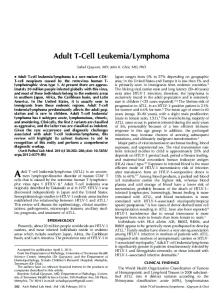
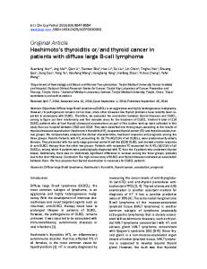
spiratory insufficiency were present, it was proposed following consultation that tumor reduction and tracheotomy be performed. The pathohistological diagnosis of the tumorous mass was Lymphoma malignum diffusum centroblasticum B glandule thyreoideae – diffuse large cell B lymphoma (figure 1). Immunohistochemical analysis showed that the neoplastic cells were positive for CD20 (figure 2) and bcl-6, whilst they were negative for CD 3, CD 10, CD 23, cyclin D1, TdT and bcl-2. Post-operative computerized tomography of the neck, thorax and abdomen showed the remains of the tumorous mass, which went from the level of the hyoid bone to the left, paralaryngeally and para- and retropharyngeally, right up to the level of the jugular fossa. It infiltrated the left half of the supraglottic and glottic regions of the larynx, reducing the air space to a latero-lateral diameter of 2 mm. In the distal half of the neck to the left, pathologically enlarged lymph nodes were visible on the jugular chain about 15 and 24 mm in size, which compromised and, it was suspected, also infiltrated the jugular vein internally. In view of the stage of the illness, further therapy was proposed and transfer of the patient to the Oncology, Hematology and Radiotherapy Clinic. During her hospitalization she received a second cycle of
Figure 1 Diffuse large cell B lymphoma of thyroid gland (HE, x 10)
Figure 2 Immunohistochemical analysis - CD20 positive, x 40.
Belkisa Izić et al.: Case presentation – thyroid lymphoma
chemotherapy according to the CHOP regime, with the addition of monoclonal CD 20 antibodies. On both occasions she had iatrogenic neutropenia which was treated with granulocyte growth factor and broad spectrum antibiotics. There were also raised levels of the enzymes lactate dehydrogenase, aspartate transaminase and creatinine present in the serum. Despite the therapy applied no regression of the tumorous mass was recorded, so the patient was still unable to swallow spontaneously, but was fed through a nasogastric tube. After two months from admission to hospital in the ENT clinic the illness progressed into the front of the upper mouth and gingiva of the upper jaw, there was loss of body mass and the patient died.
Discussion Our patient was treated for one year for reduced activity of the thyroid gland, that is, hypothyroidism. The medical history and negative Tg-At finding excluded the existence of Hashimoto thyroiditis. In a relatively short period of time the thyroid gland increased rapidly in size due to the infiltration of neoplastic cells in the form of diffuse nodes, with compression of the airways and resulting respiratory insufficiency. This condition demanded urgent surgical intervention, that is, total thyroidectomy with bilateral dissection of the neck. The incidence of primary thyroid lymphoma in patients with Hashimoto thyroiditis has increased significantly (7, 10). In fact lymphomas in the thyroid gland in almost all cases occur on the basis of chronic lymphocyte thyroiditis. Graff-Baker et al. believe that there is a pathophysiological connection between autoimmune disorder and thyroid lymphomas. The proposed theory supposes that chronic antigen stimulation is a secondary autoimmune disorder which leads to chronic proliferation of lymph tis-
sue, with resulting mutation leading to the development of lymphoma (9). Until recently there was concern whether the diagnosis of lymphoma could be established by analysis of a sample obtained by aspiration. Cha et al. (11) showed that this approach is successful in 63% patients with thyroid lymphoma. Aspirates of large cell lymphomas are typically hypercellular with visible individual lymphatic cells, which have a cytological appearance, similar or identical to lymphoma of other sites, and in that case it is simple to reach a diagnosis. In contrast to them, in aspirate smears of the lymphoma marginal zone a mixture is found of small atypical lymphocytes, centrocytes, monocytoid B cells, immunoblasts and plasma cells. As a result of the morphological appearance of the cytological smears, differentiating this type of lymphoma and reactive changes in the lymph node is almost impossible (3). The introduction of molecule technology has led to definite diagnosis and as a result open thyroid biopsy is no longer performed in modern diagnostics (6). Takashima et al. (12) and Daria et al. (13) described the use of chain reaction polymerase in strengthening the immunoglobulin heavy chain to establish the diagnosis of lymphoma. The presence of antigen CD-20 and heavy chain clonality confirm the diagnosis. Large cell B lymphoma in two patients aged 64 and 50 years, without a previous history of Hashimoto thyroiditis, were described by Akcala et al. (14), saying that due to the sudden increase in size of the thyroid gland and compression of the respiratory path, total thyroidectomy was performed and CHOP chemotherapy applied. Conclusion This unusual case of thyroid lymphoma is interesting because it occurred in a patient who did not have Hashimoto thyroiditis.
185
Acta Medica Academica 2011;40:182-186
The surgery was performed because of compression of the airways caused by the rapid increase in size of the thyroid gland due to infiltration by neoplastic cells. After two rounds of chemotherapy there was no regression of the disease and the patient died. Conflict of interest: The authors declare that they have no conflict of interest. This study was not sponsored by any external organisation. Authors’ contributions: Conception and design: BI and SI; Acquisition, analysis and interpretation of data: BI and AČ; Drafting the article: BI and ŠU, Revising it critically for important intellectual content: AČ and MK.
References 1. Ruiz Allison AG, Vassilpoulou-Sellin R. Endocrine Malignancies. In: Pazdur R, editor. Medical Oncology a Comprehensive Review. New York: PRR Huntington; 1993. p. 349-58. 2. Jameson JL, Weetman AP. Disorders of the Thyroid gland. In: Braunwld E et al. editor. Harrison´s Principles of Internal Medicine. New York: McGraw-Hill; 2001. p. 2060-84. 3. DeLellis RA, Lloyd RV, Heitz PU, Eng C. World Health Organisation Classification of Tumors. Tumours of Endocrine Organs. France: IARC Press; 2004. 4. Belal AA, Allan A, Kandal A, et al. Primary thyroid lymphoma: a retrospective analisis of prognostic factors and treatment outcome for localized intermediate and high grade lymphoma. Am J Clin Oncolo. 2001;24(3):299-305. 5. Kocjan G (ed). Thyroid. In: Clinical Cytopathology of the Head and Neck. London: Greenwich Medical Media; 2001. pp. 99-105.
186
6. Lerma E, Arguelles R, Rigla M, Otal C, Cubero JM, Bagué S, Carreras AM, et al. Comparative findings of lymphocytic thyroiditis and thyroid lymphoma. Acta Cytol. 2003;47(4):575-80. 7. Tupchong L, Hughes F, Harmer CL. Primary lymphoma of the thyroid: clinical features, prognostic factors, and results of treatment. Int J Radiat Oncol Biol Phys. 1986;12(10):1813-21. 8. Detweiler RE, Katz RL, Alapat C, el-Naggar A, Ordonez N. Malignant lymphoma of the thyroid: a report of two cases diagnosed by fine-needle aspiration. Diagn Cytopathol. 1991;7(2):163-71. 9. Graff-Baker A, Sosa JA, Roman SA. Primary thyroid lymphoma: a review of recent developments in diagnosis and histology-driven treatment. Curr Opin Oncol. 2010;22(1):17-22. 10. Kebapcilar L, Alacacioglu I, Comlekci A, Ozcan MA, Piskin O, Kargi A, et al. Primary thyroid lymphoma: case series with literature review. J BUON. 2009;14(2):295-9. 11. Cha C, Chen H, Westra WH, Udelsman R. Primary thyroid lymphoma: can the diagnosis be made solely by fine-needle aspiration? Ann Surg Oncol. 2002; 9(3):298-302. 12. Takashima S, Takayama F, Saito A, Wang Q, Hidaka K, Sone S. Primary thyroid lymphoma: diagnosis of immunoglobulin heavy chain gene rearrangement with polymerase chain reaction in ultrasound-guided fine-needle aspiration. Thyroid. 2000;10(6):507-10. 13. Doria R, Jekel JF, Cooper DL. Thyroid lymphoma. The case for combined modality therapy. Cancer. 1994;73(1):200-6. 14. Akcali Z, Sakalli H, Noyan Z, Kayaselcuk F, Ozyilkan O. Primary thyroid lymphoma: report of two cases. East Afr Med J. 2004; 81(7):378-80.