Ideal Measurement of Cardiac Output: Is Cardiography the Answer?
Impedance
Edward R. Franko, M.D. Joseph M. Van De Water, M.D. and Xiang Wang, Ph.D.*
NEW HYDE PARK, NEW YORK, and PHILADELPHIA, PENNSYLVANIA
Abstract The ideal measurement of cardiac output (CO) would be a system that is accurate, noninvasive, reproducible, continuous, and technician-free. Impedence cardiography (ICG) has the promise of meeting these criteria. The authors have developed a unique ICG system that analyzes the analog signals from an impedence cardiograph by an original software program. This was compared against standard thermodilution (TD) measurement of CO (CO ) in patients in the inTD tensive care and heart surgical units. Simultaneous measurements by CO TD and by CO ICG were performed in 65 patients. A good correlation was noted between COTD and CO ICG over a range of 2.4 to 9.7 L/min (r 0.73, p < 0.002). If patients with factors known to interfere with ICG were excluded, an improved correlation was found (r 0.89, p < .002). CO ICG followed a similar trend as CO TD even in these excluded patients. The reproducibility of the CO ICG was good (coefficient of variation =
=
= 0.071). found to be simple and automatic. The results show that it is reproducible and correlates well with CO . It also has the added advantages TD of being continuous and noninvasive. Factors such as arrhythmias, severe COPD, and mitral regurgitation were found to interfere with the CO ICG values. Overall, the versatility of. CO ICG gives the promise of very good noninvasive monitoring in critical care units and preoperative evaluation in the outpatient The
ICG CO
was
setting.
of
From the Department of Surgery, Long Island Jewish Medical Center, New Hyde Park, New York; and Philadelphia, Pennsylvania. Presented at the 37th Annual Meeting of the American College of Angiology, Atlanta, Georgia, October,
550
*Drexel 1990.
University
551 Introduction
Hemodynamic monitoring plays a critical role in any intensive care unit (ICU) patient and the measurement of cardiac output (CO) is an important component. Currently, the most widely accepted mode of monitoring is by thermodilution (TD) using the Swan-Ganz catheter. Its disadvantages include the need for skilled personnel, the requirement of hospital admission to the ICU, the involvement of the central circulation by direct access (risking possible infection), and the lack of continuous monitoring. The ideal measurement for measuring CO would involve a system that is accurate, noninvasive, reproducible, continuous, and technician-free. Impedence cardiography (ICG) has been found to be a noninvasive alternative for assessing cardiac function. This did not assume clinical potential until Kubicek et al were able to derive a formula to interpret the waveforms from the impedance cardiograph .’ ’2 However, this required tedious measurements of the waveforms and hand calculations, making its use impractical. With the advent of inexpensive microprocessors, once again ICG has gained clinical significance. The NCCOM-3 became the first commercially available system that was completely automatic. This was based, however, on a different theoretical approach than the original Kubicek formulation. Varying results have been obtained when this system has been compared with thermodilution (TD).3-s The objective of this study was to examine a new ICG system that uses ensemble averaging to examine the analog signals from the impedance cardiograph. The calculations 6 for CO are based on the theoretical framework of Kubicek et a1.6 Materials and Methods Simultaneous measurements of the CO by ICG (C01CG) and by TD(COTD) were determined in 65 consecutive patients from the ICU. CO was measured as the mean of three consecutive values. Thermodilution Method CO by TD was determined by injecting 10 cc of saline at room temperature into a Swan-Ganz paceport catheter. Computations were performed by a program module. This measurement was made during end expiration.
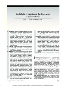
Impedance Method The impedance cardiograph was utilized to obtain the impedance values. Four disposable, 1-mil-thick, aluminum-layered electrodes were employed in a configuration seen in Figure 1. Electrode 1 was placed on the forehead. Electrode 2 was placed circumferential about the neck at the level of the thyroid cartilage. Electrode 3 was xiphisternal, and electrode 4 was 7-8 cm caudal to this. Both electrodes extended from one midaxillary line to the other on the anterior surface. Three ECG electrodes applied on the anterior chest wall in the usual fashion serve as the external trigger for the impedance signal. The impedance cardiograph generates a 110 kHz, 4mA alternating current that does not interfere with the ECG pacing signals. Changes in the voltage measured across the two inner electrodes are directly related to changes in the number of conducting ions,
552
FIG. 1. Configuration used for C01CG studies.
and hence, to volume. By using the first derivative of the AZ signal (the impedance change) and the resistivity of the blood (which is directly related to the hematocrit), the ventricular stroke volume is calculated (the Kubicek equation): SV =
p
(L/Z)2
VET
(dZ/dT)min
where, S V = the stroke volume p
=
the blood
L = the
(dZ/ d1)
length
resistivity (calculated using
a
recent
hematocrit)
between electrodes 2 and 3
slope change of the first derivative of the Z~ = the mean thoracic impedance VET = the ventricular ejection time min = the maximum
AZ
signal
technology in digital signal-processing and personal computer systems, an ensemble-averaging technique was used to analyze the impedance signals from the impedance cardiograph, and the results were employed in the Kubicek formula. The CO With the latest
was
calculated from the obtained SV measured heart rate. Results
The correlation between ICG and TD was r 0.732 with an SD of ± 0.701 and a range of 2.4 to 9.7 L/min. The scattergram can be seen in Figure 2. The majority of data points demonstrated good correlation, but in several data points, this was poor. The patients were identified who corresponded with these data points. Several patients were found to have certain factors that are known to interfere with ICG. These factors and the number of patients are listed in Table I. If these patients were excluded, Figure 3 was obtained. An improved correlation of r 0,899 (SD ± 0.590) was noted. Some of the patients in this excluded group were able to be followed up over time. The trends of ICG and TD are graphed in Figures 4 and 5. =
=
=
Discussion The
development of a theoretical model by Kubicek et al encouraged the investigation
553 of ICG. Earlier studies revealed some conflicting results.’-’° Some investigators found good correlation between ICG when compared with Fick and dye dilution methods.&dquo;&dquo; Opposing studies concluded that ICG was unreliable.’’8 Systematic overestimation of SV and CO in healthy adults and poor overall agreement in chronic and critically ill patients have been the criticisms in other reports.8~’2-’4 These objections to ICG led to reconsideration of the Kubicek formulation and to development of new models to interpret the impedance waveforms. Sramek et al proposed a new equation that differed by treating the thorax as a truncated cone instead of a cylinder, eliminating the resistivity as a hematocrit-dependent variable and substituting L (the distance between electrodes) with a relationship of L body height. &dquo;-&dquo; These modifica-
FIG. 2. CO scattergram for all
patients.
FIG. 4. CO trend in with sepsis.
patients
FIG. 3. CO scattergram
excluding specified patients.
554
FIG. 5. CO trend in
with
patients pulmonary complications.
TABLE I Factors That Interfere with ICG
tions were used to develop the first fully automatic impedance system, the NCOOM-3. Still other modifications have been implemented to the Sramek equation as well . 1,1 4,11 Comparisons of these against TD have had mixed results . 3,4,13,14,18-20 We studied a new ICG system that used the original Kubicek formula. Although a similar set of band electrodes were used, their placement was modified (Fig 1). Ensemble averaging was used as discussed elsewhere.2’ As implied by the results of several studies, p was maintained as a hematocrit-dependent variable.22-24 Band electrodes were used 6 rather than spot electrodes since the latter sacrifice accuracy and reproducibility.6 In testing the reproducibility of the two methods, the ICG system was found to be superior to TD. The coefficients of variation (SD/mean) were found to be 0.071 to 0.113, respectively. These findings correspond with those of other studies .3,13, ’9&dquo;’ The correlation coefficient of r = 0.732 also compares with those values reported elsewhere. 3-5,13,14,18 The patients whose impedance values differed significantly from TD were critically evaluated. Fifteen patients were identified with factors that have been reported to cause this type of discrepancy (Table I). Exclusion of these patients gave a much improved correlation, r 0.899. In a similar manner, Williams and Caird demonstrated improved correlation when such patients were excluded.26 Absolute impedance values will be unreliable in certain patients, and yet the cause for this can almost always be detected . 26,27 We were =
555
able to give a rational explanation for those patients who demonstrated a poor correlation. Most importantly, such patients can be easily identified. Unlike TD, which estimates CO over a period of time, impedance values are determined on a beat-to beat basis. Factors that interfere with measurements by ICG in this manner have been observed in a number of patients. Multiple studies have demonstrated that irregular cardiac rhythms, tachycardia, and pacemakers have either caused errone13,t8,26 ous results or made the results unobtainable,3-5, because the calculations of CO are dependent upon accurate determination of the heart rate, which, in turn, relies upon proper R-wave recognition. This problem was seen in 3 patients in this study. Other cardiac anomalies, eg, valvular insufficiency, left-to-right shunts, and bundle branch blocks, can also cause
poor
correlations.26
Some investigators have reported abnormal waveforms in some patients. Right bundle branch block has been found to cause a bifid appearance of the dZ/dT waveform.26 Enghoff and Lovheim had to exclude about 25070 of the patients from their study because the ICG signals were uninterpretable.2g Some patients have been noted to have multiple peaks and valleys in their dZ/dT signals.’~ Four patients in this study demonstrated this phenomenon with unusual signals having three to four peaks in each dZ/dT waveform. The etiology of this is uncertain. Sepsis and hypotension have been found to cause inaccurate COs by ICG. Since the impedance detects only the pulsatile component of flow, it has been suggested that a major portion of flow may be continuous in these patients.&dquo; Bernstein proposed that the current entering the thoracic aorta was partially diverted to the newly perfused skin, which reduces the impedance CO values.&dquo; One patient with sepsis was noted to have a significant underestimation of the CO by ICG as compared with TD. Another patient with a history of multiple myocardial infarctions was observed to have overestimation of the CO by ICG. Dampened dZ/dT waveforms were observed in both of these patients, but the Z was very low only in the latter patient. The cause for this pattern was unclear. Changes in impedance have been observed with increased pulmonary fluid.29-3’ Early detection of pulmonary edema before the presentation of clinical signs could prevent the difficulty in treating this. However, this phenomenon also appears to impair the accuracy of estimating CO by ICG.27 Impedance changes have been found to be linearly related to blood volume changes but not to be specific for changes in thoracic fluid volume.32,33 In a similar fashion, chronic obstructive pulmonary disease (COPD) has been noted to interfere with ICG possibly by causing unusually elevated Zo values.26,27,32 This distortion of CO values because of pulmonary fluid overload or pulmonary disease was observed in 4 patients in this study. Similar to other studies, the Zo values were high, causing °
diminished
C01CG
measurements.
During low-flow states, inaccurate correlations have been observed between TD and ICG.4~’3 A similar problem was encountered in 2 patients in this study. The value obtained by TD was underestimated by impedance measurements. It has been reported, however, that TD does have a biological error of 150/o to 20070.34 Furthermore, studies have shown that TD will overestimate the CO at flows less that 3.5 L/min.35 Thus, this may not actually represent an error of ICG measurement but rather a fault of TD.
556 Technical difficulties were encountered in several circumstances: (1) twisting of the electrodes tape or improper contact with the alligator clips: (2) inadequate contact of the electrode tape because of tracheostomies, abdominal dressing, colostomies, diaphoresis, or thoracic dressings (including thoracostomy tubes): or (3) artifacts from laryngeal or pharyngeal &dquo;noise.&dquo; These problems associated with ICG have been reported by other authors .1,14, &dquo;&dquo;9 The equipment was also noted to be cumbersome in relation to the limited space in most ICUs. The impedance cardiograph has not been &dquo;updated&dquo; with the more advanced bioengineering circuitry, which could probably reduce its size considerably. Because of the difficulty with band electrodes, spot electrodes have been employed by the NCOOM-3. This would help to alleviate some of the observed technical problems. However, difficulty with obtaining good signals and some of the other same problems have been encountered with the use of spot electrodes. 6’’3’’g Because of this and the theoretical notion of a less parallel current field, we continued to utilize band electrodes as originally described by Kubicek but with a different configuration.6 A modification with spot electrodes is currently being investigated because of the impractical nature of band electrodes. The continuous and noninvasive nature of ICG provides the ability to monitor CO changes during titration with drug and fluid therapies . 4,18,19 Studies have shown that C01CG closely followed the trend of CoTD after ionotrope infusion . Goldstein et al observed the hemodynamic responses to several pharmacologic and physiologic manipulations in healthy volunteers.’9 The differential changes in SV and CO by TD were also observed using ICG. This agreement demonstrates the ability of ICG to follow the trend for any changes in CO measured by TD.’9 This principle was used to evaluate the patients in Table I. Each of these patients (who had been excluded in Fig. 3) was found to have a factor that caused a poor correlation between ICG and TD. The trend in CO for both ICG and TD was followed in 4 patients. Similar to the studies above, the ICG values were observed to closely follow the trend of those measured by TD. Although the 2 patients with sepsis represented in Figure 4 had absolute CO values that significantly differed, the changes in CO by TD were matched by ICG. In Figure 5, the elevated Zo values caused the absolute TD measurements to be underestimated by ICG, and yet, again, the trends were amazingly similar. This allows any patient’s ICG values to be calibrated against TD when the absolute measurements differ.
557 Conclusions
compared with TD and good agreement was obtained. Cerarrhythmias, sepsis, and low-flow states can cause underestimation or overestimation by ICG. These patients can, however, be readily identified. It was demonstrated that the absolute CO values may differ, but the trend of CO measurements by TD is closely followed by ICG values. Thus the ICG values for most of these patients could be calibrated against TD. Several technical aspects will have to be improved before this system can have practical use in the ICU. Overall, this new ICU system may not totally replace the Swan-Ganz catheter but has the definite potential for noninvasive
A new ICG system was tain factors such as COPD,
CO measurement. Edward R. Franko, M.D.
Department of Surgery Long Island Jewish Medical Center New Hyde Park, NY 11042 References 1. Kubicek WG, Karnegis JN, Patterson RP, et al: Development and evaluation of an impedance cardiac output system. Aerosp Med 37:1208-1212, 1966. 2. Kubicek WG, Kottke, FJ, Ramos MU, et al: The Minnesota Impedance Cardiograph—theory and applications. Biomed Eng 9:140-144, 1974. 3. Salandin V, Zussci C, Gabriele R, et al: Comparison of cardiac output estimation by thoracic electrical bioimpedance, thermodilution, and Fick methods. Crit Care Med 6:1157-1158, 1988. 4. Spinale FG, Reines HD, Crawford FA: Comparison of bioimpedance and thermodilution methods for determining cardiac output: Experimental and clinical studies. Ann Thoric Surg 45:421-425, 1988. 5. Introna RPS, Pruett JK, Crumrine RC, et al: Use of transthoracic bioimpedance to determine cardiac output in pediatric patients. Crit Care Med 16:1101-1105, 1988. 6. Wang X, Sun HH, Adamson D, et al: An impedance cardiography system: A new design. Ann Biomed Eng 17:535-566,. 1989. 7. Judy WV, Langley FM, McCowen KD, et al: Comparative evaluation of the thoracic impedance and isotope dilution methods for measuring cardiac output. Aerosp Med 40:532-536, 1969. 8. Keim HJ, Wallace JM, Thurston H, et al: Impedance cardiography for determination of stroke index. J Appl Physiol 41:797-799, 1976. 9. Naggar CZ, Dobnik DB, Flessas AP, et al: Accuracy of the stroke index as determined by the transthoracic electrical impedence methods. Anesthesiology 42:201-205, 1975. 10. Denniston JC, Maher JT, Reeves JT, et al: Measurement of cardiac output by electrical impedance at rest and during exercise. J Appl Physiol 40:91-95,
1976.
11. Kinnen E: Cardiac output from transthoracic impedance variations. Ann NY Acad Sci 170:747-752, 1970. 12. Miller JC, Horvath SM: Impedance cardiography. Psychopathology 15:80-91. 1978. 13. Bernstein DP: Continuous noninvasive real-time monitoring of stroke volume and cardiac output by thoracic electrical bioimpedance. Crit Care Med 14:898-901, 1985. 14. Donovan KD. Dobb GJ. Woods WPD. et al: Comparison of transthoracic electrical impedance and thermodilution methods for measuring cardiac output. Crit Care Med 14:1038-1044, 1986. 15. Sramek BB: Noninvasive technique for measurement of cardiac output by means of electrical impedance. Proceedings of the Fifth International Conference on Electrical Bioimpedance, Tokyo, Japan 1981, pp 39-42. 16. Sramek BB, Rose KM, Miyamoto A: Stroke volume equation with a linear base impedance model and its accuracy, as compared to thermodilution and magnetic flow meter techniques in humans and animals. Proceedings of the Sixth International Conference on Electrical Bioimpedance, Zudar, Yugoslavia, 1983, p 38. 17. Bernstein DP: A new stroke volume equation for thoracic electrical bioimpedance: Theory and rationale. Crit Care Med 14:904-909, 1986. 18. Appel PL, Kram HB, Mackabee J, et al: Comparison of measurements of cardiac output by bioimpedance and thermodilution in Severely ill surgical patients. Crit Care Med 14:933-935, 1986. 19. Goldstein DS, Cannon RO, Zimlichman R, et al: Clinical evaluation of impedance cardiography. Clin Physiol 6:235-251, 1986.
558 20.
Tremper KK: Continuous noninvasive cardiac output : Are we getting there? Crit Care Med 15:278-279,
1987. 21. Muze M, Ebert TJ, Tristani FE, et al: Determination of cardiac output using ensemble averaged im-
pedance cardiograms.
J
Appl Physiol 58:200-205,
1985. 22.
Quail AW, Traugott
FM: Effects of
changing
hematocrit, ventricular rate, and myocardial inotropy on the accuracy of impedance cardiography. Clin Exp Pharmacol Physiol 8:335-341, 1981. 23. Mohaptra SN, Costeloe KL, Hill DW: Blood resistivity and its implications for the calculation of cardiac output by the thoracic electrical impedance technique. Intensive Care Med 3:63, 1977. 24. Demeter RJ, Toth PD, Hawk T, et al: The use of noninvasive impedance to determine cardiac output: Factors affecting its accuracy. Am J Noninvasive Cardiol 2:112-118, 1988. 25. Gastfriend RJ, Van De Water JM, Leonard ML, et al: Impedance cardiography—current status and clinical applications. Am Surg 52:636-640, 1986. 26. Williams BO, Caird FI: Accuracy of the impedance cardiogram in the measurement of cardiac output in the elderly. Age Ageing 14:277-281, 1985. 27. Penny BC: Theory and cardiac applications of electrical impedance measurements. CRC Crit Rev Biomed Eng 13:227 = 281, 1986.
28.
Enghoff E, Lovheim O: A comparison between the transthoracic electrical impedance method and the direct Fick and dye dilution methods for cardiac output measurements in
man.
Scand J Clin Lab Invest
39:585-590, 1979. 29. Berman IR, Scheetz WL, Jenkins EB, et al: Transthoracic electrical impedance as a guide to intravascular overload. Arch Surg 102:61-64, 1971. 30. Pomerantz M, Delgado F, Eiseman B: Clinical evaluation of transthoracic electrical impedance as a guide to intrathoracic fluid volumes. Ann Surg 171:868-891, 1970. 31. Van De Water JM, Watring WG, Linton LA, et al: Prevention of postoperative pulmonary complications. Surg Gynecol Obstet 135:229-233, 1972. 32. Van De Water JM, Mount BG, Barela JR, et al: Monitoring the chest with impedance. Chest 64:597-603, 1973. 33. Van De Water JM, Miller IT, Milne ENC, et al: Impedance plethysmography: Noninvasive means of monitoring the thoracic surgery patient. J Thorac Cardiovasc Surg 60:641-647, 1970. 34. Levett JM, Replogle RL: Thermodilution cardiac output: A critical analysis and review of the literature. J Surg Res 27:392, 1979. 35. van Grondelle A, Ditchey RV, Groves BM, et al: Thermodilution method overestimates low cardiac output in humans. Am J Physiol 245:H690-695, 1986.