The Autonomic Nervous System 25/07/05 Basic Anatomy and Physiology Dr Oliver Pratt, Specialist Registrar Dr Carl Gwinnutt, Consultant Department of Anaesthesia, Hope Hospital, Salford M6 8HD, UK. e mail:
[email protected] Many anaesthetic procedures and drugs used in anaesthetic practice have a direct influence on the autonomic nervous system. It is therefore essential that the anaesthetist should have a basic understanding of its structure and function. Before reading the tutorial, see what you already know about this subject by answering the following questions: 1. 2. 3. 4. 5.
What is the autonomic nervous system and what are its functions? How do sympathetic nerves get from the CNS to the end organs? How do parasympathetic nerves get from the CNS to end organs? What are the transmitter substances in the autonomic nervous system? What types of receptors are there in the autonomic nervous system?
What is the Autonomic Nervous System? Many bodily functions proceed without any conscious supervision from our central nervous system (CNS). For example, we don’t have to remember to digest our food after a meal, or sweat when too warm. These functions are controlled subconsciously, with a degree of automaticity, by a branch of the nervous system - The Autonomic Nervous system (ANS). The ANS can thus be thought of as the regulatory system, that partly or wholly controls most of the body’s organ systems and homeostatic mechanisms. In general, ANS effects are involuntary, relatively rapid, neuronal reflexes. The afferent input to the reflex arc varies and can be from: i)
The Autonomic Nervous System - for example the tachycardia in response to hypotension, mediated by baroreceptors, or -
ii)
The Central Nervous System – for example the “vaso-vagal response” to impending cannulation in a needle-phobic patient.
The efferent limb of neuronal autonomic reflexes consists of specific primary autonomic nerves that synapse in autonomic ganglia, with secondary or “postganglionic” fibres. These postganglionic fibres mediate the desired response at the effector organ. The “effector limb” of the ANS is subdivided in to 2 separate divisions – the sympathetic, and parasympathetic nervous systems. These two divisions differ in both structure and function as will be seen later. In general the sympathetic nervous system can be thought of as preparing the body for “fight or flight”. In the cardiovascular system, increased inotropic and chronotropic drive lead to increased cardiac output and blood flow is routed toward vital organs and skeletal muscle. There is an overall increase in CNS stimulation, and respiratory drive is increased. Visceral activity is decreased. The parasympathetic nervous system in contrast, increases the activity of the abdominal viscera. The cardiovascular system is depressed - reducing heart rate and cardiac output, and routing blood flow toward visceral beds. The respiratory system and CNS are also depressed.
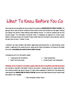
Structure of the Autonomic Nervous System In addition to its close functional relationship to the central nervous system, the ANS shares a close anatomical proximity. In the sympathetic nervous system, the ganglia are fused to form the sympathetic chain, which lies adjacent to the spinal column throughout most of its length. Preganglionic sympathetic fibres have cell bodies in the intermediolateral horn of grey matter in the spinal cord between T1 & L2. These fibres emerge from the spinal cord in the primary ventral rami of the spinal nerves and pass to the sympathetic chain via the white rami communicantes. In the sympathetic chain the fibres will synapse, giving rise to unmyelinated post-ganglionic fibres that rejoin the spinal nerves via the grey rami communicantes. Some preganglionic fibres however ascend or descend to other levels of the sympathetic chain prior to synapsing. In general therefore, sympathetic preganglionic fibres are short, and postganglionic fibres tend to be longer. Parasympathetic preganglionic fibres leave the CNS in both cranial and sacral nerves; the so-called “cranio-sacral outflow”. Cranial fibres arise from specific parasympathetic brainstem nuclei of cranial nerves III, VII, IX, and X. The fibres travel with the main body of the cranial nerves to ganglia that tend to be more distant from the CNS and close to the target organ. Consequently, in contrast to the sympathetic nervous system, preganglionic fibres tend to be long, whereas postganglionic fibres will be shorter. Sacral preganglionic fibres emerge from the CNS via the ventral rami of nerves S2-S4 and form the pelvic splanchnic nerves, which pass to ganglia close to the effector organs. The basic structure of the ANS is illustrated in the diagram below.
One can imagine, that given the anatomical differences between the 2 divisions, anaesthetic interventions may have a greater or lesser effect on the sympathetic or parasympathetic nerves. A good example of this can be seen during spinal anaesthesia. A spinal block will temporarily halt input to the sympathetic afferents at the affected levels, leading to vasodilatation and loss of sweating in the affected dermatomes. If the block is allowed to spread to the levels supplying cardiac sympathetic fibres (T1-T4/5), there will be a loss of both inotropic and chronotropic drive to the heart and progressive hypotension. The parasympathetic supply to the heart coming from the vagus nerve will be unaffected by the spinal block, leading to unopposed parasympathetic stimulation and a bradycardia.
Physiology of the ANS In order to understand the functions of the ANS, and the possible targets for pharmacological manipulation, it is necessary to have a basic knowledge of the neurotransmitters and receptors, which are integral to the ANS. As with all neuronal systems, the effects of the ANS are mediated by the release of neurotransmitters. Preganglionic fibres of both the sympathetic and parasympathetic nervous systems secrete acetylcholine – thus nicotinic receptors (see below) predominate in the autonomic ganglia. Sympathetic postganglionic fibres are mostly adrenergic in nature – i.e. secreting noradrenaline and occasionally adrenaline. The effect of postganglionic nerve stimulation will depend upon the receptors present at the effector site – usually alpha and beta adrenoreceptors. The effects are terminated by noradrenaline re-uptake in to the nerve terminals. A special case within the sympathetic nervous system is the nerve to the adrenal medulla. This nerve does not synapse within the sympathetic chain and hence is strictly still “preganglionic” when it reaches the adrenal medulla and consequently secretes acetycholine. The adrenal medulla, which can be thought of as a modified autonomic ganglion, in turn secretes adrenaline in to the systemic circulation. Parasympathetic postganglionic fibres release acetylcholine. Most effects are mediated via muscarinic receptors and actions are terminated as acetylcholine is hydrolysed by acetylcholinesterase within the synaptic cleft.
Neurotransmitters bind with specific receptors at target cells to produce their effects. Different receptor subtypes exist in each of the divisions of the ANS, and the intracellular response in the target cell and hence the target organ, is specific to the receptor type. Within the sympathetic nervous system, effects are generally mediated by adrenoreceptors. In the parasympathetic system effects are mediated generally by muscarinic acetylcholine receptors. A further special case is that of sympathetic postganglionic fibres supplying sweat glands. These fibres secrete acetylcholine and exert their effects through muscarinic receptors.
ANS Neurotransmitters & Receptors
ANS efferent outflow
GANGLIA Preganglionic cholinergic fibres Postganglionic Nicotinic receptors
SYMPATHETIC POSTGANGLIONICS Mainly release Noradrenaline ACh at Adrenal medulla & Sweat Glands Adrenal medulla releases Adrenaline
PARASYMPATHETIC POSTGANGLIONICS All release Acetylcholine
ADRENORECEPTORS Alpha 1&2 Beta 1&2
ACh RECEPTORS Musacarinic M1-M5 Nicotinic
Adrenoreceptors Adrenoreceptors are subdivided into alpha, and beta receptors. Each of these classes is further divided into subgroups – alpha 1&2, and beta 1&2.
Alpha Receptors Alpha receptors are G-protein linked receptors. Alpha-1 receptors act via the G-protein subgroup Gz and phospholipase C to increase cytosolic calcium levels. This leads to mainly excitatory effects – such as smooth muscle contraction. Alpha-1 receptors are widespread in the peripheral vascular tree and stimulation causes vasoconstriction,
increased systemic vascular resistance and diversion of blood flow from the peripheries to the vital organs. Within the ANS, alpha-2 receptors are largely presynaptic. They act via the G-protein subgroup Gi, inhibiting adenyl cyclase, reducing cytosolic cyclic AMP and calcium. They may also have a direct action – activating potassium channels and causing membrane hyperpolarization. The net effects of these responses are to downregulate, or at least reduce the sympathetic response. Alpha-2 receptors are also present in parts of the CNS – particularly the locus coerulus in floor of the forth ventricle. Their function appears to be linked to the thalamus, reticulospinal tracts and vasomotor centre – activation causing analgesia, drowsiness and hypotension.
Beta Receptors Beta receptors are again G-protein linked receptors. Beta stimulation leads to increased activity of adenyl cyclase that in turn increases intracellular cyclic AMP. Two major subgroups of beta receptors exist – beta-1 and beta-2. Traditional teaching tells us that beta-1 receptors are “cardiac”, whereas beta-2 receptors are more widespread. This is probably an oversimplification, both types of beta receptor can be found in the heart and at many other sites. The beta receptor population is rather “fluid” in nature – receptors can be down or up regulated in terms of number and function. A good example of this is seen in cardiac failure, where reduced receptor density is observed in cardiac muscle. Clinically, beta-1 stimulation leads to increased heart rate and positive inotropy. Renin release from the juxtaglomerular apparatus is stimulated leading to activation of the renin/angiotensin/aldosterone axis. Beta-2 stimulation causes relaxation of bronchial and uterine smooth muscle, vasodilatation in some vascular beds (eg skeletal muscle, pulmonary, coronary) and some degree of positive inotropy & chronotropy.
Acetylcholine Receptors Acetylcholine receptors are named according to the agonist that they responded to in early experiments. Those activated by nicotine were named “Nicotinic” receptors, whereas those that responded to muscarine were named “Muscarinic”.
Nicotinic receptors Nicotinic receptors are ion channels, that when stimulated by acetylcholine, allow a flow of cations into the cell causing depolarization. They are found in all autonomic ganglia. Acetylcholine receptors at the motor end plate of the neuromuscular junction are historically nicotinic, but their structure differs slightly from those of the ANS.
Muscarinic receptors Muscarinic receptors mediate the majority of effects caused by parasympathetic postganglionic fibres. Like adrenoreceptors, they are G-protein linked receptors and are further divided by structure and location into subtypes M1 – M5. M1 receptors are found on gastric parietal cells and stimulate acid secretion. M2 receptors are found in the heart
and have negatively chronotropic effects. M3 receptors promote smooth muscle contraction in the gut, and promote lacrimal secretion. M4 receptors cause adrenaline release from the adrenal medulla in response to sympathetic stimulation, and M5 receptors are thought to have CNS effects.
Summary •
The autonomic nervous system is instrumental in the control of most of the body’s organ systems, via a series of neural reflexes
•
The afferent limb of these reflexes can be from the ANS or CNS.
•
The efferent limb will be mediated by the sympathetic or parasympathetic divisions, which are functionally and structurally distinct.
•
The observed physiological effect will depend upon which neurotransmitter and receptor types are involved.
•
Within the ANS there are many targets for pharmacological manipulation. These will be discussed in a further tutorial.
Dr Oliver Pratt, Specialist Registrar Dr Carl Gwinnutt, Consultant Department of Anaesthesia, Hope Hospital, Salford M6 8HD, UK. e mail:
[email protected] Tutorial Of The Week
The Autonomic Nervous System Part II - Basic Pharmacology In the first of our autonomic nervous system tutorials, we saw that: •
The autonomic nervous system (ANS) reflexes are instrumental in the control of most of the body’s organ systems
•
The afferent limb of these reflexes can be from the ANS or central nervous system (CNS). The efferent limb is mediated by the sympathetic (SNS) or parasympathetic (PNS) divisions, which are functionally and structurally distinct.
•
The observed physiological effect will depend upon which neurotransmitter and types of receptors are involved.
In the normal, resting situation equilibrium exists between sympathetic and parasympathetic activity. Drugs that stimulate or inhibit activity of either the parasympathetic or sympathetic division affect this balance.
Terminology Some common terminology is used to describe the action of drugs on the autonomic nervous system. Sympathomimetics are drugs with similar actions to the postganglionic fibres of the SNS. They resemble adrenaline (epinephrine) in their actions and are also referred to as adrenergics. (Sometimes called sympathetic agonists). Sympatholytics are drugs that oppose the actions of the postganglionic fibres of the SNS. They are also referred to as antiadrenergics or sympathetic (adrenergic) antagonists. Both groups are subdivided further depending on their actions on either alpha or betareceptors. Parasympathomimetics are drugs that stimulate postsynaptic muscarinic receptors. Their actions resemble acetylcholine and they are also referred to as cholinergics or parasympathetic agonists. Parasympatholytics are drugs that oppose the actions of the PNS at the muscarinic receptors by blocking the actions of acetylcholine. They are also referred to as anticholinergics, parasympathetic antagonists or occasionally vagolytics. The autonomic effects of a drug may be the primary intended action – for example the sympathomimetic actions of dobutamine or secondary effects – for example the parasympathomimetic action of repeated doses of succinylcholine.
In the remainder of this tutorial, we will give an overview of the major groups of drugs that act on the autonomic nervous system and examine a few “special cases” - drugs that are commonly used in anaesthetic practice.
Pharmacology of the Sympathetic Nervous System In our first tutorial, we saw that most of the effects of the sympathetic nervous system are mediated by catecholamines (most commonly noradrenaline) acting at alpha or betaadrenoreceptors. All adrenoreceptors are similar in structure and belong to the family of G-protein-coupled receptors. •
Alpha-1 receptors activate phospholipase C and have their actions mainly by increasing release of intracellular calcium.
• •
Alpha-2 receptors inhibit adenylate cyclase, reducing cAMP formation. Beta-1 and -2 receptors stimulate adenylate cyclase, increasing cAMP formation.
The main actions of these receptors includes: •
Alpha-1: vasoconstriction, gut smooth muscle relaxation, salivary secretion, glycogenolysis in the liver, contraction of gut sphincters and uterus.
•
Alpha-2: vasodilatation (central), vasoconstriction (peripheral), gut smooth muscle relaxation.
•
Beta-1: positive inotropy and chronotropy.
•
Beta-2: vasodilatation in muscle, gut and kidneys, bronchodilatation, pupillary dilatation, glycogenolysis.
Drugs with agonist or antagonist effects at both types of adrenoreceptor are commonly encountered in anaesthetic practice, and the most important are discussed below.
Sympathomimetics These drugs can be classified or grouped in a number of different ways, but perhaps the easiest is to classify them according to their actions on adrenoreceptors. They either work by directly stimulating alpha and/or beta-receptors, or indirectly by stimulating the
release of noradrenaline (norepinephrine) by acting presynaptically. A further class of drugs, the phosphodiesterase inhibitors have a postsynaptic action.
Alpha-1 receptor agonists Ephedrine: an indirectly acting sympathomimetic. It is taken up into presynaptic nerve terminals, thereby displacing noradrenaline resulting in alpha mediated vasoconstriction. Ephedrine also has a direct beta agonist effect increasing heart rate and cardiac output, the overall effect increasing blood pressure. These actions last for 10-15 minutes and repeated doses have a gradually decreasing effect (tachyphylaxis). Commonly used to treat the hypotension associated with subarachnoid (spinal) block. Amphetamine: causes CNS stimulation by releasing and blocking uptake of neurotransmitters. Also has peripheral indirect sympathomimetic activity causing acute rises in blood pressure. Currently, minimal therapeutic uses but derivatives of amphetamines are used as recreational drugs, for example ecstasy. Phenylephrine acts directly almost totally on alpha-1 receptors causing vasoconstriction and increasing blood pressure, coronary and cerebral perfusion pressure. Heart rate usually slows due to reflex bradycardia. Cerebral and coronary blood flow is minimally affected. Used to treat hypotension associated with spinal and epidural anaesthesia and topically to provide vasoconstriction in the eye or nose before surgery. Large topical doses have been reported to cause significant CVS side effects including cardiac arrest. Metaraminol: predominantly a direct alpha agonist causing peripheral vasoconstriction. Also has indirect sympathomimetic actions caused by the release of noradrenaline and adrenaline. When used in the treatment of acute hypotension, a baroreceptor mediated bradycardia is frequently seen. Methoxamine: pharmacologically similar to phenylephrine, but much longer acting. Large doses may have a beta antagonist effect causing a bradycardia. Xylometazoline, oxymetazoline: topically active vasoconstrictors used on mucous membranes, with low systemic absorption.
Alpha-2 receptor agonists Alpha-2 receptors are found in the presynaptic membranes of adrenergic synapses and are widely distributed throughout the body including the CNS. They can be subdivided into three subtypes; alpha 2A (sedation, analgesia and sympatholysis), alpha 2B (vasoconstriction) and alpha 2C (CNS actions). Despite being agonists, their actions are generally more like sympatholytic drugs, but they are included here on the basis of their receptor activity.
Clonidine: a potent alpha-2 adrenergic agonist acting on the receptors in the spinal cord. When given orally or iv results in dose dependent sedation, reduces the dose of induction drug needed, reduces the MAC of volatile anaesthetics and provides a degree of analgesia. It increases haemodynamic stability during surgery, at recovery from anaesthesia and may reduce cardiac morbidity in high-risk cases. It reduces shivering and oxygen consumption at recovery. When given epidurally, clonidine increases the quality and duration of block and provides a degree of postoperative analgesia. This practice has proved particularly popular in caudal epidurals in children. When used with local anaesthetics in spinals, it increases the duration and quality of block, but may increase the degree of hypotension. The need for a urinary catheter is reduced when compared to the use of intrathecal opioids. Clonidine has been used in critical care for sedation, analgesia for invasive procedures and to assist in reducing drug withdrawal symptoms after prolonged sedation. Dexmedetomidine: has an even greater affinity for alpha-2 receptors than clonidine. Many effects are similar to clonidine but there is less clinical experience.
Beta receptor agonists The main drugs in this group are the naturally occurring catecholamines, adrenaline (epinephrine) and noradrenaline (norepinephrine). Adrenaline is an agonist at alpha and beta receptors, noradrenaline has predominantly alpha agonist actions with minor beta-1 agonist activity. They will not be considered further here, but details of some of the catecholamines can be seen in appendix 1. Isoprenaline: the first synthetic beta receptor agonist for clinical use, stimulating both beta-1 and -2 receptors. Usually given as an infusion because of its short duration of action. Used mainly to treat bradyarrhythmias and as a bronchodilator. Now largely replaced as a bronchodilator by beta-2 selective drugs because of the risk of cardiac arrhythmias. Salbutamol: predominantly a beta-2 agonist used in the treatment of asthma, both intravenously and by inhalation. Also slows peristalsis and causes muscle tremor in large doses. Alternatives include terbutaline, and salmeterol and formoterol that are longer lasting. Ritodrine: a beta-2 agonist used as a uterine relaxant (tocolytic) to prevent premature labour. Given i.v. initially followed by oral maintenance therapy. Salbutamol is also used for the same effect.
Phosphodiesterase Inhibitors Phosphodiesterase is the enzyme responsible for breakdown of the cAMP produced by beta receptor activation. Inhibition of phosphodiesterase leads to accumulation of cAMP,
which acts to amplify a beta-mediated sympathetic nervous system response. Various subtypes of phosphodiesterase are predominant in different cells and tissues and a number of different drugs exist which will predominantly inhibit the various subtypes. Whilst a full description is beyond the scope of this tutorial, 2 of the most common drugs are mentioned below. Theophylline/aminophylline: Theophylline is a methylxanthine derivative, which is a non-specific inhibitor of all the phosphodiesterase subtypes. Aminophylline is a mixture of theophylline and ethylenediamine, which improves solubility and hence allows intravenous administration. These drugs are predominantly used as bronchodilators, but other actions include weak positive inotropy, peripheral and coronary vasodilation, and a degree of CNS stimulation. Theophyllines have a narrow therapeutic index, and dosage must be guided by monitoring of serum levels. Enoximone: a selective inhibitor of phopshodiesterase subtype III, enoximone is a potent inotrope, which also causes marked peripheral vasodilation. It is particularly effective in patients with a high background sympathetic tone, such as that seen in heart failure. Extensive first-pass metabolism means enoximone is only effective when given intravenously.
Commonly used sympathomimetic drugs, routes given and doses Route given
Dose given (average adult) Bolus Infusion
Alpha agonists Ephedrine
Phenylephrine
Metaraminol Methoxamine Clonidine
IV IM Topically IV IM Topically IV IM IV IM O IV Epidural Intrathecal
3-6mg 10-20mg 0.5% solution 0.25-0.5mg 2.0-5.0mg 0.25-1.0% (nasal) 0.25-0.5mg 2.0-5.0mg 1.0-2.0mg 5-10mg 2-4 microgram/kg 1-4 microgram/kg 1-2 microgram/kg 1.0 microgram/kg (max)
IV IV Inhaled (nebulised) IV IM O
1-2 microgram 0.25mg 2.5-5.0mg
0.5-5.0mg/min
25-180 microgram/min
2-5 microgram/kg/min
Beta Agonists Isoprenaline Salbutamol Ritodrine
2-4 microgram/min 3-20 microgram/min 50-350 microgram/min
10mg 10mg
Sympatholytics These drugs block either alpha or beta adrenergic receptors. It is also possible to have a sympatholytic action by blocking the sympathetic ganglia; only one drug is available to achieve this clinically.
Alpha receptor antagonists (alpha blockers) Phenoxybenzamine: a non-specific, irreversible alpha-1 and alpha-2 antagonist. The alpha-2 blockade leads to increased noradrenaline release and a beta-receptor mediated compensatory tachycardia. Consequently non-specific alpha-blockers are often given concurrently with a beta adrenoreceptor antagonist to block this effect (see below). Mainly used orally to induce hypotension, particularly in the management of phaeochromocytoma. Phentolamine: has similar CVS effects to phenoxybenzamine but the alpha blockade is shorter acting and reversible with alpha-agonists. Tolazoline: mainly used as a pulmonary vasodilator to treat persistent pulmonary hypertension in neonates. Prazosin and Doxazosin: selective alpha-1 antagonists which cause vasodilation and hypotension, but a lesser degree of compensatory tachycardia. Mainly used as adjuncts in the control of hypertension.
Beta receptor antagonists (beta blockers) Since they were first synthesized over 50 years ago, “beta blockers” have evolved into a large family of drugs. The ones below are used to illustrate the key features of the differences between members of this family. Propranolol: relatively non-specific antagonist, blocking both beta-1 and beta-2 receptors. Decreases heart rate, blood pressure and cardiac output. Increases airway resistance in patients with asthma and COPD. Inhibits glucose metabolism and blocks sympathetic mediated “warning sings” of hypoglycaemia in diabetics. May adversely affect lipid profile. Its main use now is in the control of thyrotoxicosis, treatment of essential tremor, migraine and control of the somatic manifestations of stress. Atenolol, metoprolol: due to their predominance in blocking beta-1 receptors present in the myocardium they are often called “cardioselective”. In reality, although cardioselective beta-blockers have a predilection for beta-1 receptors, at higher doses they become less specific, blocking both beta-1 and 2 receptors. Beta-1 receptor blockade reduces heart rate, cardiac output, blood pressure and thus myocardial oxygen demand. In addition, a lusitropic action (an increase in diastolic time) increases coronary blood flow. Mainly used in the treatment of hypertension and ischaemic heart disease. Oxprenolol: a relatively non-specific antagonist, blocking both beta-1 and beta-2 receptors but also with some partial beta-2 agonist action (intrinsic sympathomimetic activity). Thought to be therapeutically beneficial. Mainly used as an antihypertensive.
Esmolol: a relatively specific beta-1 antagonist, esmolol is metabolised by red cell esterases and hence has a very short half-life. Its peak effects occur within 6-10 minutes of being given, but have almost completely disappeared by 20 minutes. Esmolol is only available for intravenous use. Useful for rapid control of hypertensive episodes such as those associated with tracheal intubation and for treatment of supraventricular tachycardias. Sotalol: a non-specific beta antagonist, which also has class III antiarhythmic activity. Used for prophylaxis of paroxysmal supraventricular tachycardias and ventricular ectopics. May induce torsade des pointes in susceptible patients. Labetalol: a non-specific alpha and beta receptor antagonist. When given intravenously, the ratio of beta to alpha activity is approximately 7:1. Ideal for the control of acute hypertensive episodes, or for hypotensive anaesthesia.
Evidence has been published which suggests peri-operative beta blockade reduces the incidence of cardiac morbidity and mortality in high cardiac risk patients undergoing major surgery. The individual drugs, patient groups, and duration of beta blockade are still debated.
Ganglion blockers This group of drugs acts by inhibiting the postsynaptic actions of acetylcholine at autonomic ganglia. Trimetaphan: the only drug in this group still in clinical use today. It causes profound peripheral vasodilatation and hypotension, reduces cardiac output and coronary and renal blood flow. It is very short acting and usually given by slow intravenous infusion to induce controlled hypotension. Causes a reflex tachycardia and tachyphylaxis is common. Not surprisingly, it also causes some degree of non-depolarizing neuromuscular block.
Commonly used sympatholytic drugs, routes given and doses Route given
Dose given (average adult) Bolus Infusion
Alpha antagonist Phenoxybenzamine
O IV IV
1-2mg/kg daily
O IV O IV
Esmolol
40-320mg/day 1mg in 1 min; max 5mg 25-100mg/day 1mg/min, max 10mg 50 micrograms
Labetalol
5-20mg, max 200mg
Phentolamine
1mg/kg (over 2hr) 1-5mg
Beta antagonist Propranolol Atenolol
0.15mg/kg over 20 min 50-200 microgram/kg/min 2mg/min
Ganglion blocker Trimetaphan
IV
2-4mg/min
Pharmacology of the Parasympathetic Nervous System Most of the effects of the parasympathetic nervous system are mediated through acetylcholine released from post-ganglionic nerve terminals and acting at muscarinic receptors. Muscarinic receptors are membrane-spanning protein structures which function as ion-channels. Several subtypes of muscarinic receptor have been identified, numbered M1-M5. •
M1 receptors stimulate gastric acid secretion
•
M2 receptors are thought to mediate the negative chronotropic effects of parasympathetic stimulation
•
M3 receptors mediate lacrimal and salivary gland secretion, and may also be involved in gut smooth muscle contraction.
•
M4 and M5 subtypes are thought to be present mainly in the CNS, though M4 receptors may also mediate adrenaline secretion from the adrenal medulla.
Muscarinic receptors are the main target of drugs that act directly on the parasympathetic nervous system. In general though, both agonists and antagonists show little specificity for the different subtypes of muscarinic receptor.
Parasympathomimetics Muscarinic Receptor Agonists Direct acting muscarinic agonists are relatively infrequently used in clinical practice, but they are still used in a few specific cases. Pilocarpine: a semi-synthetic alkaloid, which is a probably the most widely used direct muscarinic agonist. Pilocarpine is mainly used topically in the treatment of glaucoma, but has been used systemically to treat xerostomia. Systemic effects include hypotension and bradycardia, bronchoconstriction, bronchorrhoea and sialorrhoea.
Acetyl-cholinesterase Inhibitors As anaesthetists, we most commonly see parasympathomimetic actions of drugs following administration of the cholinesterase inhibitors during reversal of neuromuscular blockade. Hence it is appropriate to discuss this group of drugs (also known as anti-cholinesterases) here. By non-specifically inhibiting the breakdown of acetylcholine in synapses, anti-cholinesterases will cause the desired improvement in neuromuscular transmission, but also widespread muscarinic receptor activation. This manifests itself as relative bradycardia, hypersalivation, increased gut smooth muscle activity and meiosis. These effects are usually counteracted by concurrent administration of a muscarinic antagonist, such as atropine or glycopyrrolate (see below). Neostigmine: the most commonly used anti-cholinesterase. Neostigmine is a carbamate ester, which reversibly inhibits acetyl cholinesterase by itself acting as a substrate. During this process, the acetyl cholinesterase molecule becomes carbamylated, and consequently inactive. Regeneration of the active enzyme occurs, but much slower than following hydrolysis of acetylcholine. A single dose of neostigmine has a peak effect at 7-10 minutes, and the effect will last for up to 1 hour. Neostigmine is available in a preparation combined with glycopyrrolate to minimise the muscarinic side effects. Pyridostimine: shares its mode of action with neostigmine. It has a slower onset of action, and a longer duration of action than neostigmine. It can be given orally, and is consequently useful in the treatment of myaesthenia gravis. Edrophonium bromide: or “Tensilon” is most commonly used in the diagnosis of myaesthenia gravis. It is a reversible competitive inhibitor of acetyl-cholinesterase, which prevents acetylcholine gaining access to the active site of the enzyme. The onset and
duration of action of edrophonium are both significantly shorter than neostigmine (duration approximately 10 minutes).
Commonly used parasympathomimetic drugs, routes and doses Route given
Dose given (average adult) Bolus Infusion
Muscarinic Agonists Pilocarpine
Top (eye) Oral
Titrated to effect 5mg TDS
N/A
IV Oral Oral IV
50-70mcg/kg 15-30mg qid 0.3-1.2g in divided doses 2mg then 8mg (Tensilon test) 500-700mcg/kg (reversal)
N/A
AntiCholinesterases Neostigmine Pyridostigmine Edrophonium
N/A N/A
Parasympatholytics Muscarinic Receptor Antagonists Muscarinic receptor antagonists are the drugs with a direct action on the parasympathetic nervous system, which are most frequently used in the anaesthetic setting. They are used both intra-operatively and as premedication, to treat symptomatic bradycardia and to reduce oropharyngeal and respiratory secretions. The most commonly used agents are atropine, glycopyrrolate and hyoscine. These are all competitive, reversible, non-specific antagonists, which produce dose dependant effects. At low dose, reduced salivation, sweating & bronchial secretion predominate. At higher doses, pupillary dilatation and tachycardia are seen. Still higher doses reduce gut motility, gastric secretion & bladder muscle function.
Atropine: this naturally occurring tertiary amine is derived from the deadly nightshade plant (Atropa Belladonna). It is lipid soluble and can therefore cross the blood brain barrier, causing central as well as peripheral effects. Peripheral effects tend to predominate and include tachycardia, bronchodilation and inhibition of sweating and salivation. Atropine is commonly used in an intravenous dose of 10-20mcg/kg to treat bradycardia during anaesthesia. A dose of 3mg will produce complete vagal blockade in an adult, but such a dose is generally restricted for use in resuscitation algorithms. Smaller doses are used as an anti-sialogogue. Atropine has been linked to post-operative confusion in the elderly, probably a result of its central actions. Hyoscine: is also a tertiary amine, and is particularly lipid soluble. Central effects thus tend to predominate – including drowsiness, amnesia and analgesia. It is also a powerful anti-sialogogue. These properties have seen hyoscine used frequently as premedication, but perhaps its most common use today is as an anti-emetic. It is particularly common in many proprietary travel sickness remedies. Glycopyrrolate: a synthetic quaternary amine. Glycopyrrolate is strongly ionised and therefore unable to cross the blood-brain barrier. Consequently it has no central effects, and is thus an alternative to atropine in the elderly patient. It is a more potent antisialogogue than atropine, but has slightly weaker cardiac effects. This makes it an ideal agent to be used in conjunction with the anti-cholinesterases, when salivation can be controlled without precipitating potentially harmful tachycardia. Ipratropium: a synthetic derivative of atropine, administered by nebulisation, or metered dose aerosol inhaler in the treatment of asthma. Competitive inhibition at muscarinic receptors on bronchial smooth muscle cells reduces bronchospasm, and there may also be a secondary effect reducing mast cell degranulation (which is muscarinic receptor mediated). Some systemic absorption may lead to mild tachycardia and increased cardiac output.
Pirenzepine: an unusual drug, in that it is a relatively specific inhibitor of M1 receptors. Pirenzepine reduces gastric acid secretion and reduces gut smooth muscle activity. It is used in the treatment of peptic ulcer disease, but it’s use is not widespread.
Commonly used parasympatholytic drugs, routes and doses Route given Atropine
Glycopyrrolate Hyoscine
Ipratropium
IM/IV
Oral IM/IV SC/IM Oral Transdermal Aerosol Nebulised
Dose given (average adult) Bolus Infusion 0.015-0.02mg/kg 3mg for total vagal blockade 0.2 – 0.6mg 0.2-0.4mg S/C 0.6-2.4mg per 0.2-0.6mg (pre-med) 24hrs to reduce 0.3mg qid (nausea) Releases 1mg/72hr patch secretions in palliative care setting 20-80mcg qid 100 – 500mcg qid
Summary o A balance exists between sympathetic and parasympathetic outflow. Many common pathologies and surgical procedures can affect this balance, leading to disturbances in the function of organ systems. o Drugs can contribute to autonomic disturbance, but drugs can also be used to redress the balance. o Drugs which act on the ANS, tend to be agonists or antagonists at one or more of the various receptors, and this determines their clinical effects. o Some drugs have slightly different modes of action (such as the anticholinesterases or phosphodiesterase inhibitors) but still have marked effects on the ANS.
Appendix 1: Commonly Used Inotropes Predominantly beta 1 & 2 effects at low dose Increasing alpha effects at higher dose Useful “rescue inotrope” in resuscitation situations Alpha & beta effects at very low dose. Alpha effects quickly Noradrenaline predominate as dosage increases. Useful in vasodilatation (sepsis) Potent beta 1 agonist. Some beta 2 mediated vasodilatation is often seen, but occasionally matched by alpha mediated vasoconstriction. Dobutamine Useful in low cardiac output states. Beta 1 and dopamine-receptor agonism predominates at low dose. Increasing alpha effects seen as dose escalates. Dopamine Useful “rescue inotrope”. Renal protection probably does not occur. Potent beta 2 agonist. Some dopamine receptor agonism. Positive inotropy and peripheral/splanchnic vasodilatation seen. Dopexamine Thought by some to offer a degree of renal/gut protection – but remains a contentious issue. Non-selective beta agonist. Used most commonly in treatment of severe bradycardia Isoprenaline Adrenaline
Autonomic nervous system tutorial:
A. Questions to be answered 1. What are the major divisions of the nervous system and how does one classify the autonomic nervous system? 2. What are the anatomical divisions of the autonomic nervous system and what are the pathways involved in autonomic innervations of various organs? Where are the autonomic ganglia located? 3. What are the major neurotransmitters involved in autonomic neurotransmission? Where are these transmitters found? How are the levels and activity of these transmitters regulated at the level of the synapse 4. What are the major receptors found in the autonomic nervous system and where are they located? What are the major tissues affected by the autonomic nervous system and in which organs are they found? 5. What are the second messenger systems involved in autonomic neurotransmission and how do these second messengers effect autonomic target organs? 6. How can drugs affect the autonomic nervous system and which drugs are used to mimic autonomic effects?
B. List of readings:
1. World anaesthesia tutorial of the week: Autonomic nervous system tutorials 1 and 2 (see attached) 2. Chapter 17, W.F. Ganong. Autonomic nervous system 3. Chapters 6-10, Basic and clinical Pharmacology, B.G. Katzung, 8th edition. C. Typical examination question Describe the mechanism of action of adrenergic receptors in the airways, heart, eye and blood vessels, with particular emphasis on the second messengers stimulated or inhibited by these receptors and how these second messengers affect smooth muscle, cardiac muscle and glandular tissue function