International Journal of Electronics Communication and Computer Engineering Volume 6, Issue 6, ISSN (Online): 2249–071X, ISSN (Print): 2278–4209
Augmented Reality for the Study of Human Heart Anatomy Matina Kiourexidou
Konstantinos Natsis
Panagiotis Bamidis
Nikos Antonopoulos
Department of Anatomy, Medical School, Aristotle University of Thessaloniki, Greece
Department of Anatomy, Medical School, Aristotle University of Thessaloniki, Greece
Lab of Medical Physics, Medical School, Aristotle University of Thessaloniki, Greece
Media Informatics Lab, School of Journalism and Mass Communication, Aristotle University of Thessaloniki, Greece
Efthymia Papathanasiou
Markos Sgantzos
Andreas Veglis
Department of Anatomy, Medical School, Aristotle University of Thessaloniki, Greece
Department of Anatomy, Medical School, University of Thessaly, Biopolis, 41110, Larissa, Greece
Media Informatics Lab, School of Journalism and Mass Communication, Aristotle University of Thessaloniki, Greece
Abstract – Augmented reality is increasingly applied in medical education mainly because educators can share knowledge through virtual objects. This research describes the development of a web application, which enhances users' medical knowledge with regards to the anatomy of the human heart by means of augmented reality. Evaluation is conducted in two different facets. In the first one, the authors of this paper evaluate the feasibility of a three-dimensional human heart module using one investigator under the supervision of an expert. In the second, evaluation aims at identifying usability issues by means of the cognitive walkthrough method. Three medical students (naive users) are called upon three target tasks in the web application. Task completion is appreciated in the light of the standard set of cognitive walkthrough questions. Augmented reality content miss hits are revealed by means of the first evaluation in an effort to enhance the educational utility of the three-dimensional human heart. Cognitive walkthrough provides further improvement points, which may further enhance usability in the next software release. The current piece of work constitutes the pre-pilot evaluation. Standardized methodologies are utilized in an effort to improve the application before its wider piloting to proper student populations. Such evaluations are considered important in experiential learning methods aiding online education of anatomy courses. Keywords – Augmented Reality, Cognitive Walkthrough, E-Learning, Heart Model, Interactive Systems, Three Dimensions, Web Application.
I. INTRODUCTION
creates a unique result, since the user can see through the human body, to its anatomy [11]. Furthermore, an application that uses AR, which was named "Miracle" and was created by Tobias Blum. Another AR education tool for anatomy named “BARETA” is a platform that combines AR with 3D models to provide the student with stimulation for touch as well as sight [12]. Also a pilot program was set to explore the distance healthcare education [13]. Overall a debate has been arisen with advocates of the traditional curriculum, on the one hand, and of the innovative curriculum, on the other hand. Many important reasons are responsible for the increasing popularity of online distance learning, particularly in higher education, such as the increased number of participants and, in many cases the ability of the participant to attend the course at a convenient time [5][14]-[15]. E-learning, as an instruction delivered through digital devices, includes numerous types of media (text, audio, images, animation and streaming video) and has broad applications not only in anatomy courses but in medicine generally [16]. Visualization Cardiac AR is a derivative of virtual reality and enables the user to combine the virtual with the natural environment [17] this technology integrates computer generated 3D models into the real word and constitutes a potential learning tool. In the School of Medicine of the Aristotle University of Thessaloniki, Greece, educational innovations in anatomy teaching have been regarded with skepticism for one main reason: historically, the most fruitful eras for anatomy was when dissection was not banned the known paradigm of Andrea Vesalius [18]. This fact is evident towards the necessity of anatomy dissection. To replace anatomy dissection is probably not feasible, but educational technology has a potential to supplement the traditional curriculum [19]. The main scope of the present article is to describe and demonstrate the development of a free educational web application that, with the use of AR, will interact and provide the user with anatomical knowledge for the human heart. An envisaged breakthrough of the present paper is that the aim for use of this web application, target at the introduction of new technological techniques and tools that may reinforce the education processes when it comes to anatomy teaching in Greek universities. The latter
Many educators and developers all over the world research about the factors that influence a virtual school and students’ efficiency learning [1]. Furthermore, the growing interest for research on alternative methods of education in Medical Schools [2], led to the use of videos [3] and podcasts [4] during the e-learning process [5]. The traditional anatomy curriculum has been challenged since the early 80s [6] and educational technology has attracted great interest with the introduction of many innovations such as virtual anatomy courses and dissections [7], threedimensional (3D) atlas [8] and "mARble" application, which uses augmented reality (AR) for iphones [9]. AR enriches the user experience and describes the appearance of a digital element generated with a computer [10]. AR Copyright © 2015 IJECCE, All right reserved 658
International Journal of Electronics Communication and Computer Engineering Volume 6, Issue 6, ISSN (Online): 2249–071X, ISSN (Print): 2278–4209 becomes an issue of concern, when the decreased teaching capacity of academic staff (due to teaching hour limitations which is maximized during the recent recession times by staff number decreases) is seen in parallel with the increased students’ number in Greek universities. Thus, the introduction of new educational methods that may alleviate the above burden and constitute a swift solution to these problems sounds like a golden match. Furthermore, it should be taken under consideration that an actual procedure of anatomy of a heart, for educational purposes, encompasses many problems since the same process cannot be repeated several times as there is a lack of cadavers [10]. The use of the application allows any possible errors to be reversible, and enables the independence of movements of the trainee during the course [20]. Simultaneously, the repetition and experimentation by the user will assist the educational procedure that could be also achieved outside the classroom. The ultimate objective of the current piece of work is to evaluate, based on objective data, a 3D heart model that has been developed during the creation of an e-learning web application environment combined with AR for the study of the human heart. To achieve this purpose the following hypothesis will be tested: The creation of an elearning web application environment for the study of human heart (3D) in combination with AR is feasible, in order to be used as an educational tool for undergraduate medical students through the participation on a traditional topographical and descriptive course on human anatomy. Having developed a good working prototype of the application, a second hypothesis concerns its usability. Is it feasible to apply a proper/standard usability method in order to evaluate existing errors in the design and overall "look-and-feel" of the application? The latter is undertaken with an aim to fulfill the successful run of operating scenarios of use as well as user satisfaction. So, the current paper is organized as follows: in the next section the development of the web application and the methodological issues about the evaluation method are discussed and analyzed. Then the following two sections focus on the results of the development of the web application and the evaluation process. The last section discusses the findings and the future extensions of this work.
II. MATERIAL AND METHODS A. Development tools The interactive web application is only for computer use. Many external code libraries were used: FLARToolkit, Flex SDK, Papervision3D, for the 3d model we used the “blender” program an open source graphics and animation software [21], for the marker a draw program and the FLAR Marker Generator. The marker has a black square as outline and another white square inside it
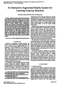
[9]. Hypertext Markup Language (HTML) used for the creation of the web application (Figure 1). The image of the application you can see in the figure 2. To use the application needs a computer, a web camera, an Internet connection and a marker were employed [9].
B. Evaluation 1) 3D model In order to evaluate the 3D model of the human heart, a checklist was designed in Table 1, which summarizes the educational goals of a traditional descriptive course of the human heart. The textbooks that have been studied are Gray’s anatomy [2] and Netter’s Atlas of Human Anatomy [22]. The English terminology conforms to Terminilogia Anatomica [23]. One investigator under the supervision of the senior author has completed the evaluation process of the 3D human heart.
2) Cognitive Walkthrough The type of assessment chosen and used for this application is called cognitive walkthrough (CW), a method conducted in a laboratory environment. There is always a researcher requesting for some particular tasks (scenarios of use/operation) so that the user's perception of the application capacities is evaluated. In other words, CW is conducted in order to understand and prove the extent to which users are able to respond as well as the type of problems they might face while running/operating the application/system [24]-[25]. In fact, the participant should conduct actions so that the evaluator will be able to observe the mistakes; these are jotted down in a list so as for them to be corrected later on. To put it differently, the main objectives are the system usability evaluation and the discovery of design errors so that they can be solved [26][27]. Small participant numbers are usually involved in CW. For instance, three students participated in Dutt’s [28] CW experiment, while two graduate students took part in Hertzum’s [29] experiment in order to evaluate applications. All in all, in CW, one user carries out the tasks while an expert evaluates the usability of the system’s design.
2a) Cognitive Walkthrough Samples in this study In the present study three (3) male students from the School of Medicine (third year students) were employed for evaluating the application with the CW method; Provision of full anonymity and confidentiality was kept throughout, by following the ethical guidelines of Aristotle University of Thessaloniki (the study was approved by the bioethical committee of the Medical School; 151/3.3.2015). All three participants were regarded as computer literate for this application as they all had significant experience in using computers and Internet and since they were users for over 5 years. However, none of them had previously used a system with AR before. Three evaluation categories/scenarios were created: 1) Do the users believe that this action is available, 2) Do the users know how to conduct this action, 3) Is the reaction to the system obvious.
Fig.1. The process of creating the application with AR Copyright © 2015 IJECCE, All right reserved 659
International Journal of Electronics Communication and Computer Engineering Volume 6, Issue 6, ISSN (Online): 2249–071X, ISSN (Print): 2278–4209
III. RESULTS A. Development The web application (Figure 2) was created in both Greek and English Languages. The main menu of the web application is divided in three categories: main page, heart and contact. On the first page, the user can see a welcoming text accompanied by an introductory video on how to use the web application. The heart category is subdivided in six subcategories corresponding to the four surfaces, base and apex of the human heart. Rotation of the virtual human heart is feasible at approximately 40 degrees to the right and left. Five views have been designated in the 3D creation software that have been termed anterior, inferior, right, left, base and apex view corresponding to the description of the heart. The 3D object has been rotated as if looking the heart right directly in front of each surface, the base and apex. Under the menu, information has been added with regard to the proper use of the AR application. At the lower part of the web application, there is a large blank space where the computer’s camera is activated, and once the user places the marker in front of it, the virtual model appears. The last category on the main menu is a contact category from where the user is able to communicate via email with the creator of the application. The checklist that has been designed for the evaluation process is shown in Table 1.
Fig.2. Interaction with augmented reality web application The introductory course of a traditional descriptive course of the heart has as its main aim to describe the heart as a 3D object. Geometric models and various views (based on the geometric model) of the same object have been traditionally used by anatomists to achieve this purpose. Since the heart is described as a pyramid six subheadings have been used in the design of the checklist corresponding to the four surfaces, base and apex. Under its subheading the anatomical terms corresponding to certain anatomical structures that the student must identify, were included. In an anterior view of the virtual heart the anterior surface consists largely of the right atrium. The left atrium is totally hidden by the ascending aorta and the pulmonary trunk. A rather large segment of the left auricle is depicted. In contrast to the anatomical pattern the
ventricular region is depicted disproportionately. The two ventricles are almost equally sized. The anterior interventricular sulcus is, thus, displaced to the right. The right surface of the heart is rounded and formed by the right atrium. The intrathoracic part of the inferior vena cava is depicted along with the superior vena cava. The sulcus terminalis is not depicted and a distinction between the true atrial and the venous components of the right atrium cannot be made. Due to the disproportionate depiction of the ventricles the left surface of the heart includes a rather large part of the right ventricle. A small part of the left atrium and auricle are visible but the right atrium and auricle are in contrast to the normal anatomical pattern not depicted. The coronary sulcus is not discernible and by rotating the virtual object is not circumscribed. The apex of the heart is anatomically positioned as the apex of the left ventricle. The inferior surface of the heart does not consist chiefly by the left ventricle and, therefore, the posterior interventricular groove is displaced to the left. The base of the virtual heart consists mainly by the left atrium. The posterior part of the right atrium is not visible. The bifurcation of the pulmonary trunk is hidden by the aortic arch. The four pulmonary veins are depicted anatomically but the right atrium is hidden by the oversized left atrium. The superior and inferior vena cavae are visible. An advantage of a virtual heart is that the gross appearance of the heart can be adjusted to conform to a more in colour realistic view of the heart. In color representations as presented in standard anatomy atlases reflect the functional role and the realistic appearance. The traditional technique of embalming using formalin results in a gross appearance that does not reflect the leaving body. This technique, however, introduces the student to imaging techniques where the physician must identify structures in a black and white scale (x-ray, CT). Threedimensional reconstructions have been implemented on CT data as an attempt to facilitate diagnosis and supplement the visualization of the structure under investigation for pathology. Three-dimensional CT reconstructions have been proposed to be used as educational tools but this technique has many limitations as highlighted by Radetzki et al. [30]. The creation of the 3D heart as performed in this study along with the evaluation method proposed offers the advantage of modifying the object. Based on the results of our research an interactive webpage can be created to supplement the traditional descriptive course. This form of application has the advantage that the student can access it from home. Factors such as the resolution of the camera, or the computer monitor have an impact on the in full colour appearance but they do not cause deformation of the virtual object. The final product will be used more than 5 times in each semester, once the application will become operational. The web application will be utilized for teaching small groups of 10 to 15 students. However the user will be able to study the book at his home individually.
Copyright © 2015 IJECCE, All right reserved 660
International Journal of Electronics Communication and Computer Engineering Volume 6, Issue 6, ISSN (Online): 2249–071X, ISSN (Print): 2278–4209 Table 1: Surface of the heart and great vessels Base of heart
Shown Not shown
Left atrium
Right atrium
X
X
Bifurcation of Posterior Coronary Crux of the Pulmonary Superior Inferior vena pulmonary interventricular sulcus heart veins vena cava cava trunk sulcus X X X X X X X
Left ventricle Apex of the heart
Anterior Shown (Sternocostal) Not shown surface
Diaphragmati Shown c (Inferior) Not shown surface
Right pulmonary surface
Left pulmonary surface
X
Shown Not shown
Shown
Left auricle
Right atrium auricle
Right ventricle
Left ventricle
Χ
Χ
Χ
Χ
Coronary Ascending sulcus aorta Χ
Anterior Pulmonary interventri trunk cular sulcus Χ Χ
Χ Right ventricle
Left ventricle
Χ
Χ
Coronary sulcus
Posterior interventricular sulcus
Χ
Χ Terminal groove
Right atrium
Inferior vena cava
Superior vena cava
Χ
Χ
Χ Χ
Not shown
Shown
Left ventricle
Left atrium
Auricle
Χ
Χ
Χ Χ
Not shown
B. Evaluation 1) 3D model
2) Cognitive Walkthrough
The overall performance of the evaluation process of the 3D human heart is presented below in Table.
Task 1: Users can find Anterior surface Yes No Do the users believe that this action is available? Do the users know how to conduct this action? Is the reaction to the system obvious?
Coronary sulcus
The tasks and the results arising from the CW procedure are included in Table 2:
Table 2: Cognitive Walkthrough Task 2: Download Marker from web application Yes No Do the users believe that this action is available? Do the users know how to conduct this action? Is the reaction to the system obvious?
Since AR had not been previously used, it was something new for the users and thus not so familiar to them. The user’s evaluator reported the following problems during the research: There was a short delay initially because the users’ were unfamiliar with the application. Another problem was that one of the users did not understand how to use marker and print it at first, but
Task 3: Download Marker from web application Yes No Do the users believe that this action is available? Do the users know how to conduct this action? Is the reaction to the system obvious?
finally the task it was completed. Researchers added a text mentioning, “Please, see the introductory video related to the use of the application on the first page” along with the usage steps of the application in order to solve these problems. The fact that users easily found the anterior surface and they understood how to use AR was really positive.
Copyright © 2015 IJECCE, All right reserved 661
International Journal of Electronics Communication and Computer Engineering Volume 6, Issue 6, ISSN (Online): 2249–071X, ISSN (Print): 2278–4209
IV. DISCUSSION The aim of the anatomy subjects in the laboratory is to educate students on the human body anatomy. Furthermore, the anatomy terminology is mentioned through this process. All the above mentioned in the medical curriculum have succeeded in a combination with lectures and laboratory exercising with the use of cadavers. The student until now may only borrow anatomical models from the laboratory in order to have the chance to study without a supervisor. With this application, the medical lectures, laboratory exercises and the anatomical models, the targets of medical curriculum are accomplished. Because anatomical models and academic lessons cannot study at home the student is capable now to use the special web application and interact with 3D model and as result to be taught the heart anatomy with immersion. This could be done in the University laboratory with teamwork or in private. The web application that was created for this research is at an initial stage and for this reason is not tested in classes. The present study has obviously got certain limitations. Apart from the anatomical component, the development of an interactive webpage integrates the informatics component as well. However, the anatomical component must first be addressed according to the authors’ opinion. A pilot study will be conducted to evaluate the informatics component but after the final correction of the virtual human heart. Online information is abundant but is not always reliable. Although students’ opinions must be considered with regard to education, a scientific approach, with regard to the evaluation of virtual objects, is to objectively evaluate them with the objective data described in standard anatomy textbooks and by experts. During the creation of the 3D model the evaluation process used, contributed to the refinement of the model, although inaccuracies still remain. The web application will not be activated before the correction of the inaccuracies. We propose this simple method of evaluation for every technology application in anatomy in an attempt to provide a certification form in 3D objects that they conform to the standard anatomy description and can be used for educational purposes. This is also an attempt to dissuade students from using online resources that are not intended for educational purposes. The CW part of the investigation identified just a couple of design pitfalls too. It is expected that proper dressing of the application with appropriate user manual and handles will solve these problems. However, as shown in the corresponding results section, the fact that users easily found the anterior surface and they understood how to use AR was really positive in the current phase of development.
relatively new tool for the education of young physicians and it is a difficult process altogether. However, what the current piece of research shows is that it is feasible and doable. The long-term aim is obviously not to replace the anatomy on the cadaver per se, thereby limiting the exposure of medical to actual basic medical science objects skills, but rather to enable it continuity beyond the boundaries of a typical classroom/amphitheatre, thereby enhancing self directed learning, life-long learning and distance education [31]-[32]. The latter are all contemporary demands of medical education in a slowly but surely changing landscape equipped with niche technologies [33].
REFERENCES [1]
[2]
[3]
[4]
[5]
[6]
[7]
[8]
[9]
[10]
[11]
[12] [13]
[14]
V. CONCLUSION [15]
Considering all of the above, it is understood that the design and the implementation of the educational application using AR and incorporating the threedimensional model of the heart of the human body, is a
[16]
F. S. Motlagh & J. A. Pour, “Designing a model for developing students' needs skills of high schools for using virtual learning,” International Journal of Electronics Communication and Computer Engineering, 2014, 3(6), pp. 1444-1448. S. Standring, Gray's anatomy: the anatomical basis of clinical practice, expert consult (4st Ed). Spain: Churchill Livingstone, 2008. K. Romanov & A. Nevgi, “Do medical students watch video clips in e-Learning and do these facilitate learning?,” Med Teach, 2007, vol. 29, pp. 484-488. S. Shantikumar, “From lecture theatre to portable media: students’ perceptions of an enhanced podcast for revision,” Med Teach, 2009, 31, pp. 535-538. D. A. Back, N. Haberstroh, A. Antolic, K. Sostmann, G. Schmidmaier & E. Hoff, “Blended learning approach improves teaching in a problem-based learning environment in orthopedics - a pilot study,” BMC Medical Education, 2014, 14, p.17. M. A. Aziz, J. C. McKenzie, J. S. Wilson, R. J. Cowie, S. A. Ayeni & B. K. Dunn, “The human cadaver in the age of biomedical informatics,” Anat Rec (New Anat), 2002, 269, pp. 20-32. H. Petersson, D. Sinkvist, C. Wang & O. Smedby, “Web-based interactive 3D visualization as a tool for improved anatomy Learning,” Anat Sci Ed, 2009, 2, pp. 61-8. J. Li, L. Nie, Z. Li, L. Lin, L. Tang & J. Ouyang, “Maximizing modern distribution of complex anatomical spatial information: 3D reconstruction and rapid prototype production of anatomical corrosion casts of human specimens,” Anat Sci Ed, 2012, 5, pp. 330-9. U. Von Jan, C. Noll, M. Behrends & U. Albrecht, “mARble augmented reality in medical education,” Biomed Tech 2012, 18, pp. 67-7. H. Hazidar & R. Sulaiman, “Visualization cardiac human anatomy using augmented reality mobile application,” IJECCE, 2014, 5(3), pp. 497-501 T. Blum, V. Kleeberger, C. Bichlmeier & N. Navab, “Mirracle: an augmented reality magic mirror system for anatomy education,” Virtual Reality Short Papers and Posters, 2012, pp. 169-170. R. Thomas, N. John & M. Delieu, “Augmented reality for anatomical education,” J Vis Commun Med, 2010, 33, pp. 6-15. M. Schoonheim, R. Heyden, J.M. Wiecha, “Use of a virtual world computer environment for international distance education: lessons from a pilot project using Second Life,” BMC Medical Education, 2014, 14, p. 36. S. B. Issenberg, W. C. McGaghie, E. R. Petrusa, G. D. Lee & R. J. Scalese, Features and uses of high-fidelity medical simulations that lead to effective learning: a BEME systematic review. Med Teach 2005; 27:10-28. C. Nikendei, P. Weyrich, J. Jünger & M. Schrauth, “Medical education in Germany,” Med Teach, 2009, 31, pp. 591-600. P. Bamidis, S. Konstantinidis, C. Papadelis, E. Perantoni & C. Styliadis, “An e-learning platform for aerospace medicine,” Hippokratia, 2008, 12, pp. 15-22.
Copyright © 2015 IJECCE, All right reserved 662
International Journal of Electronics Communication and Computer Engineering Volume 6, Issue 6, ISSN (Online): 2249–071X, ISSN (Print): 2278–4209 [17]
[18] [19]
[20]
[21] [22] [23]
[24]
[25]
[26]
[27]
[28]
[29]
[30]
[31]
[32]
[33]
X. Zhong, P. Boulanger & N. Georganas, “Collaborative augmented reality: A prototype for industrial training,” Proceedings of 21st Biennial Symposium on Communication, Kingston, Canada, 2002. J.M. Ball, Andreas Vesalius, the reformer of anatomy. Medical Science Press: St. Louis, 1910. S.N. Biasutto, L.I. Caussa & L.E. Criado del Rio, “Teaching anatomy: cadavers vs. computers?,” Ann Anat, 2006, 188, pp. 187-190. B. Papachristou, “Evaluate the contribution of augmented reality in education,” Proceedings of 3rd Conference on Informatics in Education, Athens, Greece, 2011. Blender, Program blender. Available: www.blender.org. F. H. Netter, Atlas of Human Anatomy: with Student Consult Access (5st Ed). Saunders Elsevier: Philadelphia, 2010. Federative Committee, Terminologia Anatomica: International Anatomical Terminology. Thieme Medical Publishers: New York, 2002. C. Wharton, J. Rieman, C. Lewis, & P. Polson, The cognitive walkthrough method: A practitioner’s guide. In usability inspection methods. Edited by Nielsen, J., and Mack, R. New York: John Wiley & Sons, Inc., 1994. P.G. Polson, C. Lewis, J. Rieman, C. Wharton, “Cognitive walkthroughs: a method for theory-based evaluation of user interfaces,” International Journal of Man-Machine Studies, 1992, 36, pp. 741-773. C. Lewis, P. Polson, C. Wharton, & J. Rieman, “Testing a walkthrough methodology for theory-based design of walk-upand-use interfaces,” Proceedings of the ACM CHI’90, Seattle, USA, 1990, pp. 235-242. M. Fossum, A. Ehnfors Mfruhling, A Fruhling & A. Ehrenberg, “An evaluation of the usability of a computerized decision support system for nursing homes,” Appl Clin Inform, 2011, 19(2), pp. 420-36. A. Dutt, H. Johnson, P.Johnson, Evaluating evaluation methods. In People and Computers IX. Edited by Cockton G, Draper SW, Weir GRS. Cambridge: Cambridge University Press; 1994, pp. 109-121. M. Hertzum & N.E. Jacobsen, “The Evaluator Effect: A Chilling fact about usability evaluation methods,” International Journal of Human-Computer Interaction, 2001, 13, pp. 421-443. F. Radetzki, T. Mendel, H. Noser, D. Stoevesandt, M. Rollinghoff, N. Gutteck, K.S. Delank & D. Wohlrad, “Potentialities and limitations of a database constructing threedimensional virtual bone models,” Surg Radiol Anat 2013, 35, pp. 963-8. E. Dafli, P. Antoniou, L. Ioannidis, N. Dombros, D. Topps & P. D. Bamidis, “Virtual patients on the semantic web: a proof-ofapplication study,” J Med Internet Res, 2015, 17(1), p. e16. D. Schwarz, P. Štourač, M. Komenda, H. Harazim, M. Kosinová, J. Gregor, R. Hůlek, O. Smékalová, I. Křikava, R. Štoudek & L. Dušek, “Interactive algorithms for teaching and learning acute medicine in the network of medical faculties MEFANET,” J Med Internet Res, 2013, 15(7), p. e135. P.E. Antoniou, C.A. Athanasopoulou, E. Dafli, P.D. Bamidis, “Exploring design requirements for repurposing dental virtual patients from the web to second life: a focus group study,” J Med Internet Res, 2014, 16(6), p. e151.
Natsis Konstantinos is Orthopaedic Surgeon and Professor of Anatomy, in the school of Medicine of Aristotle University of Thessaloniki, Greece. Director of the Laboratory Descriptive Anatomy of the Department of Medicine of the Faculty of Health Sciences, Aristotle University of Thessaloniki. He taught undergraduate and postgraduate courses in several Universities. Publications in International Journals: 143, Abstracts in International and Greek Conferences: 579, Publications in Conference Proceedings: 93, Abstract Publications: 229, Publications in Greek Journals: 41, Lectures: 251, Organizing Committee on Conferences: 49, Translation Scientific Books: 23, Curator Medical Books 29, Publications Reports: 306.
Bamidis Panagiotis is Assistant Professor of Medical Education Informatics is leading the working group of Medical Education Informatics at the Medical School of the Aristotle University of Thessaloniki. He has been the co-ordinator of large European projects as well as the principal investigator for a number of national and international funded projects. His research interests are within technology enhanced learning in Medical Education and Affective and Physiological Computing and HCI and Affective Neurosciences. In 2009, he was awarded the Prize of the AUTH Research Committee for the Best Track Record in funded research projects among AUTH young academic staff.
Antonopoulos Nikos is Ph.D. Graduate from the Aristotle University of Thessaloniki. He received his B.Sc. in Information Technology and Telecommunications at the Higher Technological Educational Institute of Larissa, MSc in Cultural Informatics and Communication at the Aegean University. His research interests include online media, humancomputer interaction, education, social networking, digital marketing and usability. Website: www.antonopoulos.info
AUTHOR’S PROFILE Kiourexidou Matina is Ph.D. Candidate of the Laboratory Descriptive Anatomy of the Department of Medicine of the Faculty of Health Sciences, Aristotle University of Thessaloniki. Graduate of the Management Department of Cultural Heritage and New Technologies with a specialization in Cultural Informatics. In the future course attended postgraduate Cultural Informatics and Communication direction: Design of Digital Cultural Products. She has excellent knowledge of the creation of multimedia applications, interactive applications, website and three-dimensional models. Website: www.kiourexidou.gr
Copyright © 2015 IJECCE, All right reserved 663