Research
www. AJOG.org
IMAGING
Fetal magnetic resonance imaging in isolated diaphragmatic hernia: volume of herniated liver and neonatal outcome Kevin C. Worley, MD; Jodi S. Dashe, MD; Robert G. Barber, RN; Stephen M. Megison, MD; Donald D. McIntire, PhD; Diane M. Twickler, MD OBJECTIVE: We sought to use magnetic resonance (MR) imaging
RESULTS: Fifteen CDH fetuses underwent MRI at a median gestational
(MRI) to estimate percentage of fetal thorax occupied by lung, liver, and other abdominal organs in pregnancies with congenital diaphragmatic hernia (CDH).
age of 29 weeks. Liver herniation was found in 93%. When the liver occupied ⬎ 20% of the fetal thorax, neonatal deaths were significantly increased. Percentages of lung and other herniated organs were not associated with outcome.
STUDY DESIGN: This was a retrospective study of pregnancies with isolated CDH referred for MRI between August 2000 and June 2006. Four regions of interest were measured in the axial plane by an investigator blinded to neonatal outcome, and volumes were then calculated. The percentages of thorax occupied by lung, liver, and all herniated organs were then compared with neonatal outcomes.
CONCLUSION: In our MR series of isolated CDH, neonatal deaths were
significantly increased when ⬎ 20% of the fetal thorax was occupied by liver. Key words: Congenital diaphragmatic hernia, magnetic resonance imaging, prenatal diagnosis
Cite this article as: Worley KC, Dashe JS, Barber RG, et al. Fetal magnetic resonance imaging in isolated diaphragmatic hernia: volume of herniated liver and neonatal outcome. Am J Obstet Gynecol 2009;200:318.e1-318.e6.
B ACKGROUND AND O BJECTIVE Predicting neonatal outcome in the setting of antenatally diagnosed congenital diaphragmatic hernia (CDH) remains problematic. The overall mortality in affected offspring is approximately 50-60% despite advances in the respiratory treatment of critically ill neonates.1-3 In fetuses without associated major anomalies or aneuploidy, perina-
From the Departments of Obstetrics and Gynecology (Drs Worley, Dashe, McIntire, and Twickler), Radiology (Dr Twickler), and Surgery (Mr Barber and Dr Megison), University of Texas Southwestern Medical Center, Dallas, TX. Presented at the 27th Annual Clinical Meeting of the Society for Maternal–Fetal Medicine, San Francisco, CA, Feb. 5-10, 2007. Received March 25, 2008; revised Aug. 7, 2008; accepted Oct. 3, 2008. Reprints: Kevin C. Worley, MD, Department of Obstetrics and Gynecology, University of Texas Southwestern Medical Center at Dallas, 5323 Harry Hines Blvd., Dallas, TX 753909032.
[email protected]. 0002-9378/$36.00 © 2009 Mosby, Inc. All rights reserved. doi: 10.1016/j.ajog.2008.10.008
318.e1
tal mortality ultimately depends on the degree of pulmonary hypoplasia and the severity of the pulmonary vascular alterations with resultant pulmonary hypertension.4 Consequently, the antenatal assessment of these isolated cases of diaphragmatic hernia has focused on the estimation of residual lung tissue and the identification of other markers of disease severity, including fetal liver herniation into the thorax. The evaluation of the residual contralateral lung in CDH fetuses has been studied extensively using both ultrasound and magnetic resonance imaging (MRI). Although calculations of residual fetal lung volumes have been made with both ultrasound and MRI, these measurements have not consistently predicted neonatal survival.1,4-8 Perhaps the most widely used estimation of residual fetal lung tissue is the sonographically measured lung-to-head ratio (LHR). Since its introduction by Metkus et al9 in 1996, the usefulness of the LHR as a predictor of postnatal outcome has been confirmed in a number of studies.10-13 The LHR is most predictive of outcome when used in the subset of fetuses who have intrathoracic liver herniation,12
American Journal of Obstetrics & Gynecology MARCH 2009
a variable that some believe is the single most important determinant of survival.14 Unfortunately, however, contemporary prenatal predictors of neonatal outcome are somewhat limited. Although the LHR is reassuring when it falls in the good prognosis range, a substantial number of fetuses will have ratios in the indeterminate range, and importantly, some fetuses with LHRs in the lowest range are now surviving.15 Furthermore, although liver herniation is generally regarded as a poor prognostic factor, there are certainly varying degrees of hepatic involvement, and few studies have attempted to quantify the amount of herniated liver to refine the distinction of liver “up” vs “down.”7,16 Fetal MRI is being increasingly used as an adjunct to ultrasound to better characterize suggested anomalies and improve antenatal diagnosis. With the advent of the single-shot fast spin echo sequence (SSFSE), which minimizes the artifact generated by fetal motion, highquality MRI of the fetus can now be readily obtained. Although MRI has been used extensively to further evaluate fetal central nervous system anomalies, the applications of fetal MRI have ex-
Imaging
www.AJOG.org panded to include a variety of sonographically identified anomalies, including diaphragmatic hernias.17 MRI, for example, has been demonstrated to be the most accurate method of determining liver position in fetuses with diaphragmatic hernia and can be used to differentiate this lesion from other chest masses if the diagnosis is in question.18 The purpose of this study was to use MRI to quantify the contents of the fetal thorax in an effort to refine the prognosis in severe cases of isolated CDH. Specifically, we sought to use volumetric measurements to estimate the percentage of the fetal thorax occupied by liver, stomach, bowel, and lung and to correlate these variables with postnatal outcome. We hypothesized that correlating these measurements with not only neonatal death but also with markers of serious neonatal morbidity may improve prediction of outcome in severe isolated cases of CDH.
M ATERIALS AND M ETHODS Patient selection This study was retrospective and observational in design. We examined singleton pregnancies with isolated CDH, initially identified by ultrasound, referred for MRI between August 2000 and June 2006. In each case, T2-weighted SSFSE MRI was performed to better evaluate the lesion and characterize any associated anomalies. Fetuses with an antenatal diagnosis of aneuploidy or additional anomalies that precluded surgical correction were excluded from this analysis. Our fetal MRI department has become a referral center for further anatomic evaluation of selected severe fetal anomalies, including diaphragmatic hernia. Fetal MRI is not the primary method of evaluating these anomalies, but rather, is requested by maternal-fetal medicine specialists after the initial diagnosis is made to evaluate for the presence of additional abnormalities and assist in refining the diagnosis, so as to better counsel families and anticipate needs at delivery. The MRI examination was performed at the time of initial referral, and as such, we evaluated images from studies performed across a range of gestational ages.
As a referral center with capability for extracorporeal membrane oxygenation (ECMO), we ascertained severe cases of CDH and included all pregnancies for which delivery and neonatal care occurred at our institution. This study was granted exempt status by our institutional review board.
MRI technique MRI was performed using a 1.5-T Signa magnet (General Electric Medical Systems, Milwaukee, WI). A 15-second localizer 3-plane gradient echo T2weighted sequence was performed to assess the orthogonal planes relative to the fetal position. A SSFSE sequence, with the repetition time (TR) set as long as necessary to virtually eliminate T1 weighting and complete the required acquisition, was used to obtain images. The parameters used were: TR range/effective time of echo range, 30,000-98,000/ 50-100; field of view, 12-36 cm; matrix, 256 ⫻ 128 or 512 ⫻ 256; bandwidth, 31.2 or 62.5 kHz; average number of excitations, 0.5; and slice thickness, 3-7 mm. Variation in slice thickness depended on the specific magnet in which the examination was performed, and in all but 2 examinations, the slice thickness was 5 mm. A surface coil was wrapped around the mother’s pelvis and centered over the fetal region of greatest interest. No maternal sedation was given because, with acquisition times of 1.0-1.5 s/slice, fetal sedation was not necessary.19 Time needed for a complete acquisition sequence varied from 40-90 seconds, depending on image quantity required. On average, 7 MR sequences (range, 5-9) were acquired with a total average scanning time of 9-12 minutes. The orthogonal MR planes were oriented in relation to the fetal position. The entire MR study, including the setup, was completed within 30 minutes. Volumetric measurements and outcomes The images were evaluated by a single investigator (D.M.T.) who was blinded to clinical information and neonatal outcomes. Liver position was considered “up” if any portion was in the chest above the normal level of the diaphragm
Research
and “down” if the liver was completely within the abdomen. From a representative sequence of images, 4 regions of interest (ROI) were measured in the axial plane: areas of total thorax (excluding spine), both lungs, liver, and remaining herniated organs (small and large bowel, stomach). Axial-plane images were used because they were easily identified and allowed for circumferential measurements of the fetal thorax to be made. In each sequence, all slices in which lung tissue could be identified were included. Mediastinal structures such as the fetal heart, great vessels, and thymus were identified and included in the measurements of the total thorax but were excluded from the other measurements. Volumes were calculated as the summation of areas multiplied by slice thickness. To correct for differences in gestational age, thoracic volume was used as the denominator, and the percentage of thorax occupied by lung, liver, and other herniated organs was calculated. These values were then compared with neonatal outcomes. Neonatal outcome variables of interest were abstracted from hospital charts. These included the use of high-frequency oscillatory ventilation (HFOV); ECMO; the type, if any, of surgical correction undertaken (ie, primary closure vs Gore-Tex mesh [W.L. Gore and Associates, Inc, Flagstaff, AZ] patch closure); and neonatal survival to hospital discharge. Fetal growth restriction was defined as birth weight below the third percentile for gestational age, based on nomograms derived from our hospital population and adjusted for maternal ethnicity and infant sex.20
Neonatal treatment Infants were initially transferred to our neonatal intensive care department for stabilization and were subsequently transferred to our adjoining children’s hospital if they were potential candidates for ECMO. In general, the approach to ventilation was to support them using the least aggressive method feasible to avoid damaging the residual lung tissue. The decision to place an infant on ECMO was at the discretion of the pediatric intensive care attending and was
MARCH 2009 American Journal of Obstetrics & Gynecology
318.e2
Research
Imaging
www.AJOG.org
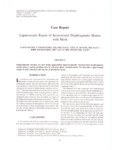
FIGURE 1
Left-sided diaphragmatic hernia with liver involvement at 38 weeks’ gestation
A, Coronal image demonstrating fetal stomach (arrow), right lung (L), thymus, (T), and approximate level of representative axial sections (yellow line). Axial sections show outlines of following regions of interest: B, total thorax, C, right lung, and D, herniated liver. Worley. Fetal magnetic resonance imaging in isolated diaphragmatic hernia: volume of herniated liver and neonatal outcome. Am J Obstet Gynecol 2009.
based on failure of HFOV, defined as an inability to maintain an arterial pH ⬎ 7.2 or an oxygen saturation ⬎ 90%. Surgical correction of the diaphragmatic hernia was performed once the infant was stabilized. The decision to use a patch or close the defect primarily was based on the location and size of the defect and was made by the pediatric surgery attending at time of operation.
Statistical analyses Statistical analyses included Fisher’s exact tests for comparisons of categorical data, Wilcoxon rank sum test for continuous measures, and receiver operating characteristic (ROC) curves and area under the curve (AUC). Fisher’s exact test and the Wilcoxon rank sum test were chosen because of the size of the statistical sample, as cell sizes of the contingency table might be small and the statistical normality of the continuous measures could not be assured with sufficient power. Statistical analyses were performed using software (SAS, Version 9.1; SAS Institute, Cary, NC) and significance levels (P values) ⬍ .05 were considered statistically significant.
R ESULTS A total of 18 fetuses with CDH underwent MRI evaluation. Three fetuses were ultimately found to have additional ma318.e3
jor anomalies and were excluded from further analysis. Of the remaining 15 fetuses, the median gestational age at the time of the MRI study was 29 weeks (range, 22-38). Eleven fetuses had a leftsided diaphragmatic hernia, 4 were right sided, and all but 1 of the 15 (93%) had evidence of liver herniation by MRI. In each study, the volumes of total thorax, residual lung (including the ipsilateral lung, when present), liver, and all remaining herniated abdominal contents could easily be generated (Figure 1). Selected neonatal outcomes are presented in Table 1. There were no stillbirths in the group and, as shown in Table 1, the majority of the infants were delivered in good condition. Although 1 infant was delivered at 31 weeks, the remaining deliveries occurred at or beyond 35 weeks’ gestation. There were 8 survivors, yielding an overall survival of 53%. Two infants had evidence of growth restriction and both survived. Eighty percent (n ⫽ 12) of the infants required HFOV during the initial stabilization, and 40% (n ⫽ 6) subsequently required ECMO. Of the neonates who were placed on ECMO, there was only 1 survivor. Four infants underwent primary surgical closure of the defect, and these infants all survived. Another 4 infants were too critically ill for surgery and died prior to
American Journal of Obstetrics & Gynecology MARCH 2009
an attempt at correction. Six of the remaining 7 infants required patch closure as they had insufficient remaining diaphragmatic tissue around the defect, and TABLE 1
Selected neonatal outcomes Characteristic Gestational age at delivery (wks)a
Result 37.6 ⫾ 2.5
........................................................................................................... a
Birthweight (g)
2807 ⫾ 674
...........................................................................................................
Birthweight ⬍ third percentile
2 (13)
...........................................................................................................
Mode of delivery
..................................................................................................
Vaginal
3 (20)
..................................................................................................
Cesarean
12 (80)
.........................................................................................
Repeat
6 (40)
Dystocia
3 (20)
Fetal distress
2 (13)
Fetal anomalies
1 (7)
5-min Apgar score ⬍ 4
0 (0)
5-min Apgar score ⬍ 7
2 (13)
pH ⬍ 7.0
1 (7)
......................................................................................... ......................................................................................... ......................................................................................... ........................................................................................................... ........................................................................................................... ........................................................................................................... ...........................................................................................................
Intubation in delivery room
10 (67)
........................................................................................................... a
Data presented as mean ⫾ SD and N (%).
...........................................................................................................
Worley. Fetal magnetic resonance imaging in isolated diaphragmatic hernia: volume of herniated liver and neonatal outcome. Am J Obstet Gynecol 2009.
Imaging
www.AJOG.org
FIGURE 2
Association of volumetric measurements with neonatal death Specificity (True positive)
1.0 Liver/Thorax AUC: 0.91+0.08
0.8
(Liver+Other)/Throax AUC: 0.71+0.15
0.6 0.4
Lung/Thorax AUC: 0.64+0.15
0.2 0.0
0.0
0.2
0.4
0.6
0.8
1.0
1 - Specificity (False positive)
Receiver operating characteristic curves of sensitivity and false-positive rates for volume ratios of liver/thorax, lung/thorax, and all herniated organs (liver, stomach, and bowel)/thorax in predicting neonatal death. Only liver/thorax volume was significant predictor, P ⫽ .01. AUC, area under curve. Worley. Fetal magnetic resonance imaging in isolated diaphragmatic hernia: volume of herniated liver and neonatal outcome. Am J Obstet Gynecol 2009.
half of these survived to hospital discharge. Information regarding the type of surgical correction used was not available for 1 infant. The relative volumes of the fetal thorax were then compared with neonatal outcomes. The volume of liver within the fetal chest was significantly associated with an increased risk of neonatal death, AUC 0.91 ⫾ 0.08 (Figure 2). Based on visual inspection of the ROC curve, the most useful marker of neonatal outcome was identified to be 20% of the fetal thorax occupied by liver. Specif-
ically, if ⱖ 20% of the thorax was occupied by liver, the neonatal mortality was 86% (95% confidence interval [CI], 42100%) compared with 13% (95% CI, 0-53%) when ⬍ 20% of the thorax was occupied by liver, P ⫽ .01. Selected infant outcomes by degree of liver herniation are presented in Table 2. Neither fetal lung volumes nor the volumes of all other herniated abdominal organs were associated with neonatal survival. The need for HFOV, ECMO, or more extensive surgical correction with a patch were not significantly associated with residual lung volumes or the proportion of the fetal chest occupied by liver or other abdominal contents. The mean percentages of the fetal thorax occupied by these organs comparing lethal and nonlethal groups are shown in Table 3. Using the Wilcoxon rank sum test, a significant increase was noted in the percentage of the fetal thorax occupied by liver in the lethal group compared with the nonlethal group. There were no statistically significant differences in the mean volumes of lung, other herniated abdominal organs (excluding liver), and all herniated abdominal organs (including liver). In both groups, stomach and bowel occupied the largest percentage of the thorax and, taken in total, the abdominal organs occupied more than half of the volume of the fetal thorax. Evaluating the 4 fetuses with rightsided diaphragmatic hernias revealed a trend that was similar to the entire cohort. Specifically, there were 2 survivors
TABLE 2
Selected infant outcomes by degree of liver herniation Liver/thorax < 20% N ⴝ 8 (%)
Liver/thorax < 20% N ⴝ 7 (%)
P value
High-frequency ventilation
5 (63)
7 (100)
.20
ECMO
2 (25)
4 (57)
.31
Patch repair of CDH
3 (38)
3 (43)
1.00
Neonatal death
1 (13)
6 (86)
.01
.............................................................................................................................................................................................................................................. .............................................................................................................................................................................................................................................. a .............................................................................................................................................................................................................................................. ..............................................................................................................................................................................................................................................
CDH, congenital diaphragmatic hernia; ECMO, extracorporeal membrane oxygenation. Data presented as N (%). a
Patch data available for 14 infants.
..............................................................................................................................................................................................................................................
Worley. Fetal magnetic resonance imaging in isolated diaphragmatic hernia: volume of herniated liver and neonatal outcome. Am J Obstet Gynecol 2009.
Research
in this group, and the percentages of thorax occupied by liver in these 2 were 8% and 11%, respectively. By contrast, the percentage of liver occupied by the thorax in the nonsurviving offspring with right-sided CDH was 25% and 35%, respectively.
C OMMENT In this study, we demonstrated that volumetric measurements of the fetal thorax can easily be made using fast spinecho acquisition sequence MRI. It has previously been shown that estimating fetal lung volumes with MRI can be done quickly in multiple planes with good intraobserver and interobserver agreement, both in the setting of normal fetal lungs and in the setting of CDH.6,21,22 It has also been demonstrated that reliable measurements of fetal lung volume are not dependant on variations in slice thickness.22 Using the same technique employed to generate lung volumes with MRI, we were also able to calculate the volumes of total thorax, liver, and other herniated gastrointestinal organs. We evaluated images in the axial plane and were easily able to delineate the ROI based on differences in signal intensity and characteristic appearance and location of the tissues within the fetal thorax. Although these distinctions were most clear when the study was performed in the third trimester, clear delineation could also be made in fetuses with less mature organ systems in the mid-late second trimester. Another important finding was that the degree of liver herniation was significantly and positively associated with neonatal death. Although fetuses with liver “up” into the diaphragmatic defect are generally regarded as having a worse overall prognosis, few studies have used MRI to quantify the extent of liver herniation.7,16 Walsh et al7 calculated a liver/diaphragm ratio and detected a significant difference between survivors and nonsurvivors, but they were unable to identify a cut-off value for prediction of outcome. Kitano et al16 also examined the liver/diaphragm ratio in 9 fetuses with left-sided diaphragmatic hernias and, although there were no statistical
MARCH 2009 American Journal of Obstetrics & Gynecology
318.e4
Research
Imaging
www.AJOG.org
TABLE 3
Percentage of fetal thorax occupied by lung and herniated organs in lethal and nonlethal cases Organ
Nonlethal Nⴝ8
Lethal Nⴝ7
Lung
15.1 (4.1)
11.6 (3.8)
.15
Liver
12.3 (8.3)
28.8 (8.8)
⬍ .01
Other abdominal organs
40.5 (7.0)
30.3 (11.2)
.05
All herniated organs
52.8 (9.4)
59.1 (8.7)
.16
P value
.............................................................................................................................................................................................................................................. .............................................................................................................................................................................................................................................. a .............................................................................................................................................................................................................................................. b
..............................................................................................................................................................................................................................................
Data presented as percent mean (⫾ SD). a
Small and large bowel and stomach. b Liver and other abdominal organs.
..............................................................................................................................................................................................................................................
Worley. Fetal magnetic resonance imaging in isolated diaphragmatic hernia: volume of herniated liver and neonatal outcome. Am J Obstet Gynecol 2009.
differences between survivors and nonsurvivors, none of the neonates with ratios ⬍ 0.2 survived. Our findings support those of these 2 studies; specifically, more extensive liver herniation appears to confer a worse prognosis and decreased likelihood of survival. With our data, however, we were able to suggest a cut-off value, and when ⱖ 20% of the fetal thorax was occupied by liver, neonatal deaths were significantly more likely. Our study was limited by its small sample size and retrospective design. We were not able to demonstrate an association between the volumetric measurements and neonatal interventions such as HFOV, ECMO, or the need for a patch repair of the diaphragmatic defect. Although some recommend routinely using high-frequency ventilation as a part of a lung-protective strategy in the liver “up” subset of CDH,16 the use of ECMO is generally reserved for infants with more severe pulmonary hypoplasia or pulmonary hypertension who have failed both conventional ventilation and HFOV. In our study, only 1 infant survived after being placed on ECMO, suggesting that such therapy is a marker of disease severity. Predicting which offspring will require ECMO is, therefore, of great importance. In our analysis, twice as many neonates were placed on ECMO when at least 20% of the fetal thorax was occupied by liver, but this difference was not significant due to the small absolute numbers. A larger similar study may be able to detect such a differ318.e5
ence, and these volumetric measurements may predict outcomes that we did not identify in our relatively small cohort. Hedrick et al,14 for example, analyzed the pregnancy courses and outcomes of 89 patients with isolated leftsided CDH and determined a significant and dramatic increase in the need for ECMO in fetuses with liver “up” compared with those with liver “down” (80% vs 25%, respectively). They found liver position to be the best predictor of both need for ECMO and survival, emphasizing the need for refinement in the classification of liver “up” such that quantification of the degree of liver herniation may improve the ability to predict outcome. We present in this report a novel technique of using MRI to quantify the contents of the fetal thorax in the setting of isolated CDH. Liver position continues to be recognized as one of the most important prognostic variables in fetuses with diaphragmatic hernias, but determination of the position of the liver, especially when only a portion of the liver is displaced into the fetal thorax, is difficult with ultrasound and largely depends on the level of experience of the examiner.16 As such, MRI examinations are being increasingly requested in the setting of CDH to confirm the diagnosis and accurately ascertain liver position. In our study population, all but 1 fetus had evidence of liver herniation, suggesting that this was a particularly severe cohort of diaphragmatic hernia cases. It
American Journal of Obstetrics & Gynecology MARCH 2009
is in these patients, however, that prognostic refinements are badly needed, as perinatal survival in the subset of fetuses with liver “down” has been reported to be ⬎ 90% in centers with experience caring for such infants.14 Although prospective studies are needed, our results suggest that quantifying the degree of liver involvement may provide us with a prognostic tool that would better equip us to counsel patients in the setting of severe isolated CDH. f REFERENCES 1. Gorincour G, Bouvenot J, Mourot MG, et al. Prenatal prognosis of congenital diaphragmatic hernia using magnetic resonance imaging measurement of fetal lung volume. Ultrasound Obstet Gynecol 2005;26:738-44. 2. Colvin J, Bower C, Dickinson JE, Sokol J. Outcomes of congenital diaphragmatic hernia: a population-based study in Western Australia. Pediatrics 2005;116:e356-63. 3. Ruano R, Aubry MC, Barthe B, Dumez Y, Zugaib M, Benachi A. Three-dimensional sonographic measurement of contralateral lung volume in fetuses with isolated congenital diaphragmatic hernia. J Clin Ultrasound 2008;36: 273-8. 4. Deprest J, Jani J, Cannie M, et al. Prenatal intervention for congenital diaphragmatic hernia. Curr Opin Obstet Gynecol 2006;18: 255-367. 5. Ruano R, Benachi A, Joubin L, et al. Threedimensional ultrasonographic assessment of fetal lung volume as prognostic factor in isolated congenital diaphragmatic hernia. BJOG 2004;111:423-9. 6. Paek BW, Coakley FV, Lu Y, et al. Congenital diaphragmatic hernia: prenatal evaluation with MR lung volumetry–preliminary experience. Radiology 2001;220:63-7. 7. Walsh DS, Hubbard AM, Olutoye OO, et al. Assessment of fetal lung volumes and liver herniation with magnetic resonance imaging in congenital diaphragmatic hernia. Am J Obstet Gynecol 2000;183:1067-9. 8. Gaillot T, Ferry M, Beuchee A, Pladys P, Betremieux P. Magnetic resonance imaging measurement of fetal lung volume does not match postnatal survival. Arch Dis Child Fetal Neonatal Ed 2007;92:F78. 9. Metkus AP, Filly RA, Stringer MD, Harrison MR, Adzick NS. Sonographic predictors of survival in fetal diaphragmatic hernia. J Pediatr Surg 1996;31:148-51. 10. Harrison MR, Keller RL, Hawgood SB, et al. A randomized trial of fetal endoscopic tracheal occlusion for severe fetal congenital diaphragmatic hernia. N Engl J Med 2003; 349:1916-24. 11. Laudy JA, Van Gucht M, Van Dooren MF, Wladimiroff JW, Tibboel D. Congenital diaphrag-
Imaging
www.AJOG.org matic hernia: an evaluation of the prognostic value of the lung-to-head ratio and other prenatal parameters. Prenat Diagn 2003;23:634-9. 12. Jani J, Keller RL, Benachi A, et al. Prenatal prediction of survival in isolated left-sided diaphragmatic hernia. Ultrasound Obstet Gynecol 2006;27:18-22. 13. Lipshutz GS, Albanese CT, Feldstein VA, et al. Prospective analysis of lung-to-head ratio predicts survival for patients with prenatally diagnosed congenital diaphragmatic hernia. J Pediatr Surg 1997;32:1634-6. 14. Hedrick HL, Danzer E, Merchant A, et al. Liver position and lung-to-head ratio for prediction of extracorporeal membrane oxygenation and survival in isolated left congenital diaphragmatic hernia. Am J Obstet Gynecol 2007; 197:422.e1-4.
15. Arkovitz MS, Russo M, Devine P, Budhorick N, Stolar CJ. Fetal lung-head ratio is not related to outcome for antenatal diagnosed congenital diaphragmatic hernia. J Pediatr Surg 2007;42: 107-10. 16. Kitano Y, Nakagawa S, Kuroda T, et al. Liver position in fetal congenital diaphragmatic hernia retains a prognostic value in the era of lungprotective strategy. J Pediatr Surg 2005;40: 1827-32. 17. Levine D. Obstetric MRI. J Magn Reson Imaging 2006;24:1-15. 18. Hubbard AM, Crombleholme TM, Adzick NS, et al. Prenatal MRI evaluation of congenital diaphragmatic hernia. Am J Perinatol 1999;16: 407-13. 19. Reddy UM, Filly RA, Copel JA; Pregnancy and Perinatology Branch, Eunice Kennedy
Research
Shriver National Institute of Child Health and Human Development, Department of Health and Human Services, National Institutes of Health. Prenatal imaging: ultrasonography and magnetic resonance imaging. Obstet Gynecol 2008;112:145-57. 20. McIntire DD, Bloom SL, Casey BM, Leveno KJ. Birth weight in relation to morbidity and mortality in newborn infants. N Engl J Med 1999;340:1234-8. 21. Rypens F, Metens T, Rocourt N, et al. Fetal lung volume: estimation at MR imaging–initial results. Radiology 2001;219:236-41. 22. Ward VL, Nishino M, Hatabu H, et al. Fetal lung volume measurements: determination with MR imaging– effect of various factors. Radiology 2006;240:187-93.
MARCH 2009 American Journal of Obstetrics & Gynecology
318.e6